Lacrimal apparatus. The eyes are protected and lubricated by tear fluid, or simply tears. The organs of the lacrimal apparatus produce this fluid and drain its excess into the nasal cavity.
The lacrimal apparatus is a system of organs that secrete tear fluid and drain its excess from the eyeball. The lacrimal gland produces fluid, the outflow of which occurs through the lacrimal puncta.
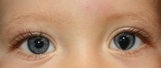
The eyes remain moist due to the continuous production of small amounts of tear fluid by the tear glands. The liquid contains a very strong antibacterial substance lyso-zyme. The main part of the liquid (approximately 1 ml daily for each eye) evaporates. The remaining tear fluid is drained through the nasolacrimal canal into the nasal cavity.
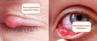
Inflammatory diseases of the lacrimal gland
In ophthalmology, several ailments associated with disturbances in the functioning of the lacrimal gland have been recorded.
- Dacryocystitis is an inflammatory process that occurs at the top of the nasolacrimal duct due to its obstruction
- Sjögren's syndrome is an autoimmune connective tissue disorder that occurs against the background of pronounced dysfunction of the secretory activity of the glands, and is a common cause of excessive dry eyes.
- Dacryoadenitis is an inflammation that affects the tissue of the lacrimal gland. It can be both acute and chronic. It does not often occur on its own; it is often a complication of various diseases.
- Canaliculitis – inflammation of the tear ducts caused by infection
- Mikulicz's disease - originates from a pathology of the immune system, causes swelling and an increase in the size of the lacrimal and salivary glands
Return to contents
Causes of inflammation
| Infection most often occurs through the entry of viral particles into the circulatory system. This route is called hematogenous. |
The cause of inflammation can be general ailments such as influenza, scarlet fever, throat diseases, and mononucleosis. Dirt or local suppuration that occurs near the tear duct is also a negative factor.
Symptoms of damage to the lacrimal gland
External signs of the disease are very clearly visible even with a normal superficial examination, and it is almost impossible not to notice them, much less ignore them. Especially when you consider that very often the disease is accompanied by unpleasant sensations.
- The pain appears in the inner corner of the eye and intensifies when pressed.
- Redness of the mucous membrane and swelling are noticeable.
- Decrease or increase in the amount of fluid secreted
- Dryness or, on the contrary, increased lacrimation
- Burning and itching
In addition to local visual manifestations, there are also general ones inherent in all infectious diseases. Namely:
- Increased body temperature
- Headache,
- Fever,
- Weakness,
- Nausea.
- Enlarged lymph nodes
- There are complaints from patients about difficulties arising when raising the eyelid
- Fuzzy picture, split image.
Possible deviations in the anatomy of the lacrimal gland
The effect of dry eyes is eliminated with the help of drops.
The anatomical structure of the lacrimal gland is a clear structure that works smoothly and continuously, so any, even minor deviations and disturbances greatly undermine the entire activity of the gland.
Pathologies of this gland can occur as a result of previous illness, various injuries to the eye and eyelids.
One of the possible deviations may be reduced secretory function of the lacrimal gland. Reduced secretion leads to insufficient production of the necessary tear fluid, which in turn causes drying of the surface of the eye, microcracks in its surface layer, and injuries to the cornea.
With such a deviation, eye diseases and decreased vision inevitably occur, accompanied by very unpleasant and painful sensations and redness. This phenomenon can occur as a result of various diseases, and not only eye diseases, but also due to injuries to the lacrimal gland and chemical exposure.
The second variant of deviation is the opposite: increased secretory function of the lacrimal gland. This deviation is often observed with various injuries to the nose and eyes. In addition to injuries, large amounts of tear fluid can cause diseases that result in blockage of the lacrimal canaliculi.
In addition to acquired abnormalities of the lacrimal gland, congenital anatomical abnormalities are sometimes observed. Congenital abnormalities of the lacrimal gland include:
- Absence of tear ducts;
- Anatomical deviations of any structural parts and units in the lacrimal gland;
- Congenital disorder of secretion.
The first variant of congenital abnormalities is a very rare occurrence, and, as a rule, this fact is discovered in the first days after the birth of the child. Anatomical congenital disorders of any part of the lacrimal gland are also not a very common occurrence, and the degree of damage may vary.
Secretion disorders in the first days after birth are detected quickly enough, which allows doctors to provide the necessary assistance and treatment to the child.
Functions of the lacrimal gland
- Moisturizing the surface of the cornea
- Elimination of xerophthalmia, that is, dry eye syndrome resulting from fatigue and visual strain
- Delivery of nutrients to the cornea. In particular, lysozyme is an enzyme that destroys bacterial cells, and beta-lysine is a whey protein that has antimicrobial activity.
- Protection from the harmful effects of microorganisms
- Cleansing the surface of the eyeball from external contaminants, dust, and foreign objects
- Tears released in stressful situations or under the influence of emotions control the sudden release of hormones and help stabilize the mental state.
Tear film
| The tear film is a multicomponent substance distributed over the surface of the eyeball. It helps the substances released during the functioning of the gland to fully realize their functions. |
This means protection from exposure to atmospheric oxygen and from excessive drying out. When blinking, part of the secretion settles on the front convex side of the eyeball, while the water component of the flow mechanically cleanses the cornea.
The tear film has a complex structure and consists of three layers that never mix:
- Internal . Adjacent directly to the cornea. Its main component is the mucous substance mucin. Allows fluid to be distributed freely and evenly throughout the epithelium by reducing the surface tension of the film.
- Average. Formed by auxiliary small lacrimal glands, it has a liquid base. Is the thickest of all. Cleanses the surface of the eye and serves as a conductor for nutrients.
- Outer. Otherwise called lipid or oil, it consists of fats. Prevents the evaporation of the water layer, and also maintains the smoothness of the film and promotes its better distribution.
Return to contents
Lacrimal apparatus of the eye
The lacrimal apparatus of the eye consists of the lacrimal gland ( glandula lacrimalis
), lacrimal canaliculi (
canaliculi lacrimalis
), lacrimal sac (
saccus lacrimalis
), nasolacrimal duct (
ductus nasolacrimalis
).
The lacrimal gland is located in the fossa of the same name in the frontal bone and produces a complex, slightly alkaline liquid (tear), which has pronounced bactericidal properties, as well as moisturizing the conjunctival sac and cornea. Tears produced by the lacrimal glands are a clear, slightly alkaline liquid. It contains 98% water, and the rest is protein, sugar, sodium, potassium, mucus, fat and the bacteriostatic enzyme lysozyme. In human tissues, lysozyme is localized in lysosomes. Lysozyme is secreted into biological fluids and intercellular substance by macrophages. This enzyme catalyzes the hydrolysis of complex amino acids in the cell wall of bacteria, which causes their dissolution (lysis) and subsequent death. The structure of the lacrimal gland is complex alveolar-tubular; its ducts, ductuli exeretorti
(about 12) open into the upper fornix of the conjunctiva.
When the eyelids are closed, the tear flows along the lacrimal stream ( rivus lacrimalis
) - the depressions on the back edges of the eyelids.
When the eyes are open, tears flow from the lateral corner of the eye to the medial corner due to blinking movements. The lacrimal lake ( lacus lactimales
) is located in the medial canthus. Tears from the lacrimal lake are absorbed through two lacrimal canaliculi (superior and inferior) and enter the lacrimal sac. It is located in the fossa of the same name on the medial wall of the orbit. The muscle fibers encircle the lacrimal sac in the form of a loop and, with blinking movements of the eyelids, either compress it or expand it, facilitating the removal of tears into the nasolacrimal duct. The nasolacrimal duct (Ferrein) is a continuation of the lacrimal sac downwards and is located in the bone canal of the same name, opening into the anterior section of the lower nasal meatus. When any part of this duct system is blocked or there is overproduction of tear fluid, tears flow down the face.
The organ of hearing and balance consists of 3 parts: the outer, middle and inner ear ( auris externa, media, interna
)
.
VESTIBULO-COCHLEARE ORGANUM VESTIBULO-COCHLEARE
(
ORGANUM STATUS ET AUDITUS
)
The outer ear consists of the auricle ( auricula
) and external auditory canal (
meatus acusticus externus
).
The boundary between the outer and middle ear is the eardrum ( membrana tympani
)
The auricle is made up of cartilage, which is covered on all sides by skin. The arched outer edge is called the helix
), the antihelix (
anthelix
) is located parallel to the helix. Anterior to the external auditory canal is the tragus (
tragus
), and on the lower border of the antihelix there is an antitragus (
antitragus
).
There is no cartilage in the lower part of the auricle; this section consists of adipose tissue and is called the lobulus
. Recently, the method of auriculodiagnosis and auriculotherapy has become widespread. The method is based on the principle of projecting organs onto the auricle.
The external auditory canal is S-shaped and consists of cartilaginous and bone parts. Its internal opening is closed by the eardrum ( membrana tympani
). The skin of the external auditory canal is characterized by the presence of hairs and special cerulinous glands that produce sulfur. The structure is a bone-fibrous formation.
The eardrum is a thin cone-shaped membrane in the center of which the navel ( umbo)
).
It is the boundary between the outer and middle ear. Its upper, unstretched part is called pars flaccida
.
The rest of the part is stretched - pars tensa
.
Rice. 25. Outer, middle and inner ear, right
(frontal cut through the external auditory canal).
The middle ear is represented by the tympanic cavity ( cavitas tympanica
ossiculi auditus
), mastoid cells (
cellulae mastoidea
) and auditory (Eustafian) tube (
tuba auditiva
located in it .
The tympanic cavity is the space of the temporal bone between the outer and inner ear, in which the auditory ossicles are located. The tympanic cavity is connected to the nasopharynx through the auditory tube. The shape of the tympanic cavity is an irregular cube with six walls, which got their name from the anatomical formations adjacent to them. Lateral wall – paries membranaceus
formed by the eardrum, which is a weakly translucent membrane, 1 mm thick.
It is usually divided into quadrants: anterior-superior, anterior-inferior, posterior-superior and posterior-inferior. The medial wall faces the labyrinth of the inner ear and is called the labyrinthine wall - paries labyrinthicus
.
In the center of this wall there is a bony protrusion - a promontory ( promontorium
), which is formed by the lateral wall of the cochlea dome.
There are grooves on the surface of the promontory, which, as they deepen, form bone canals. plexus tympanicus
pass through these canals .
The upper wall is formed by the structure of the pyramid of the temporal bone of the same name and is therefore called the tegmental wall - paries tegmentalis
.
It is represented by a thin plate in which there are cracks (digestions), thanks to which the structures of the dura mater come into contact with the mucous membrane of the tympanic cavity. The inferior wall projects onto the jugular fossa and is therefore called paries jugularis
.
The lower edge of the tympanic membrane is located above the bottom of the tympanic cavity, forming a depression - recessus hypotympanicus
, in which fluid can accumulate during inflammatory diseases.
The tympanic nerve, inferior tympanic artery and vein pass through the floor of the tympanic cavity. The anterior wall – paries caroticus
– separates the tympanic cavity from the internal carotid artery and corresponds to the canal of the same name in the temporal bone.
The upper part of the anterior wall is occupied by the mouth of the auditory tube, with a diameter of 5 mm; Below is the canal of the tensor tympani muscle. The anterior wall contains tubules containing nerve fibers and vessels originating from the plexus caroticus internus
.
At the back, the tympanic cavity communicates with the cells of the mastoid process and therefore the posterior wall is called mastoid - paries mastoideus
.
It contains the bony pyramidal eminence eminentia pyramidalis
, inside which the stapedius muscle
m is located.
stapedius .
Outside this elevation there is an opening for the drum string ( chorda
tympani). With prolonged sluggish otitis, infection may spread into the air cells of the mastoid process, which leads to the development of mastoiditis.
Inside the tympanic cavity there are auditory ossicles: the malleus
), anvil (
incus
) and stirrup (
stapes
), connected to each other by movable miniature joints.
The joint between the incus and the malleus is called the incus-malleola joint ( articulatio incudo-malleolaris
), which has a thin capsule.
The articulation of the incus with the stirrup is distinguished by a large range of movements - the incus-stapedia joint ( articulatio incudo-stapedia
), which is supported by two ligaments - the posterior and superior.
Their function is the one-way transmission of air vibrations from the surface of the eardrum to the base of the stapes, which in turn closes the window of the vestibule ( fenestra vestibuli
).
The base of the stapes is covered with cartilage, which is connected to the cartilaginous edge of the oval window through the annular ligament. The annular ligament, firstly, closes the gap and, secondly, ensures mobility of the stapes. The mechanical transmission of sound vibrations is carried out thanks to two muscles. The first is the tensor tympani muscle m.
tensor tympani .
This muscle retracts the handle of the malleus, straining the eardrum. This muscle is innervated by a branch of the same name from the third branch of the trigeminal nerve. The second muscle is the stapedius m.
stapedius is attached to the posterior leg of the stapes at the head.
This muscle is a functional antagonist of the previous one, innervated by n.
facialis , which gives off a small branch –
n.
stapedius .
The auditory or Eustachian tube connects the tympanic cavity with the nasopharynx and thus balances the pressure in the tympanic cavity with atmospheric pressure. It consists of bone ( pars ossea
) and cartilaginous (
pars cartilaginea
) parts.
Its length is 3.5-4 cm. The tubal tonsils ( tonsila tubaria
) are located at the pharyngeal openings of the auditory tube, and the mucous surfaces of the tube are in contact and the tube opens only when swallowing, which is recommended to do during air travel.
The inner ear consists of a bone ( labyrinthus osseus
) and membranous labyrinth (
labyrinthus membranaceus
). Moreover, the membranous labyrinth is located inside the bone labyrinth, repeating its shape. Endolymph circulates inside the membranous labyrinth, and perilymph circulates between the membranous and bony labyrinths.
The bony labyrinth is located inside the pyramid of the temporal bone and consists of 3 parts: the bony vestibule - vestibulum osseum
;
bony semicircular canals – canales semicirculares ossei
;
bone snail – cochlea osseum
.
The central part of the labyrinth is the vestibule. It is divided from the inside by the bony crest of the vestibule into 2 pockets: spherical ( recessus sphericus
) and ellipsoidal (
recesus ellipticus
), into which 5 openings of the semicircular canals open.
On the outer wall of the vestibule there are 2 windows: the window of the vestibule ( fenestra vestibuli
), it faces the tympanic cavity and is closed by the base of the stapes, and the window of the cochlea (
fenestra cochleae
).
It is covered by a secondary tympanic membrane ( membrana tympani secundaria
), which dampens vibrations of the perilymph of the scala tympani.
The three semicircular canals are located in three mutually perpendicular planes: anterior, posterior and lateral ( canales semicirculares anterior, posterior et lateralis
).
Each channel has an arc and 2 legs. One leg of each semicircular canal is dilated and called the ampulla ( crura ossea ampullaria
).
The anterior and posterior canals form a common stalk ( crus osseum commune
), and the lateral canal forms a simple stalk (
crus osseum simplex
). Thus, the semicircular canals open into the vestibule with five openings.
The bony labyrinth of the cochlea is a bony tube wrapped in 2.5 turns around its axis or rod ( modiolus
).
The cavity of the rod is a canal - canalis modiolus
.
Inside the spiral canal of the cochlea there is a bony spiral plate ( lamina spiralis ossea
), which, together with the basement membrane, divides its cavity into two parts: the vestibule staircase (
scala vestibuli
) - located above the bony plate and the scala tympani (
scala tympani
), which is the lower staircase.
The membranous labyrinth is located inside the bone labyrinth, basically repeating its shape, but its walls consist of connective tissue. It has 3 parts: the membranous vestibule ( vestibulum membranacei
);
membranous semicircular ducts ( ductuli semicircularis membranacei
);
webbed snail ( cochlea membranacei
) or cochlear duct (
ductus cochlearis
).
The membranous labyrinth of the vestibule includes the utricle ( utriculus
) and sac (
sacculus
).
The utricle is located in an elliptical pocket, and the saccule is located in a spherical one. They are connected to each other by the utero-sac duct ( ductus utriculosaccularis
). 5 openings of the membranous labyrinth of the semicircular canals open into the posterior wall of the uterus.
On the inner surface of the utricle and sac there are spots - macula utriculi
and
macula sacculi
. They are receptors of the vestibular nerve and consist of hair cells of the sensitive vestibular epithelium surrounded by supporting cells. It is believed that the receptors of the utricle and sac perceive gravity and linear acceleration, i.e. provide balance to the body at rest.
In the membranous labyrinth of the semicircular ducts (anterior, posterior and lateral), a special place is occupied by the receptors of the ampullary legs, represented by sensitive crests with neuroepithelial cells that perceive angular acceleration and are organs of dynamic balance, i.e. provide balance to a body moving in space.
The membranous labyrinth of the cochlea includes the cochlear duct, which lies in the scala vestibuli, has a triangular shape and is limited by 3 walls. The upper wall is the vestibular (Reisner's) membrane. The inferior wall is the basement membrane on which the organ of Corti is located. The lateral wall is represented by the periosteum of the bony canal of the cochlea and is lined with a special epithelium of the stria vascularis, the capillaries of which produce endolymph.
The organ of Corti is located on the basement membrane and contains sensory hair cells surrounded by a network of supporting cells. These cells are covered by the nerve fibers of the spiral ganglion ( ganglion spirale
), located at the base of the cochlear shaft, forming the first neuron of the auditory pathway (for the auditory pathway and the balance path, see the description of the VIII pair of cranial nerves).
Orbit and auxiliary apparatus of the eye
penetrate into the orbit through the superior orbital fissure within the ring of Zinn (Fig. 2.1.9).
The superior branch of the oculomotor nerve is located in the muscular infundibulum medial to the optic nerve (Fig. 2.7.2, 2.7.3). This location is noted only up to the point where the nerve enters the superior rectus muscle of the eye (at a distance of 15 mm
from the apex of the orbit). It then gives the two terminal branches to the levator superioris.
The inferior branch of the oculomotor nerve divides into three branches. One branch goes down and forward and penetrates the inferior rectus muscle. The second goes under the optic nerve and penetrates the internal rectus muscle of the eye.
The longest third branch, spreading along the orbit, penetrates the inferior oblique muscle. This branch gives off vertical parasympathetic fibers to the ciliary ganglion located slightly higher. The exact location of parasympathetic fibers in the nerve is unknown, but it is assumed that they lie superficially on the superior-inner side of the nerve [129].
Abducens nerve (p.
abducens
(VI). The sixth cranial nerve penetrates the orbit through the superior orbital fissure, located inside the ring of Zinn. Moreover, it lies between the optic nerve and the external rectus muscle (Fig. 2.7.3). The abducens nerve is included in the internal surface external rectus muscle at the border of the transition of the posterior third of the muscle to the anterior two-thirds.
Trochlear nerve (n.
trochlearis
(IV). The fourth cranial nerve penetrates the orbit through the superior orbital fissure and is located slightly outside the ring of Zinn and inside the frontal nerve (Fig. 2.7.3). It is directed forward and medially under the roof of the orbit and penetrates the superior oblique muscle. The course of the nerve can be observed only by excision of the periosteum of the upper wall of the orbit.
Surgical approaches to various parts of the orbit
Having presented the basic information regarding the structure of the bone formations of the orbit, its soft tissues, and the structure of the eyelids, it seems necessary to remind the reader about the main surgical approaches to the contents of the orbit.
It was shown above that in various parts of the orbit (subperiosteal surgical space, orbital space outside the muscular infundibulum, space located inside the muscular infundibulum, episcleral surgical space located between Tenon’s capsule and the eyeball) one or another type of pathological process predominates. Most often these are tumors
of various origins and degrees of malignancy. Depending on the location of the tumor, various surgical approaches to one or another part of the orbit have been developed (Fig. 2.7.5). In this case, the surgeon has to dissect and manipulate various soft formations of the orbit during surgery, which predetermines the need to know the topography of the tissues of the orbit. We list the main approaches to orbital structures.
Rice. 2.7.5. The main surgical approaches to the orbit (according to
Wright, Steward, 1978): 1
- lateral orbitotomy according to Kronline; 2
— transconjunctival orbitotomy (the incision can be made in the limbus or behind the eye);
3
— extraperiosteal orbitotomy;
4
— transseptal orbitotomy; 5—medial orbitotomy
- The medial approach
(medial orbitotomy) is used in cases where the pathological process is localized on the nasal side of the orbit. This approach is best for visualizing the optic foramen. It can expose the inner wall of the orbit, which makes it possible to diagnose the pathology of the ethmoid sinus (ethmoiditis, mucocele, tumors, etc.) and perform the necessary manipulations. - An incision in the eyebrow area.
The scar after the incision in the eyebrow area is not visible. Most often it is produced during the development of tumors in the area of the lacrimal gland or arteriovenous aneurysms. - Lateral transconjunctival approach.
After dissection of the external rectus muscle and removal of the eyeball, this approach allows free access to the muscular infundibulum. For this reason, it is most preferable when performing manipulations in the posterior part of the orbit. - Medial transconjunctival approach.
This approach involves resection of the internal rectus tendon and removal of the eyeball. In this way, the approach to the muscle funnel is achieved. The disadvantage of the lateral and medial transconjunctival approaches is the small space created as a result of the operation.
Anatomical structure
The lacrimal apparatus of the eyeball is a complex system of organs through which tears and tear fluid are secreted and drained. This section contains a gland that produces fluid. The glandular tissue constantly works, due to which the surface of the eyes is constantly lubricated. A special feature of the lubricating composition is the presence of a unique antibacterial substance, lysozyme. Every day, approximately 1 ml of the composition evaporates from each eye, thus leaving the bulk of the lubricant. The remaining fluid is drained through the nasolacrimal duct into the nasal cavity.