Content
- The need for an MRI
- At what age is the examination performed?
- Progress of the procedure
- Features of the procedure Closed (capsule) tomograph
- Open tomograph
- Anesthesia and drug-induced sleep
- MRI with contrast
- Brain
How to prepare for the procedure?
Another question that worries parents is: “How to prepare a child, especially a preschooler, for an MRI of the brain and other parts of the body?” First of all, you should stop eating before the procedure. For infants - at least a couple of hours; for older babies, the period increases to 4-5 hours. Why is it important? The fact is that anxiety - and it is inevitable - in some cases provokes vomiting. If an MRI is planned under anesthesia, the child should not eat since the evening of the previous day. Parents should also pay attention to the clothes they dress their child in. The presence of metal parts (rivets or zippers, etc.) is not allowed. If a child wears earrings or a cross, they also need to be removed.
The need for an MRI
MRI at an early age is prescribed to identify various pathologies of the brain, spinal cord, soft tissues, internal organs and bone structures. Using the method, you can obtain clear images in a three-dimensional projection, which is not always possible with ultrasound, x-rays and CT. The technique is especially valuable in cases where it is necessary to diagnose diseases of the blood vessels of the brain, eyes and heart.
MR tomography visualizes soft tissue better than other diagnostic procedures, so it is indispensable in identifying damage to tendons, ligaments, and muscles. The procedure is prescribed for children with suspected oncological or inflammatory processes.
At what age is the examination performed?
Most clinics agree to perform the procedure only on patients over 5 years of age. For newborns and young children, the study is performed only in cases of extreme necessity (when there is a threat to life). Scanning for infants is also prescribed when alternative examination methods (ultrasound, X-ray and CT) do not provide complete information about the disease.
Age restrictions are imposed due to the patient's need to stay in the tomograph capsule for a long time. If you neglect this rule, you will get low-quality images from which the doctor will not be able to make a correct diagnosis. At the same time, MR radiation is absolutely safe for children and women during pregnancy.
In what other cases is MRI performed on newborns? The list of indications includes:
- birth injuries;
- suspicion of dysfunction of the nervous system;
- pathologies of an infectious nature;
- poor sleep and appetite in the child.
Progress of the procedure
If a child needs an MRI, parents should follow some recommendations:
- Get a referral for the procedure from a doctor (this is especially true for young children). For children, MRI is performed only under anesthesia or medicated sleep. Scanning is not recommended without serious reasons. Only a specialist, after visually examining the child and obtaining laboratory tests, will be able to understand whether the child needs the procedure or not.
- Choose a suitable clinic. The service is provided free of charge, but to receive it you often need to wait in line. If there is a suspicion of a rapidly progressing disease, then parents are advised to contact a paid clinic that has a pediatric anesthesiologist and resuscitator.
- Pay attention to the equipment used in the clinic. Many children find it more comfortable to be in an open type tomograph, as it makes less noise. One of the parents or medical personnel may be near the child at this moment.
It should be noted that a child cannot always be examined using open tomographs. They have little power compared to capsule-type devices and produce lower-quality images. Low-power tomographs will not help doctors diagnose a child with a malignant tumor or cerebrovascular disease in the early stages of development. When choosing a tomograph for examining a child, parents should consult a doctor.
Below we will discuss information about the stages of a conventional MRI, with contrast and anesthesia. The nuances of preparing children of different ages and diseases detected through scanning will also be outlined. At the end of the article, the most prescribed studies for a child are indicated.
Indications for MRI in children
- Neoplasms in the brain, abdominal cavity, kidneys.
- The need to confirm the preliminary diagnosis.
- Developmental anomalies of the musculoskeletal system and nervous system.
- It is impossible to establish pathology in any other way due to the patient’s age.
Nervous system
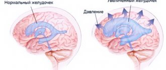
For diseases of the brain (vascular aneurysms, cysts, hemorrhages, hydrocephalus) and nervous system, the child is often prescribed an MRI with contrast or a conventional procedure. The first method is somewhat more expensive, but allows you to get a more accurate and detailed result. The contrast agent is an indicator of pathological conditions of brain tissue.
MRI for epilepsy in children
The study helps to identify the disease at an early stage and understand what factors provoked it. Parents should pay attention that MRI for epilepsy in children is most often performed under anesthesia, so as not to provoke a seizure.
Osteoarticular system
Thanks to magnetic resonance imaging, specialists have the opportunity to monitor the condition of joints with developmental anomalies, as well as monitor recovery processes after injuries.
Soft tissues and internal organs
The MRI method allows timely detection of various diseases that affect the organs of the digestive or respiratory system. It is more informative compared to ultrasound and radiography, and parents choose this method also because of its safety for health, because there is no radiation exposure with MRI.
Features of the procedure
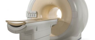
4.1. Closed (capsule) tomograph
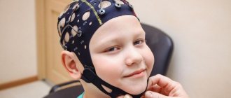
An MRI tomograph is a closed installation with a movable table surrounded by magnets and emitters. The child is placed on the table with his back down and his limbs are secured with clamps. This is necessary to avoid accidental movements during diagnosis. Headphones are placed on the little patient's head to minimize the unpleasant sounds generated by the device. The specialist takes a series of layer-by-layer images, which are then printed onto tape or stored on information media.
The duration of the procedure, depending on the area being examined, ranges from 15-40 minutes. Modern capsule devices are equipped with monitors on which cartoons are broadcast during the study. This will make it easier for the child to maintain a still position.
4.2. Open tomograph
If a child is allowed to undergo examination on open-type tomographs, then parents need to familiarize themselves with the basic specifics of the procedure. Unlike capsule-type devices, in open-loop devices the magnetic field is generated only from 2 sides, which reduces the information content and clarity of the images.
The advantages of such a study for children include:
- the ability to be accompanied by a doctor or close relative during the scan;
- providing comfort to patients who suffer from spinal injuries;
- the ability to take medications during the procedure;
- low noise level.
4.3. Anesthesia and drug-induced sleep
Depending on the age of the child and his ability to remain in one position for a long time, the issue of the need for anesthesia is decided. Doctors can offer parents several options for putting a child into medicated sleep: superficial (using special masks) and deep (using the injection of medications into a vein). Some diseases require a combination of superficial and deep anesthesia.
The question of how to introduce a child into medicated sleep is decided by the parents together with the resuscitator who will be responsible for this procedure. Indications for the use of general anesthesia include:
- children's age up to 5-6 years;
- claustrophobia;
- hyperactivity;
- examination of brain structures (this condition is mandatory for both children and adults);
- psychological disorders;
- patient's fear of the procedure.
Diagnosis can be carried out without medicated sleep in children who are old enough and are able to maintain a motionless position for a long time. Anesthesia is not required for examination of internal organs and bone tissue.
4.4. MRI with contrast
To diagnose certain pathologies, the child is given a contrast agent intravenously. The procedure is painless for the little patient. The contrast allows doctors to better see blood vessels and small tumors. The drugs used during the scanning process rarely cause allergic reactions. Before administering it, the laboratory technician will ask the parents if the child has had negative reactions to food and medications.
A contrast-enhanced procedure requires more preparation than a regular MRI. The child's parents must adhere to the following rules:
- Exclude gas-forming foods (cabbage, carbonated drinks, fresh baked goods, confectionery) from the subject’s diet. The rule must be followed before examining the abdominal organs. Gases in the intestines distort the information content of images and prevent doctors from examining pathological processes.
- Do not feed the child for 6-8 hours before the scan and do not give water for 2 hours. This rule must be observed so that after an MRI with contrast, a small patient does not experience any adverse reactions from the gastrointestinal tract - nausea, vomiting, defecation disorders.
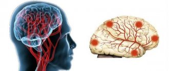
Why do an MRI of a child's brain?
The method helps to identify serious and dangerous changes occurring in the skull. They are difficult to diagnose using standard methods (ultrasound, x-ray), so medical specialists give preference to tomography in this matter.
Disorders detected using magnetic resonance imaging:
- inflammatory processes in the meninges of an infectious nature;
- formation of malignant and benign tumors;
- injuries resulting in the formation of hematomas or bleeding;
- pathologies of blood vessels and nerve fibers;
- diseases of intracranial structures (for example, pituitary gland).
Preparing the child
It is worth considering in more detail the main stages of preparation for a standard MRI procedure for children of various ages. Activities will differ for older and younger children. In the first case, parents need:
- Consult the child about how the diagnosis will be carried out and how to behave during it (not move, listen to the doctor’s recommendations).
- Give your child a sedative if he or she feels anxious before the test.
- Do not let your child sleep before the procedure, as a tired state will make it easier for him to maintain a calm position.
- Provide the child with loose clothing that he can easily take off before the examination and put on after it. It is important that no metal jewelry (earrings, brooches, rings) remains on the little patient’s body, as they negatively affect the authenticity of the images.
- Do not offer your child water or food less than 4 hours before the scan. If he cannot calm down and maintain a motionless position, then anesthesia will be required, which is carried out only on an empty stomach.
After the standard procedure, the child is immediately sent home. If MRI was performed under anesthesia, then parents will have to wait some time before the child wakes up. After drug-induced sleep, the patient can remain in a sleepy state for up to 24 hours.
Children under 7 years of age can also undergo the procedure without anesthesia. In this case, parents should try to tire the child so that he can maintain a calm position longer. To do this you need:
- Eliminate 1 or 2 naps during the day (it all depends on the age of the person being studied and his activity).
- Feed and drink the child 15 minutes before the procedure (if it is known for sure that it will be performed without anesthesia).
- Walk with your baby in the fresh air for 1.5-2 hours.
- Dress the baby so that there are no metal elements on his clothes and body.
Should anesthesia be administered during an MRI of a child’s brain?
Clinic specialists argue the need for artificial medicated sleep due to the inability of younger preschool children to understand the meaning of the procedure. It is needed for an accurate examination, during which the patient needs to maintain a calm body position. Kids won’t be able to do this; they are mobile and active. Those over 5 years old are able to follow the requests of parents and diagnosticians. They do not require anesthesia.
Anesthetic substances are used under a mask. Medication-induced sleep is controllable, therefore it is used for patients with problems in the respiratory and cardiovascular systems. Modern anesthetics are quite safe and are eliminated through the lungs during breathing. Inhalation anesthesia is done together with an intravenous injection of anesthesia. First intravenous, then add a mask. It is administered to the baby while he is lying on the mobile table of the device. His parents are present with him at this time. After falling asleep, they leave the room. While the examination is underway, the patient’s vital signs are monitored by an anesthesiologist-resuscitator: he monitors breathing, pulse, and oxygen levels in the blood.
If the person being examined is inactive, but is worried, he will be given a sedative to calm him down.
MRI under anesthesia
If a child requires anesthesia, a doctor specializing in this procedure - an anesthesiologist - is invited to the children's clinic. The doctor places a mask on the patient’s face or selects intravenous medications. The specialist calculates the dosage of the drug based on the child’s body weight and his individual characteristics. The anesthesiologist remains with the child throughout the entire examination period (in the next room) to monitor his vital functions.
Propofol-based medications are used as anesthetics in children, differing in:
- short-term effect;
- low degree of toxicity;
- low risk of side effects;
- no effect on the respiratory system;
- rapid elimination from the body.
Propofol-based drugs do not affect the central nervous system, so they are safe even for infants. In rare cases, after anesthesia, a child may experience side effects: allergic reactions, drowsiness, frequent mood swings.
Parents should prepare their child for an MRI with anesthesia in advance to reduce the risk of side effects:
- Attend a preliminary consultation with an anesthesiologist to identify any allergic reactions to medications in your baby.
- Do not allow older children to eat for 8 hours before the procedure, and for younger children – 4-6 hours.
Risk of MRI for a child
MRI scanning has no risk for the child, since the parts of the device do not touch the body of the small patient, and he does not feel pain during the scan.
The patient's body is not exposed to x-rays, which makes it possible to perform several procedures in 1 day or a short period of time. Side effects may occur after administration of a contrast agent or anesthesia. In the first case, reactions manifest themselves from the gastrointestinal tract, in the second - from the nervous system.
Negative actions after MRI with contrast and anesthesia are most often associated with patients’ failure to comply with preparation rules and ignoring contraindications.
In addition to MRI, young patients may be offered other types of examinations - X-rays, ultrasound or CT. The latter type of diagnosis is not inferior in information content to MRI, so people ask the question: what is best for the child? The answer depends on the type of disease. For example, pathologies of bone tissue are better visualized using a computed tomograph. Some of the advantages of MRI over CT include:
- Better visualization of soft tissues.
- Possibility of controlling the development of cancer tumors.
- Clear visualization of vessels and small capillaries.
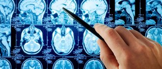
What does an MRI show?
Most often, children are prescribed 4 main studies using MRI: brain, spine, pelvic and abdominal organs, bones and joints.
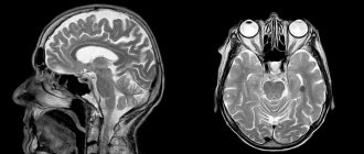
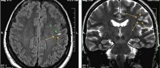
8.1. Brain
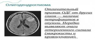
Indications for brain examination in children: skull injuries, suspicion of benign and cancerous tumors, heart attack or ischemia, severe infectious and parasitic diseases, frequent vomiting.
Using a scan, the doctor can detect demyelinating changes in the brain that occur against the background of severe poisoning, infectious diseases or ischemia.
MRI signs of demyelinating pathologies:
- hyperintense signal on T1 image;
- the presence of destruction of brain tissue (about 20%) on a T1-weighted image;
- the length of the changed areas is no more than 5 mm.
Tomography also reveals infectious processes in the brain:
- Encephalitis is a nonspecific increase in signal intensity on a T2 image.
- CNS abscess – hypo- or isointensity of the signal on the T1 image and hypointensity of the signal on the T2 WI.
- Parasitic diseases. Each type of pathology has its own MRI signs. With toxoplasmosis, small nodules are visualized in the cerebral hemispheres and ganglia.
8.2. MRI of the spine
The main indications for scanning the spine in a child are birth injuries, abnormal development of bone structures, congenital torticollis, and falls from a height.
During the scan, the structures of the spine are comprehensively studied, including nearby soft tissue, nerve endings and blood vessels.
MRI can reveal:
- congenital anomalies in the structure of the spinal column;
- narrowing of the spinal canal;
- osteoporosis;
- destruction of vertebral discs;
- tumors of bone structures and metastases.
When making a diagnosis, the doctor is guided by knowledge of anatomy, standard indicators and the results of previous examinations.
8.3. MRI of joints
MRI of bone tissue for children is prescribed for suspected:
- tumor processes in cartilage and bone tissues;
- sports injuries;
- infectious joint damage;
- displacement of implants;
- hemarthrosis.
From the images, the doctor can examine all the affected bone structures and assess the extent of the spread of the pathological process to nearby tissues.
Tomography visualizes:
- Bone tissue and the processes of inflammation and degeneration present in them. You can also examine in detail the presence of cracks and cysts in the structures.
- Cartilaginous tissue. Using tomography, doctors are able to determine the degree of cartilage wear, microscopic damage and ruptures.
- Ligamentous and tendon tissues, as well as their sprains resulting from injuries.
- Areas of articular joints that are not visible on x-rays.
What are mothers worried about if their child is scheduled for a brain MRI?
Sometimes mothers, even having received a doctor’s recommendation to do an MRI of the brain, do not take their child for examination. You won’t find any number of myths about MRI on the forums! The fears some mothers have about this procedure are completely unfounded. Let's look at the comments of experts regarding opinions widespread on the Internet about the dangers of the MRI procedure.
| Myth | Doctor's comment |
| MRI is dangerous, it involves radiation | During a magnetic resonance examination, the patient’s body is not exposed to radiation |
| during MRI the brain is “demagnetized” | MRI has no effect on brain activity |
| my child is weak and will not tolerate the procedure well | MRI does not affect your well-being and has no negative consequences |
| the child is too young for such serious procedures | since MRI has no contraindications, the study can be performed even on newborns |
| my child will be afraid of the device | MRI of the brain in children creates more curiosity than fear |
Thus, doubts about the advisability of performing MRI of the brain in children are completely unjustified. Especially when you consider that a correct diagnosis is half the success in treatment, and raising a healthy and happy child is the most important thing for a mother.