We will talk about multicystic pathology of the fetal kidneys.
For many reasons, but quite rarely - in approximately 1% of cases of all renal pathologies of the fetus, multicystic kidney dysplasia develops from approximately 6 to 10 weeks of pregnancy. The incidence of the disease is 1:1000 newborns with unilateral lesions, and 10 times less often with bilateral multicystic kidney disease (with this option, the pathology is always FATAL!)
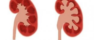
For those who want to know more precisely the mechanism of development of multicystic disease: there is no fusion of the rudiments of the mesonephrogenic and metanephrogenic blastema, as a result of which the secretory apparatus of the kidney produces urine, which has nowhere to be excreted, so it accumulates in the tubules, turning them into cysts, and instead of a normal kidney by the 2nd During the trimester of pregnancy, a conglomerate of cysts forms, similar to a bunch of grapes.
The causes of the disease are not precisely determined, but most cases are associated with complex malformations and genetic diseases.
As a result, a kidney with multicystic dysplasia does not function AT ALL (except for the segmental variant of the lesion), and is represented by numerous cysts of various sizes that do not communicate with each other.
With a unilateral process, the second kidney in approximately 40% of cases also has another type of anomaly, more often - pyeloectasia against the background of obstruction of the ureteropelvic anastomosis, but there may be rotation of the pelvis, duplication, kidney dystopia, hydronephrosis.
In case of unilateral multicystic dysplasia, the pathology is NOT ACCOMPANIED by oligohydramnios, because the second healthy kidney takes over the function of the affected one, hence the rule - if multicystic kidney disease is discovered, and everything is fine with amniotic fluid, then there is a chance of giving birth to a healthy child with a good prognosis (even if only one - the healthy kidney - functions).
And in case of anhydroamnion (i.e., critical oligohydramnios), when identifying unilateral multicystic pathology of the kidney, it is necessary to clearly make sure that it is with the other kidney, and first of all to exclude its aplasia (i.e., absence). An assistant in this case is the detection of a fetal bladder filled with urine.
Now let's get back to arithmetic:
1+1 = 1:
1 healthy kidney + 1 kidney with multicystic disease = 1 functioning kidney in a newborn child.
And let’s add the second formula for multicystic kidney disease:
2+0 = 0:
Both fetal kidneys with multicystic disease(2) + empty bladder (non-visualized bladder(0) = 0 chance of child survival, fatal pathology.
Despite the progress of prenatal diagnostics, according to various authors and centers, multicystic disease in the fetus is detected antenatally (before birth) in only 50 to 67% of cases.
And this is absolutely not clear to us, because... We have NEVER missed this kidney pathology. This is completely excluded given the colossal volume of expert ultrasound examinations of pregnancy that we perform.
This means that all these oversights of multicystic kidney disease stated in the statistics are the result of a superficial and incomplete examination of pregnant women during quick screenings.
Diagnosis and treatment of patients with polycystic kidney disease and infected cysts
Trushkin R.N., Lubennikov A.E., Pogodina K.S.
Polycystic kidney disease (PCD), or autosomal dominant polycystic disease, is the most common inherited kidney disease [1]. The main complications from cysts are: infection, hemorrhage, and malignancy is less common [1-5]. In one single-center study, cyst infection (IC) was found to be relatively rare, occurring in only 0.01 patients/episode/year. Predisposing factors were age, female gender and previous transurethral instrumentation. There was also an increase in their occurrence in patients receiving program dialysis, while at the same time after kidney transplantation such a pattern was not observed [2]. IC poses a serious threat to health and life, especially in patients with end-stage chronic renal failure (ESRD), in whom the risk of developing sepsis and death is many times higher, and therefore timely diagnosis of suppuration in the cyst is extremely important [3] . Despite its apparent simplicity, diagnosing IC is difficult for both clinicians and diagnostic services. Differential diagnosis of IR is carried out with hemorrhage and malignant neoplasm.
It is possible to speak reliably about IR only after obtaining an aspirate containing leukocytes and/or bacteria; in all other cases, the diagnosis is considered probable [4]. Laboratory examination of aspirate is the gold standard for diagnosis, however, puncture of the cyst is not always technically feasible due to the large size of the kidneys, the difficulty of visualizing IR and the risk of injury to the abdominal organs [2-5].
CLINICAL PICTURE AND LABORATORY DATA
A probable diagnosis of IR is established in the presence of: fever, pain in the projection of the kidneys/kidneys, high levels of C-reactive protein (CRP), leukocytosis, positive urine and blood cultures [4,5]. M. A. Lantinga et al. analyzed 70 papers published from 1948 to 2014, which described a total of 119 cases of IC. The authors found that the median fever was 390C, pain in the kidney projection was noted by 59-88% of patients, the median CRP was 55-228 mg/l, the median leukocytosis was 10.7-17.4x10⁹, negative urine cultures were noted in 46% of patients, blood - in 61% of patients; in 88% of cases, urine cultures corresponded to the culture of contents from the cyst [5]. Gram-negative flora is the main etiological cause leading to suppuration in the cyst; the leading position is occupied by E. coli, detected in a third of patients, which makes it possible to suspect an ascending path of infection of the cysts [3,4,5,6].
ULTRASONOGRAPHY
To date, ultrasound cannot be considered as a screening method for diagnosing IC. Firstly, this is due to the fact that blood and pus in the cyst cavity can give a similar echo picture, and secondly, the large size of the kidneys makes visualization difficult. So in the work of M. Sallee et al. Ultrasound data were positive in only 6% of patients with a verified diagnosis of IC [2].
CT SCAN
Computed tomography (CT) has low information content in the diagnosis of IC [2,5]. IC can be suspected by the presence of a thickened wall and heterogeneous hyperdense contents inside the cyst (Fig. 1 a). However, the high density contents in the cyst
observed both in the presence of a purulent substrate and in hemorrhage [7,8]. The pathognomonic CT sign of IC is a dynamic assessment: an increase in size, a change (increase) in densitometric density, wall thickening over a short time [5]. Contrast does not significantly increase the information content of CT, since an increase in the density of the cyst wall can be associated with the inflammatory process, the remaining functional parenchyma, and the presence of renal cell carcinoma [9,10]. Thus, in a retrospective 10-year study by M. Sallee et al. using CT, the diagnosis was made only in 18% of patients with proven IC [2], in the work of F. Juret et al. - only 15% [4].
MAGNETIC RESONANCE IMAGING
Magnetic resonance imaging (MRI) in T1, T2 mode (Fig. 1 c, d) is more informative compared to CT, but false-positive results reach 60% [2,4]. The presence of IR can be assumed by the increase in the MR signal from the cyst wall after administration of a contrast agent [11,12]. However, the association of gadolinium-containing substances with the development of nephrogenic systemic fibrosis limits its use in patients with chronic renal failure [5]. Other disadvantages of MRI include the long duration of the study, the influence of patient movements on the interpretation of results, limited use in patients with metal implants, and the inability to conduct the study in patients with a pacemaker.
In recent years, more and more publications have appeared indicating the high information content of MRI using diffusion-weighted imaging protocols (DW-MRI) (Fig. 1b) [13-16]. DW-MRI allows you to visualize and measure the random (Brownian) motion of water molecules. The signal intensity in a diffusion-weighted image (DWI) reflects the diffusivity of water molecules of the object under study [17]. Diffusion is difficult when the “cellularity” of the tissue increases - for example, in tumors, fibrosis, abscesses, cytotoxic edema [18-21], as well as in structures containing a high concentration of protein and hemoglobin degradation products [6]. Quantitative assessment of MR diffusion is achieved by constructing measurable diffusion coefficient (MDI) maps [22], however, at present, IDC threshold values for IR in patients with polycystic kidney disease have not been reliably determined [5]. However, diffusion MR is an informative addition to traditional sequences used in MRI. Its use allows, in some cases, to avoid the introduction of expensive contrast agents and is not associated with significant time or technical costs, increasing the diagnostic efficiency of the method [22].
Rice. 1. a - CT without contrast: increased density of the cyst, thickening of the walls, b - MRI - increased intensity on DWI from this cyst, c - T1 weighted image, d - T2 weighted image demonstrate a decrease in the MP signal
POSITRON EMISSION TOMOGRAPHY COMBINED WITH CT
According to some authors, positron emission tomography combined with CT (PET-CT) with 18-fluoro-deoxyglucose (18F-FDG) is currently considered the most sensitive method in diagnosing IR in patients with polycystic kidney disease (Fig. 2) , the sensitivity of which is 77-90% [9,23-25].
Rice. 2. PET-CT with 18F-FDG. There is hyperfixation of 18FDG in the cysts of the upper and lower segments of the left kidney, which indicates their infection; in addition, accumulation of 18F-FDG is visible in the para-aortic lymph nodes and some cysts of the right kidney. Accumulation of 18F-FDG in the stomach - physiological state
The positive aspects of this diagnostic method are: the absence of nephro- and hepatotoxicity, including in patients with initial forms of chronic renal failure, the possibility of spatial visualization, assessment of the dynamics of the degree of the inflammatory process against the background of conservative treatment, the possibility of identifying an inflammatory focus in the abdominal cavity of extrarenal localization. The last point is extremely relevant in patients with PP and fever of unknown origin. F. Jouret et al. report that in 6 patients with polycystic kidney disease after
PET-CT treatment tactics were changed. These patients were examined in connection with suspected IR, but the following were identified: cholangitis, diverticulitis of the small intestine, diverticulitis of the ascending colon, prostatitis, pyelonephritis of the transplanted kidney, inflammation in the walls of the abdominal aortic aneurysm [25]. In the publication of G. Piccoli et al. it is noted that in 4 patients IC was not confirmed, but pathological accumulation of the radiopharmaceutical in the peripancreatic lymph nodes was detected, which was associated with the presence of mesenchymal formations [9]. The disadvantages of the method are high cost, limited availability, difficulties in differentiating an inflammatory process from a malignant one, and also insufficient data to date on the likelihood of false-positive results in hemorrhage into a cyst [4,25].
TREATMENT
The vast majority of available publications note that fluoroquinolones are the drugs of choice in initial therapy, this is due to their lipophilic properties and the ability to create high concentrations inside cysts [2,26,27]. Unfortunately, conservative therapy is doomed to failure in the vast majority of cases. Thus, in 2021, M. Lintinga et al. present the results of an analysis of 60 publications since 1948, which describe the treatment of 85 patients with IR. The effect of antibacterial therapy was achieved in only 25% [26].
If a suppurating cyst is detected, its drainage, bacteriological analysis of the contents, and etiotropic antibacterial therapy are indicated. If it is impossible to drain under ultrasound guidance, it is advisable to use CT navigation taking into account previously obtained DW-MRI data. The effectiveness of this approach has been demonstrated in a number of publications [28,29].
The lack of effect from conservative treatment and minimally invasive interventions is an indication for kidney revision, and in some cases, nephrectomy [26,30].
Considering the high risk of sepsis, death, and side effects from antibacterial therapy in patients receiving program dialysis, in the absence of the possibility of draining the cyst, especially in the presence of multiple IR, nephrectomy is indicated [30].
CONCLUSION
Fever, pain in the projection of the kidneys/kidneys, high levels of leukocytosis, C-reactive protein dictate the need for diagnostic measures to detect cyst infection in patients with polycystic kidney disease. It must be taken into account that bacteriological examination of blood and urine is negative in almost half of patients. Considering the low information content of ultrasound and CT, the first step in the absence of contraindications is to carry out DW-MRI, or, if technically accessible, PET-CT.
When an IK is detected, its drainage under ultrasound guidance is indicated; in complex cases, it is advisable to use CT navigation, focusing on MRI-DWI data.
Probably, antibacterial therapy should be considered as initial treatment only in patients with PP, preserved renal function, no obvious signs of suppuration in the cyst, and without factors of complicated urinary infection.
In patients receiving dialysis treatment, nephrectomy in the presence of CPB should be considered as the primary treatment option.
LITERATURE
1. Torres VE, Harris PC, Pearson Y. Autosomal dominant polycystic kidney disease. Lancet 2007;369(9569):1287-1301.
2. Sallee M, Rafat C, Zahar JR, Paulmier B, Grunfeld JP, Knebelmann B, et al. Cyst infections in patients with autosomal dominant polycystic kidney disease. Clin J Am Soc Nephrol 2009;4(7):1183-1189. doi: 10.2215/CJN.01870309.
3. Christophe JL, van Ypersele, de Strihou C, Pirson Y. Complications of autosomal dominant polycystic kidney disease in 50 haemodialysed patients. A case-control study. The UCL Collaborative Group. Nephrol Dial Transplant 1996;11(7):1271-1276.
4. Jouret F, Lhommel R, Devuyst, Annet L, Pearson Y, Hassoun Z, et al. Diagnosis of cyst infection in patients with autosomal dominant polycystic kidney disease: attributes and limitations of the current modalities. Nephrol Dial Transplant 2012;27(10):3746-3751. doi: 10.1093/ndt/gfs352
5. Lantinga MA, Drenth JP, Gevers TJ.Diagnostic criteria in renal and hepatic cyst infection. Nephrol Dial Transplant 2015;30(5):744-751. doi: 10.1093/ndt/gfu227
6. Yukhno E.A. Trofimenko I.A. Trufanov G.E. Malignancy of endometrioid cysts in the aspect of magnetic resonance imaging: semiotics and diagnostic errors. Tumors of Female Reproductive System 2013;(3):72-80.
7. Gupta S, Seith A, Dhiman RK, Chawla YK, Sud K, Kohli HS, et al. CT of liver cysts in patients with autosomal dominant polycystic kidney disease. Acta Radiol 1999;40(4):444-448.
8. Gupta S, Seith A, Sud K, Kohli HS, Singh SK, Sakhuja V, et al. CT in the evaluation of complicated autosomal dominant polycystic kidney disease. Acta Radiol 2000;41(3):280-284.
9. Piccoli GB, Arena V, Consiglio V, Deagostini MC, Pelosi E, Douroukas A, et al. Positron emission tomography in the diagnostic pathway for intracystic infection in ADPKD and 'cystic' kidneys. A case series. BMC Nephrol 2011;12:12-48. doi:10.1186/1471-2369-12-48.
10. Keith DS, Torres VE, King BF, Zincki H, Farrow GM. Renal Cell Carcinoma in Autosomal Dominant Polycystic Kidney Disease. J Am Soc Nephrol 1994;4(9):1661-9.
11. Wood cG, Stromberg LG, Harmath CB, Horowitz JM, Feng C, Hammond NA, et al. CT and MR imaging for evaluation of cystic renal lesions and diseases. Radiographics 2015;35(1):125-141. doi: 10.1148/rg.351130016.
12. Migali G, Annet L, Lonneux M, Devuyst O. Renal cyst infection in autosomal dominant polycystic kidney disease. Nephrol Dial Transplant 2008;23(1):404-405.
13. Ichioka K, Saito R, Matsui Y, Terai A. Diffusion-weighted magnetic resonance imaging of infected renal cysts in a patient with polycystic kidney disease. Urology 2007;70(6):1219.
14. Katano K, Kakuchi Y, Nakashima A, Takahashi S, Kawano M. Efficacy of diffusion-weighted magnetic resonance imaging in detecting infected cysts in a case of polycystic kidney disease. Clin Nephrol 2011;75:24-26.
15. Takase Y, Kodama K, Motoi I, Saito K. Cyst infection in unilateral renal cystic disease and the role of diffusion-weighted magnetic resonance imaging. Urology 2012;80(5):61-62. doi: 10.1016/j.urology.2012.07.022.
16. Suwabe T, Ubara Y, Sumida K, Hayami N, Hiramatsu R, Yamanouchi M et al. Clinical features of cyst infection and hemorrhage in ADPKD: new diagnostic criteria. Clin Exp Nephrol 2012;16(6):892-902. doi:10.1007/s10157-012-0650-2. 17. Mascalchi M, Filippi M, Floris R, Fonda C, Gasparotti R, Villari N. Diffusion-weighted MR of the brain: methodology and clinical application. Radiol Med 2005;109( 3):155-97.
18. Bruegel M, Holzapfel K, Gaa J, Woertler K, Waldt S, Kiefer B, et al. Characterization of focal liver lesions by ADC sequence for detection of focal liver lesions. Invest. Radiol 2008;43:261-266.
19. Lowenthal D, Zeile M, Lim WY, Wybranski C, Fischbach F, Wieners G, et al. Detection and characterization of focal liver lesions in colorectal carcinoma patients: comparison of diffusion-weighted and Gd-EOB-DTPA enhanced MR imaging. Eur Radiol 2011;21(4):832-840. doi:10.1007/s00330-010-1977-2.
20. Miller FH, Hammond N, Siddiqi AJ, Shroff S, Khatri G, Wang Y, et al. Utility of diffusion-weighted MRI in distinguishing benign and malignant hepatic lesions. J Magn. Res. Imaging 2010;32(1):138-147. doi: 10.1002/jmri.22235.
21. Coenegrachts K, Delanote J, Ter Beek L, Haspeslagh M, Bipat S, Stoker J, et al. Improved focal liver les ion detection: comparison of single shot diffusion-weighted echoplanar and single shot T2 weighted turbo spin echo techniques. Brit J of Radiol 2007;80(955):524-531.
22. Bergen T.A. Trofimenko I.A. MR diffusion technique and its application in the study of pelvic tumors in women. Bulletin of Siberian Medicine 2012;(11):10-12.
23. Bobot M, Ghez C, Gondouin B, Sallee M, Fournier PE, Burtey S, et al. Diagnostic performance of (18)F fluorodeoxyglucos positron emission tomography-computed tomography in cyst infection in patients with autosomal dominant polycystic kidney disease. Clin Microbiol Infect 2016; 22(1):71-77. doi: 10.1016/j.cmi.2015.09.024
24. Paschali AN, Georgakopoulos AT, Pianou NK, Anagnostopoulos CD. (18) F-fluorodeoxyglucose positron emission tomography/computed tomography in infected polycystic kidney disease. World JNuclMed 2015;14(1):57-59. doi: 10.4103/1450-1147.150553.
25. Jouret F, Lhommel R, Beguin C, Devuyst O, Pearson Y, Hassoun Z, et al. Positron-emission computed tomography in cyst infection diagnosis in patients with autosomal dominant polycystic kidney disease. Clin J Am Soc Nephrol 2011;6(7):1644-1650. doi: 10.2215/CJN.06900810
26. Lantinga MA, Casteleijn NF, Geudens A, de Sevaux RG, van Assen S, Leliveld AM, et al. Management of renal cyst infection in patients with autosomal dominant polycystic kidney disease: asystematic review. Nephrol Dial Transplant. 2021. first published online January 29.
27. Suwabe T, Araoka H, Ubara Y, Kikuchi K, Hazue R, Mise K, et al. Cyst infection in autosomal dominant polycystic kidney disease: causative microorganisms and susceptibility to lipid-soluble antibiotics. Eur J Clin Microbiol Infect Dis 2015;34(7):1369-1379. doi: 10.1007/s10096-015-2361-6
28. Kale PG, Devi BV, Lakshmi AY, Sivakumar V. Use of diffusion-weighted magnetic resonance imaging in the management of infected renal cyst in polycystic kidney disease. Indian J Nephrol 2015;25(1):60-61. doi: 10.4103/0971-4065.
29. Kato S, Hotta H, Mineta M, Miyake M. Case of infected renal cyst in polycystic kidney disease. Nihon Hinyokika Gakkai Zasshi 2013;104(3):536-539.
30. Lysenko M.A., Vtorenko V.I., Trushkin R.N., Lubennikov A.E., Sysoev A.M., Sokolov A.A. Bilateral nephrectomy in patients with end-stage chronic renal failure and active chronic pyelonephritis. Urology 2016;(1): 46-52
Journal "Experimental and Clinical Urology" Issue No. 4 for 2016
Magazine
Journal "Experimental and Clinical Urology" Issue No. 4 for 2016
Comments
To post comments you must log in or register
How is dysplasia treated?
The choice of treatment methods depends on the patient's condition and the cause of the disease. Genetic abnormalities are corrected through surgery. This is not a treatment in the truest sense of the word. For aplasia, for example, an implant is placed rather than the gland being grown.
Benign dysplasia is treated both conservatively and surgically. For example, cystic mastopathy can be easily corrected with homeopathic medicines and herbs (Mastodinon).
When choosing treatment, the doctor takes into account the cause of the disease and may recommend:
- psychocorrection;
- hormonal therapy (Danazol);
- antitumor agents (Tamoxifen);
- homeopathic remedies (Mastopol);
- enzymes (Wobenzym);
- anti-inflammatory drugs (not steroids)
Drugs can be prescribed locally and orally. If there is an underlying pathology (for example, endocrine disorders), treatment of the underlying disease is indicated. For grade II-III dysplasia, the doctor can reasonably recommend removal of the affected tissue.
You often ask whether dysplasia is treated with antibiotics. Of course not. Despite the articles found on the Internet!
This group of drugs is prescribed for the presence of pathogenic microorganisms in your body. For example, for cervical dysplasia due to an STD, antibacterial therapy is indicated. For mastopathy, no, unless the cysts in your breasts have festered.
Causes
Kidney dysplasia in most cases refers to congenital pathologies based on genetic disorders.
However, a third of cases associated with the manifestation of dysplasia remain unknown, since modern medicine cannot indicate the exact cause of its occurrence. Boys are three times more likely to suffer from this severe pathology.
At the birth of a baby, in addition to dysplasia, he is diagnosed with serious pathologies of the ureters and renal pelvis.
Due to improper development of the tissues of the renal organs, the patient is determined to have a large number of cysts, multicystic.
Cysts fill with urinary fluid because a patient with dysplasia also has abnormal development of the renal tubules, leading to impaired urine flow.
At the same time, the tubes expand and provoke the formation of cysts. Unfortunately, a large number of filled cysts significantly narrow the lumen of the ureter, thereby impairing the process of urination.
In some severe cases, the urethra may be completely blocked. This leads to an increase in the size of the kidney organ and suppuration of the internal contents of the cyst.
This situation cannot go unnoticed, and therefore provokes the death of the kidney itself. If the required amount of healthy kidney tissue is available, surgery is performed, during which the cysts are resected.
This provides an opportunity to try to save the kidney. Otherwise, it must be removed to save the patient's life.
Symptoms of the disease
According to statistics, kidney dysplasia in the fetus occurs in approximately 5% of cases among all diseases of the urinary system. The greatest danger is posed by bilateral kidney damage, since in this situation there is still no effective treatment method, however, the activities of highly qualified specialists and the rapid development of medical equipment will ultimately give a positive result in the fight against pathology.
Doctors divide bilateral dysplasia into two categories:
- Hypoplastic dysplasia, as a result of which chronic renal failure quite often develops, and the functioning of the entire organism as a whole worsens. To identify this disease, doctors refer the patient to;
- aplastic dysplasia, the consequence of which is death.
Due to the fact that most children live fully and with one healthy and fully functioning kidney, however, as the child develops, the load on the organ increases significantly, which in turn can cause damage.
The main symptom of simple segmental dysplasia will be the appearance in the child of hypertension that cannot be controlled with medications.
Reasons for formation
Most cases of renal dysplasia have an unknown etiology. Many experts tend to assume that the disease is based on a genetic predisposition.
A congenital disorder is formed against the background of the influence of various negative factors on a pregnant woman:
- exposure to toxic substances (alcohol, nicotine, drugs);
- the course of infectious diseases during pregnancy, improper treatment of diseases (use of aggressive medications that have a detrimental effect on the fetus);
- diabetes mellitus, other kidney and liver diseases in the mother.
During pregnancy, all medications that a woman takes must be agreed with the attending physician. The fetus is negatively affected by drugs that lower blood pressure, antidepressants, and antibiotics.
Breast dysplasia - what is it?
Dysplasia is the abnormal development of organs and tissues of the human body. Pathology of the mammary glands is the most common. It can be expressed in hyperplasia and hypoplasia. The first is often called mastopathy. Hyperplasia, or excessive growth of glandular tissue, is characterized by the appearance of nodular seals in the chest. Refers to a fibrous type of pathology. It is this form of dysplasia that can develop into cancer, which is a good reason to consult a doctor if pain or lumps appear in the mammary gland.
Hypoplasia does not bring such global problems into a woman’s life. The only drawback is the underdevelopment of the breasts, which does not affect its functioning.
What causes kidney dysplasia?
Kidney dysplasia in children can be caused by maternal exposure to certain drugs or genetic factors. Pregnant women should consult their healthcare provider before taking any medications during pregnancy. Drugs that can cause kidney dysplasia include medications to treat seizures and medications to lower blood pressure called angiotensin-converting enzyme inhibitors (ACE inhibitors) and angiotensin receptor blockers (ARBs). Addiction to smoking, alcohol or drugs can also cause kidney dysplasia in the unborn child.
Kidney dysplasia can also have genetic causes. During kidney dysplasia in a child, an ultrasound examination may reveal the same disease in one of the parents.
Some genetic syndromes that affect other body systems. A child with kidney dysplasia may also have problems:
- with the gastrointestinal tract;
- nervous system;
- heart and blood vessels;
- muscles and skeleton;
- other parts of the urinary tract.
Urinary tract problems that lead to kidney dysplasia can also affect a normal kidney that is functioning. Over time, if these problems that the abnormal kidney can cause are not corrected, it can impair the functioning of the normal kidney and lead to kidney failure.
What types of renal dysplasia are there?
Causes and degrees of hydronephrosis of the left kidney: treatment and prognosis
Renal agenesis: Most people are born with two kidneys. But 4,000 babies (more boys than girls) come into the world with only one kidney, this is the main characteristic signal of renal agenesis. In some cases, ultrasound may show a very small second kidney that is abnormal.
Renal hypoplasia: a child is born with two normally functioning kidneys, but they are significantly small in size. Renal hypoplasia is not a hereditary disease and occurs in both boys and girls. If a child's kidneys are extremely small, their function declines as the child enters adolescence.
Renal dysplasia may be associated with abnormalities of the ureter, as the urinary tube connects the kidney to the bladder. This condition is characterized by a backflow of urine from the bladder into the kidneys.
Causes in children and adults
To date, the reasons for this anomaly are completely unclear. Kidney dysplasia belongs to congenital disorders that are formed in the intrauterine development of the fetus under the influence of various external and internal circumstances. The following reasons are identified:
- maintaining an unhealthy lifestyle of the mother (nicotine addiction, alcohol consumption, drugs);
- diabetes;
- infectious diseases suffered during pregnancy;
- hereditary factors.
Causes of kidney problems in children can be caused by taking certain medications during fetal development. Therefore, girls who are expecting a baby need to talk to their doctor about taking various types of medications. Medicines taken to lower blood pressure and calm during attacks of hysteria can cause disruption in the development of the fetus, and subsequently cause defects in newborns.
The causes should include syndromes at the genetic level that affect the body of newborns as a whole. The genetic causes listed below for renal dysplasia, if not corrected, will cause damage to a healthy organ. This may subsequently develop into kidney failure. Causes:
- deterioration of the stomach and intestines;
- pathologies of the development of the nervous system;
- cardiovascular disorders;
- musculoskeletal problems;
- dysfunction of the genitourinary system.
Causes
Microcytosis in a general blood test (microcytic anemia): what it is, causes in adults and children, symptoms, treatment and prognosis
Kidney dysplasia in most cases refers to congenital pathologies based on genetic disorders.
However, a third of cases associated with the manifestation of dysplasia remain unknown, since modern medicine cannot indicate the exact cause of its occurrence. Boys are three times more likely to suffer from this severe pathology.
At the birth of a baby, in addition to dysplasia, he is diagnosed with serious pathologies of the ureters and renal pelvis.
Kidney damage
Due to improper development of the tissues of the renal organs, the patient is determined to have a large number of cysts, multicystic.
Cysts fill with urinary fluid because a patient with dysplasia also has abnormal development of the renal tubules, leading to impaired urine flow.
At the same time, the tubes expand and provoke the formation of cysts. Unfortunately, a large number of filled cysts significantly narrow the lumen of the ureter, thereby impairing the process of urination.
In some severe cases, the urethra may be completely blocked. This leads to an increase in the size of the kidney organ and suppuration of the internal contents of the cyst.
This situation cannot go unnoticed, and therefore provokes the death of the kidney itself. If the required amount of healthy kidney tissue is available, surgery is performed, during which the cysts are resected.
https://youtube.com/watch?v=WKlx-ctR2D8
This provides an opportunity to try to save the kidney. Otherwise, it must be removed to save the patient's life.
Symptoms of the disease
If the kidneys are cold, the main symptoms and signs if the kidneys are cold in women and men
According to statistics, kidney dysplasia in the fetus manifests itself in approximately 5% of cases among all diseases of the urinary system. The greatest danger is posed by bilateral kidney damage, since in this situation there is still no effective treatment method, however, the activities of highly qualified specialists and the rapid development of medical equipment will ultimately give a positive result in the fight against pathology. Doctors divide bilateral dysplasia into two categories:
- Hypoplastic dysplasia, as a result of which chronic renal failure quite often develops, and the functioning of the entire organism as a whole worsens. To identify this disease, doctors refer the patient for a kidney biopsy;
- aplastic dysplasia, the consequence of which is death.
Due to the fact that most children live fully and with one healthy and fully functioning kidney, however, as the child develops, the load on the organ increases significantly, which in turn can cause damage.
Parents of babies need to focus their attention on symptoms such as:
- Intense pain felt in the lower abdomen;
- severe and frequent headaches;
- blood pressure exceeding normal;
- the appearance of seizures;
- increased fatigue;
- lag, in contrast to peers, in mental development;
- slow physical development;
Indirect signs of abnormalities in the proper functioning of the kidneys are susceptibility to colds, as well as the fairly frequent occurrence of cystitis with a feeling of pain when urinating. If two or more signs of the disease are detected, you must contact a urologist to avoid serious consequences.
Causes
Kidney dysplasia in most cases refers to congenital pathologies based on genetic disorders.
However, a third of cases associated with the manifestation of dysplasia remain unknown, since modern medicine cannot indicate the exact cause of its occurrence. Boys are three times more likely to suffer from this severe pathology.
At the birth of a baby, in addition to dysplasia, he is diagnosed with serious pathologies of the ureters and renal pelvis.
Due to improper development of the tissues of the renal organs, the patient is determined to have a large number of cysts, multicystic.
Cysts fill with urinary fluid because a patient with dysplasia also has abnormal development of the renal tubules, leading to impaired urine flow.
At the same time, the tubes expand and provoke the formation of cysts. Unfortunately, a large number of filled cysts significantly narrow the lumen of the ureter, thereby impairing the process of urination.
In some severe cases, the urethra may be completely blocked. This leads to an increase in the size of the kidney organ and suppuration of the internal contents of the cyst.
This situation cannot go unnoticed, and therefore provokes the death of the kidney itself. If the required amount of healthy kidney tissue is available, surgery is performed, during which the cysts are resected.
This provides an opportunity to try to save the kidney. Otherwise, it must be removed to save the patient's life.
Features and cause of dysplasia
Kidney dysplasia is a malformation of an organ that is accompanied by disturbances in the formation of the renal structure. In this case, the process may have different intensity of manifestation. Since there is a change in the structure of the kidney tissue, there is also a violation of the functional capabilities of the organ.
The reasons for the origin of all developmental anomalies of certain organs are not fully understood to date. For such diseases, only risk factors have been identified that increase the chances of the occurrence of this pathology. With renal dysplasia, the same picture is observed.
The main risk factors for the development of dysplasia are the following conditions and diseases:
- if the mother’s body was not fully prepared for pregnancy;
- various bad habits of the mother before pregnancy and during the process of bearing the baby, for example, smoking, alcohol, drugs;
- the expectant mother has diabetes;
- all kinds of abdominal injuries in a pregnant woman during pregnancy;
- serious infectious diseases of the mother;
- intrauterine infection of the fetus with rubella, measles and cytomegalovirus viruses;
- if genetic mutations occur during the formation of the embryo.
As you can see, there are many such dangerous factors. Both different factors individually and their combination can lead to disruption of the formation of kidney tissue in the fetus.
What are the symptoms of renal dysplasia in a child?
According to the observations of specialists, kidney dysplasia in the fetus accounts for about 4% of other diseases of the urinary system. A very dangerous stage is when dysplasia affects not one kidney, but two at once. In such situations, treatment is simply impossible. Bilateral kidney damage is distinguished:
2. Hypoplastic. This type of disease reduces the functioning of organs and in most cases develops kidney failure, which is chronic. In order to identify the disease, specialists use a biopsy method of these paired organs.
Signs of this anomaly:
- The child feels severe pain in the lower abdomen;
- Convulsions are observed;
- Headache;
- Blood pressure increases.
Also, a child who has been diagnosed with kidney dysplasia is significantly delayed in development compared to his peers.
Do I need treatment?
There is no specific treatment for kidney dysplasia. Therapy is symptomatic. In mild situations, the doctor chooses the tactics of dynamic observation.
More complex cases require the following treatment methods:
Treatment with antibiotics (if infection is present). Painkillers are prescribed for renal colic. Medicines to lower blood pressure. Diuretics help improve diuresis and lower blood pressure. Special low-carb diet.
If organ functions are severely impaired, hemodialysis is required. In case of total damage to the kidney, when the organ does not perform its functions, the pathology contributes to the development of persistent hypertension, then a unilateral resection is performed. It is possible to remove part of the organ or the entire kidney. Usually the operation is performed as a laparoscopy.
In the case of bilateral total dysplasia, organs cannot function normally, this has a negative impact on other organs and systems. In this situation, the only way to save the patient’s life is through a kidney transplant.