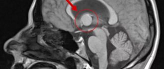
Brain stem glioblastoma
A glioma of this location cannot be removed surgically, because this would lead to disruption of the functioning of vital parts of the brain, for example, the respiratory and vasomotor centers.
As they grow, the connection between these centers and nuclei of the cranial nerves with the underlying structures is disrupted, which is characterized by the appearance of symptoms typical for this location, including arrhythmia and pathological types of breathing. In this case, the prognosis of the disease is unfavorable and survival rate is low.
Pathogenesis
Gliomas are any malignant brain tumors that form glial cells . When malignant transformation occurs, glial cells, namely astrocytes , become less differentiated, losing the characteristics of functional mature cells. In addition, their connection with neighboring cells is lost, and division becomes uncontrolled.
Thus, glioblastoma consists of poorly differentiated astrocytes, it has foci of necrosis and areas of vascular proliferation. A characteristic feature of this formation is a random arrangement of cells, polymorphism of cell nuclei and extensive perifocal edema. The tumor grows rapidly and invades the brain tissue. It is impossible to trace clear boundaries of the affected area. The neoplasm does not metastasize beyond the nervous system.
It is most often found in the frontal and temporal lobes. Very rarely, a tumor forms in the brain stem, spinal cord, or cerebellum.
In some cases, glioblastoma develops from anaplastic and low-grade astrocytomas, but most often this lesion is primary.
The tumor spreads to neuroglial cells, whose functions are related to the nutrition and protection of neurons. These cells do not reproduce in adulthood, but there are precursors of glial cells in the brain. If disturbances occur in their development, glioblastoma may develop.
A characteristic feature of glioblastoma, which determines its aggressive course, is its heterogeneity. The nature of the neoplasm differs in different patients. Moreover, even in one patient, the tumor can contain different cells that are not genetically similar, with different mutations. During the treatment process, cells mutate, actively evolve and develop new methods of survival.
Characteristic signs of glioblastoma
During the research, scientists noted that a significantly lower risk of developing glioma – by 40% – is observed in people suffering from various types of allergies . Experts explain this by the fact that in such people the immune system is in an activated state and prevents tumor growth.
Focal and cerebral symptoms
General cerebral:
• vestibular – pathological gait, dizziness,
• hypertensive-hydrocephalic – migraines, nausea, weakness.
Focal:
• speech disorder,
• memory impairment,
• loss of ability to perform complex actions.
Focal symptoms depend on location. In rare cases, against the background of headache, extensive hemorrhage and hemorrhagic stroke may occur as one of the complications of the disease.
Mental disorders
This type of deviation manifests itself in the form of general weakness, apathy, and some lethargy. In many cases, doctors detect a certain confusion in the patient’s consciousness, which is why the person reacts poorly to the events happening around him and generally does not clearly understand where he is at a particular moment.
Sometimes glioblastoma makes itself felt by paralysis of certain areas of the body or even the entire side of the body and adjacent limbs. In addition, there is often a violation of the sensitivity of the body. The so-called horizontal nystagmus may be present, manifested by transverse floating swaying to the sides.
A characteristic feature of this defect is that the patient himself does not notice these swaying movements. Also, the patient may suffer from hallucinations (usually auditory). Sometimes phantom odors occur. The probability of epileptic seizures is about 10%, which makes this defect rare.
Early stage: symptoms
Initially, the process will proceed differently: it depends primarily on the location of the tumor in the cranial cavity. However, the most typical syndrome is headache, which appears due to increased intracranial pressure. And before clear signs of the disease appear, glioblastoma of the brain must reach a fairly large size.
Other symptoms:
• visual impairment,
• drowsiness,
• periodic dizziness,
• epileptic seizures,
• numbness of the limbs.
Treatment methods for glioblastoma of the brain
The outcome of tumor treatment is determined by the stage at which it is detected and how quickly therapy is started.
In the early stages, the disease is often detected incidentally during examinations for other complaints.
Neurosurgical intervention is the most radical way to get rid of glioblastoma.
Since the tumor does not have clear boundaries and penetrates deeply into neighboring tissues, it is rarely possible to completely remove all cancer cells during surgery. This leads to relapses, which occur in approximately 80% of cases.
When localization of the lesion is difficult to reach, “Cyber Knife” is used - stereotactic radiosurgery.
A combination of surgery with radiation and chemotherapy can prevent recurrence of glioblastoma. This blocks the activity of malignant cells and prevents tumor growth.
For targeted therapy of relapses, drugs are used that destroy tumor vessels and prevent the proliferation of glial cells.
Diagnostics
Diagnosis is carried out using the following steps of additional examination and laboratory and instrumental diagnostics:
• MRI
• CT
• PET – positron emission tomography
• echoencephaloscopy
• trephine biopsy, checking for gene mutation
• lumbar puncture and cerebrospinal fluid analysis.
Recently, stereotactic biopsy has been used to obtain tissue samples. To do this, a very miniature trepanation hole is made in the bones of the skull - up to 1 mm in diameter, through which, under the control of X-ray equipment, a biopsy needle is inserted directly into the tumor focus, and material is taken for research. Determination of the localization of the pathology and subsequent insertion of the needle occurs using a stereotactic frame (frame-based) or a navigation system.
Diagnosis of glioblastoma
To make and clarify the diagnosis of glioblastoma in the presence of symptoms, specialists prescribe instrumental studies :
- MRI of the brain or spinal cord;
- magnetic resonance spectroscopy and/or perfusion;
- computed tomography;
- positron emission tomography of the brain;
- stereotactic biopsy using computer navigation systems.
For laboratory diagnostics use:
- histological typing;
- molecular test;
- immunohistochemical study;
- analysis of gene activity (methylation of the MGMT gene).
Find out the price
Find out the price
Error! Please fill in all required fields
Thank you! We will contact you shortly
✕
Surgical intervention
The course of the removal operation includes 3 main stages:
1. Visualization of process boundaries. This is possible with the use of special drugs that lead to the accumulation of protoporphyrin around the pathology. Thanks to this technique, the tumor is not only better visualized, but also slows down its spread.
2. Direct removal of tumor, including metastases. Unfortunately, due to the fact that the boundaries of glioblastoma are blurred, in some situations even parts of healthy tissue have to be removed. But this option is carefully discussed both with the patient himself and his relatives, because when some structures are removed, the consequences are worse than from the underlying disease.
3. Suturing soft flaps, suturing, aseptic dressing on the postoperative wound.
Due to the fact that it has unclear boundaries and an irregular shape, its individual elements, with all the efforts of the surgeon, can be missed and go unnoticed. For this reason, complete removal is often not possible, and there is always a high probability of relapse of the disease. In this regard, the course of surgical intervention is often supplemented with subsequent chemotherapy treatment.
Survival, life expectancy
Due to the fact that it is impossible to predict in advance how many cancer cells will remain after surgery and survive, despite subsequent treatment with cytostatics or a course of radiotherapy, the final answer can only be obtained after completing the entire course of therapy. On average, life expectancy for glioblastoma rarely exceeds 1.5 years.
Preference in treatment is given to cellular bioimmunotherapy; the latest developments in this area are supervised by Professor V.I. Skvortsova. Clinical studies of new drugs began this year, giving hope for prolonging the lives of many patients with glioma tumors.
Prognosis and survival
In advanced situations, recovery becomes impossible due to massive damage, so at this stage treatment includes courses of chemotherapy and radiation therapy. A three-month course of drugs: dendritic vaccine, LAK therapy, can significantly slow down the growth of an existing tumor, and dosed ionizing radiation can significantly reduce the total number of areas of pathological cell growth.
Diagnostic methods and treatment
The first specialist that a patient with a suspected brain tumor should visit is a neurologist. In his office, the sick person must undergo a professional examination, consisting of a series of characteristic neurological tests.
If a doctor suspects the presence of a tumor, he should write a referral for a detailed study of brain structures using non-invasive neuroimaging methods. These include magnetic resonance and computed tomography of the brain. In this case, it is preferable to use an MRI - in the resulting images, the neoplasm will look like a dense accumulation of light-colored nervous tissue.
The choice of method for studying brain structures is influenced by a large number of factors, including the patient’s tolerance of a special contrast agent. It is used to accurately determine the location of glioblastoma and study its effect on neighboring tissues. The images obtained in both cases of the study represent a layer-by-layer image of the brain in several projections. Using them, experts determine the size of the tumor and the nature of its spread in the brain.
To obtain accurate information about the histological composition of the neoplasm, stereotactic brain biopsy is used, which is an invasive procedure to obtain a sample of atypical tissue for subsequent histological examination.
The method of treatment will be determined by the following specialists: neurologist, oncologist, neurosurgeon, chemotherapist and radiation therapy doctor. In this case, the tactics of medical manipulations depend on the location of the tumor and the nature of the damage it caused.
So, for example, if the tumor is located deeper than 30 cm and spreads to both hemispheres, then surgical intervention is contraindicated for such a patient, but if other methods predictably will not bring the desired effect, then such a patient is prescribed only the use of potent narcotic drugs that will alleviate it suffering.
All treatment methods for glioblastoma of the brain are aimed at removing it or slowing its growth, since this type of cancer rarely metastasizes.
Typically, treatment for glioblastoma takes place in several stages and may include the following manipulations:
- surgical removal of the tumor;
- a course of radiation or chemotherapy;
- supportive treatment.
The most effective treatment for brain cancer is surgical excision of the tumor. It is carried out if the tumor has clearly defined boundaries and does not spread to the nerve centers responsible for performing vital processes. This approach can significantly increase the patient’s life expectancy with minimal consequences.
Until recently, glioblastoma was removed using primitive surgical methods, which did not allow the tumor to be completely removed without consequences. However, at the moment, the use of 5-aminolevulinic acid greatly simplifies this task, since it is able to highlight atypical cells, that is, now during the operation the neurosurgeon can visually highlight the border between healthy and atypical tissue.
After most of the tumor cells have been removed, the remaining glioblastoma particles are exposed to ionizing radiation. This approach prevents further spread of the tumor or significantly slows down its growth. To achieve the desired effect, the patient must undergo a 6-week course of radiation therapy. In this case, simultaneously with such treatment, the patient is prescribed chemotherapy drugs, for example, Temodal.
As the last stage, the patient is prescribed to take antitumor medications no earlier than 4 weeks after completion of phase 2 of therapy.
Many patients are concerned about the question of whether glioblastoma of the brain can be completely cured. Alas, the answer is often disappointing, since this type of tumor is practically untreatable: in most cases, the disease relapses or the patient cannot physically withstand all medical manipulations.
Prevention
The most effective method of prevention is regular attendance at preventive examinations, which make it possible to identify problems at an early stage. It is especially important to undergo annual examinations for people over 45 years of age, because with age the likelihood of this dangerous disease increases.
To reduce the risk of developing this disease, you must follow the recommendations regarding a healthy lifestyle:
- Get a full night's rest - healthy sleep and its sufficient duration are important.
- Spend time outdoors – you need to walk as much as possible every day.
- Avoid stress and emotional turmoil.
- Give up bad habits - do not smoke, do not consume excessive amounts of alcohol.
- Do not abuse spending time with various gadgets.
- Eat right - lots of vegetables, fruits and grains, be sure to have legumes, high-quality proteins, and unsaturated fats. Harmful foods - fast food, smoked meats, soda, confectionery, etc. - must be excluded.