© Author: Z. Nelly Vladimirovna, doctor of the first qualification category, especially for SosudInfo.ru (about the authors)
Ocular pressure, intraocular pressure (IOP) or ophthalmotonus, is the pressure of the fluid contained inside the eyeball on the walls of the eye. Intraocular pressure is now determined for all persons who have crossed the 40-year mark, regardless of whether the person makes complaints or not. This is due to the fact that increased eye pressure is the main prerequisite for the development of a disease such as glaucoma, which, if left untreated, leads to complete blindness.
Intraocular pressure is measured using a special tonometer, and the results are expressed in millimeters of mercury (mmHg). True, ophthalmologists of the 19th century judged the hardness of the eyeball by pressing on the eye with their fingers. In other cases, in the absence of equipment, a similar method is used today as a preliminary assessment of the condition of the visual organs.
Why is it important to know IOP?
The attention paid to such an indicator of health status as intraocular pressure is due to the role played by IOP:
- Maintains the spherical shape of the eyeball;
- Creates favorable conditions for preserving the anatomical structure of the eye and its structures;
- Maintains blood circulation in the microvasculature and metabolic processes in the tissues of the eyeball at a normal level.
The statistical norm of eye pressure, measured by tonometric method, is within 10 mmHg. Art. (lower limit) – 21 mm Hg. Art. (upper limit) and has average values in adults and children of the order of 15 - 16 mm Hg. Art., although after 60 years there is a slight increase in IOP due to the aging of the body, and the norm of eye pressure for such persons is set differently - up to 26 mm Hg. Art. (tonometry according to Maklakov). It should be noted that IOP is not particularly constant and changes its values (by 3-5 mm Hg) depending on the time of day.
It would seem that at night, when the eyes are resting, eye pressure should decrease, but this does not happen in all people, despite the fact that the secretion of aqueous humor slows down at night. Closer to the morning, eye pressure begins to increase and reaches its maximum, while in the evening, it, on the contrary, decreases, therefore, in healthy adults, the highest IOP values are observed early in the morning, and the lowest in the evening. Fluctuations in ophthalmotonus in glaucoma are more significant and amount to 6 or more mm Hg. Art.
Normal eye pressure
Source: youtube.com At a young age, in the absence of any disorders, there are usually no fluctuations in IOP.
If this happens, it is due to overstrain of the visual organs at work. Deviations may indicate disturbances in the functional functioning of the retina or optic nerve. Patients begin to complain of blurred images, headaches and discomfort in the eyes. If these signs persist for a week, it is extremely important to consult an ophthalmologist.
Normally, in people up to forty years of age, the intraocular pressure of the fundus remains normal, and then, due to the aging of the body, disorders develop, so the elderly are at risk. Women are more susceptible to ophthalmological disorders due to anatomical features.
After forty years, a jump in IOP may be associated with menopause and a surge in hormones. Normal eye pressure at age 50 ranges from 10–23 mm. Hg Art. At the age of 60, the retina in people transforms, which is why eye pressure indicators increase to 26. The norm of eye pressure in men changes more smoothly.
The following factors can affect the level of intraocular pressure:
- Times of Day;
- age;
- measuring device;
- physiological characteristics;
- emotional condition;
- chronic diseases;
- physical training;
- presence of bad habits;
- nutritional features.
The ophthalmotonus of an adult normally should not go beyond 10-23 mm Hg. Art. This level of pressure allows you to maintain microcirculatory and metabolic processes in the eyes, and also maintains the normal optical properties of the retina.
Decreased eye pressure is very rare; most eye dysfunction is associated with increased pressure inside the eye. Problems with pressure inside the eye begin more often in patients who have reached the age of forty. If you do not take timely measures to normalize it, you can get glaucoma.
It should be taken into account that IOP (intraocular pressure) may fluctuate throughout the day. For example, in the morning it is higher, and in the evening it gradually decreases. As a rule, the difference in indicators is no more than 3 mm Hg. Art.
Ophthalmotonus is corrected with the help of medications, but for a positive effect the eyes need to get used to them. In addition, medications are selected strictly individually; it happens that a patient tries several types of them, but there is no effect.
Intraocular pressure refers to the amount of tone that occurs between the membrane of the eyeball and its internal contents.
Reference!
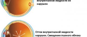
Normally, 2 cubic millimeters of fluid enter the eye every minute, and the same amount should be the volume of outflow, but with certain disorders, the difference between these two values increases.
Also, fluid can accumulate in the organs of vision, which leads to an increase in the indicator, and this can lead to such an additional problem as deformation of the vessels through which the fluid is transported.
There are three types of such violations:
- In case of transient disorders, destabilization of IOP is short-term, and it is restored without the need for treatment within a short period.
- With labile disorders, short-term pressure surges are also observed, which go away on their own, but the processes are regular.
- If the excess of the norm is constant and does not go away, they speak of a stable type of pathology. Such jumps can be dangerous, especially if there is a decrease in IOP. In such rare cases, which can be caused by injuries, infectious and endocrine diseases, dry eye syndrome may appear.
If the indicator increases, which is diagnosed more often, in the absence of treatment, compression of the optic nerve may occur, which subsequently can lead to its atrophy. Changes in pressure in the organs of vision require immediate intervention by specialists and timely treatment.
Intraocular pressure (or ocular tone) is measured in millimeters of mercury. Reference! The optimal indicator of IOP can be considered values ranging from 9 to 23 mmHg, although if the diagnosis is carried out according to Dr. Maklakov’s method, we are talking about a norm of 15-26 units.
Norms of eye pressure for different ages and in different cases In most cases, in adults, the norm of intraocular pressure remains unchanged for people of any age, and this indicator can mainly change in case of some ophthalmological diseases.
- 40 years
The average value for people aged 40 years and older is considered to be from 10 to 23 millimeters of mercury. With such indicators, all metabolic and tear-forming processes proceed in a normal natural mode.
This indicator of fundus pressure is the same for men, women and children, although in a child the indicator rarely approaches 20 units.
- 50-60 years
At the age of 50-60 years, intraocular pressure increases slightly, but this is normal, and a reading of 23-25 units is not considered pathological and does not require intervention.
Although this is already a signal that a person may develop glaucoma and other inflammatory processes, therefore, after fifty years, it is necessary to undergo an ophthalmological examination every six months.
- 70 years old
For people aged 70 years and older, a figure of 23-26 units is considered normal. What is normal eye pressure for glaucoma? Ocular pressure readings change dramatically when glaucoma occurs.
This disease can occur in one of four degrees of severity, which determines how much the indicator will increase:
- At the initial stage of the disease, IOP can fluctuate from normal to 4-5 units higher. Usually the pressure does not exceed 27 millimeters of mercury.
- In severe glaucoma, the value can be from 27 to 32 units
- At a deep stage, the pressure rises to 33 millimeters of mercury.
- When IOP exceeds 33 units, they already speak of the final stage of glaucoma.
Intraocular pressure is measured during any routine ophthalmological examination, since based on these numbers, a specialist can draw conclusions about the presence of certain ophthalmic defects, even if they do not show any symptoms.
General information about standards
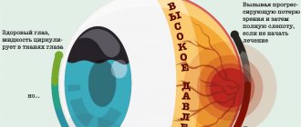
To maintain blood microcirculation in the eyes, which ensures the functioning of the retina and metabolic processes, normal pressure inside the eyes is necessary. This indicator is individual for each person and is generally considered normal when it does not go beyond the reference indicators.
Each age group has its own average parameters. Knowing them, you can understand why vision is deteriorating and what to do about it. A table of intraocular pressure values by age and measurement method will help you monitor the indicators:
- IOP in young people
Balanced eye pressure is a sign of the absence of ophthalmic diseases. At a young age without the presence of pathologies, the indicator fluctuates very rarely, most often due to eye strain at work. For everyday intraocular pressure, the norm in adults varies between 10-20 mm. mercury column.
Deviations may indicate incipient processes in the retina or optic nerve, the first signs of which are a blurred image, eye pain and headache. If symptoms last more than a week, it is better to be examined by an ophthalmologist.
- IOP after 60 years
Until the age of 40, people without ophthalmic pathologies have good vision, but then it begins to gradually deteriorate due to the aging of the body. Anatomical features are such that the eye pressure in women changes faster, and they are exposed to eye ailments more often.
Iphthalmotonus and normal eye pressure in men change more smoothly. At the age of 50, the pressure levels out and, in the absence of congenital or acquired eye diseases, reaches the normal level of 10-23 mm. mercury column. The changes are abrupt in nature and are caused by exacerbation of chronic diseases.
In women, increased pressure in the eyes occurs after 40 years of age during menopause, when the level of estrogen in the blood drops. At the age of 60, patients' retina transforms, which entails an increase in pressure to 26 mm. mercury column according to Maklakov, the occurrence of cataracts and glaucoma.
Normal for glaucoma
An upward change in IOP indicates processes of changes in blood microcirculation in the eye, and serves as a harbinger of glaucoma.
Both at the initial stage of the disease and during its progress, blood pressure readings must be taken twice a day - morning and evening to draw an objective picture.
For elderly people with end-stage disease, measurements are taken 3-4 times a day. The average norm of eye pressure in glaucoma is fixed in the range from 20 to 22 mmHg. At the last stage, the norm reaches 35 mm Hg.
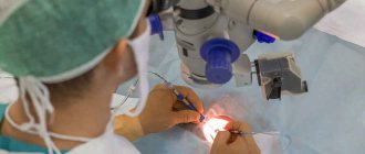
Measuring intraocular pressure
It should be noted that not all people referred for annual preventive examinations to an ophthalmologist are enthusiastic about the upcoming measurement of intraocular pressure. Women may be afraid of ruining carefully applied makeup; men will refer to the absence of any complaints about their own organs of vision. Meanwhile, measuring intraocular pressure is a mandatory procedure for people who have turned 40 or older, even if they assure the doctor that they are in perfect health.
Measuring intraocular pressure is carried out using special equipment and instruments, and in general, modern ophthalmology uses 3 main types of measuring intraocular pressure:
tonometry according to Maklakov
The above-mentioned method according to Maklakov - many patients remember it, know it and most of all dislike it, since drops are dropped into the eyes, providing local anesthesia, and “weights” are installed (for a very short time), which are quickly removed and lowered onto a clean sheet paper to leave imprints indicating the IOP value. This method is more than 100 years old, but it still has not lost its relevance;- Pneumotonometry, very similar to Maklakov tonometry, but different in that it uses an air jet. Unfortunately, this study is not particularly accurate;
- Electron diffraction is the most modern method, successfully replacing the previous two. It is used mainly in specialized institutions (not all clinics can yet afford expensive ophthalmological equipment). The method is classified as non-contact, highly accurate and safe research.
Most often in the Russian Federation and neighboring countries, Maklakov tonometry or non-contact tonometry using an electronograph are used.
Tonometry according to Maklakov
The method has been known and widely used for 120 years. It is a procedure that is carried out using a set of weights of different weights. The measuring device is made of high-quality cylindrical metal. The device is hollow inside. One end of the device is equipped with frosted glass plates with a diameter of 1 mm. The research is carried out in the following sequence:
- To avoid patient discomfort, the procedure begins with local anesthesia.
- Cylindrical weights, pre-treated with a special pigment, are placed on the cornea of the eye.
- The purpose of the weights is to apply pressure to the eyeball with varying intensities.
- Markings are applied using a special pigment.
- The imprint of the painted areas is transferred to paper.
- Indicators are measured using a ruler.
- The procedure ends with eye drops of a disinfectant solution.
Tonometry according to Maklakov has become widespread among ophthalmologists in different countries. IOP measurement using the Maklakov principle is considered to be one of the most accurate and safe technologies. But to find out your blood pressure readings, there is not always a need to go to an ophthalmologist. Let's learn how to check eye pressure at home.
A home eye pressure monitor is a small electronic device. The operating instructions are limited to recommendations for applying mechanical pressure on the eyeball. Having such a device in your home, you can easily monitor eye pressure, based on the norms, at any age: at 30, 40 and 50 years old. This will help prevent vision problems.
Increased intraocular pressure
Increased eye pressure (ophthalmohypertension) is not necessarily the result of age-related changes, as many people think.
The reasons for increased IOP can be very diverse, for example:
- Constant tension on the organs of vision, leading to fatigue;
- Atherosclerosis;
- Persistent arterial hypertension (periodic surges in blood pressure, as a rule, are not dangerous for the eyes);
- Vegetative-vascular dystonia;
- Psycho-emotional stress, chronic stress;
- Fluid retention in the body due to cardiovascular pathology;
- Intracranial hypertension often causes increased fundus pressure;
- Professional activities (wind musicians);
- Individual (strength) physical exercises;
- Medicines used locally;
- Strong tea or coffee (due to caffeine);
- Heart rhythm disturbances, respiratory arrhythmia;
- Features of the anatomical structure of the eye;
- Intoxication;
- An inflammatory process localized in the organ of vision;
- Diencephalic pathology;
- Traumatic brain injuries;
- Diabetes;
- Menopause;
- Hereditary pathology;
- Side effects of certain medications, treatment with corticosteroid hormones.
Increased intraocular pressure is often a sign of glaucoma, the risk of developing which increases markedly after 40 years.
Deviations of intraocular pressure from normal
A decrease in intraocular pressure is observed quite rarely; much more often doctors have to deal with its increase. The risk of developing pathology increases with age; most often, increased intraocular pressure is diagnosed in patients over 45 years of age.
Tonometry according to Maklakov is considered a fairly accurate and reliable technique with minimal dependence on the researcher.
High levels of intraocular pressure are observed with glaucoma, arterial hypertension, severe fatigue, heart failure, hypothyroidism, poisoning, and kidney disease.
Increased intraocular pressure can be of several types:
- transient - the indicator goes beyond the normal range for a short time, intraocular pressure usually normalizes on its own and does not require treatment;
- labile - intraocular pressure either rises or returns to normal, while the jumps are often regular;
- stable – indicates glaucoma or a high risk of its development.
Increased intraocular pressure is manifested by deterioration of vision, a reduction in the field of view, redness of the eyes, headache, pain in the eyes, flashing spots and/or rainbow circles before the eyes, and a feeling of fullness in the eyes. With such a deviation of eye pressure from the norm, compression of the optic nerve is possible; if this happens frequently, it leads to its atrophy and is fraught with complete loss of vision.
A decrease in intraocular pressure is observed with injuries, eye surgeries, low blood pressure, infectious and inflammatory processes, dehydration, retinal detachment, eyeball abnormalities, kidney diseases, endocrine pathologies.
When intraocular pressure decreases, the patient experiences eye pain, redness, dryness, irritation, a feeling of a foreign body in the eye, and rapid eye fatigue. Slightly reduced intraocular pressure is usually asymptomatic.
An experienced ophthalmologist can give a rough estimate of intraocular pressure without instruments by palpating the patient's eyeball through the eyelids.
In order to prevent deviations of intraocular pressure from the norm, it is necessary to adhere to proper nutrition, ensure adequate physical activity, give up bad habits, have sufficient sleep at night, and avoid physical and mental stress. Patients at risk are recommended to be monitored by an ophthalmologist. How many times a year you need to be examined depends on the pathology detected.
Warning symptoms of increased IOP
Increased eye pressure may not show any particular signs of trouble for a long time. A person continues to live in a normal rhythm, unaware of the impending danger, because real symptoms of a pathological eye condition appear only when the IOP changes significantly towards an increase. And here are some signs of illness that may suggest that, putting everything aside, you need to immediately visit an ophthalmologist to check your vision and measure intraocular pressure:
- Pain in the eyes, in the eyebrow area, in the frontal and temporal areas (or on one side of the head);
- “Fog” before the eyes;
- Multi-colored circles when looking at a burning lamp or lantern;
- Feeling of heaviness, fullness and tiredness of the eyes at the end of the day;
- Attacks of unmotivated lacrimation;
- Change in corneal color (redness);
- Decreased visual acuity, lack of image clarity (with glaucoma, patients often change glasses).
An increase in IOP and the development of glaucoma can be suspected if a person often changes glasses because he begins to be unable to see in the “old” ones, and also if this disease was diagnosed in close relatives.
Normal IOP in children
For children, normal intraocular pressure readings are between 10 and 20 mmHg. Art. IOP in patients under 14 years of age is measured using a tonometric study. With increased ophthalmotonus, babies become capricious, become apathetic and lethargic, and have reddening of the whites. They experience discomfort, which affects their behavior.
If elevated eye pressure is detected in children, the causes are most often dysfunction in the endocrine system, in particular, dysfunction of the thyroid gland. At the first signs of increased IOP, the child should be taken to the doctor.
For starters, drops for eye pressure
If the pathological process has not gone too far, but the risk of developing glaucoma is quite high, then treatment usually begins with a direct impact on the high level of IOP, and for this purpose the doctor prescribes eye pressure drops, which:
- Promote fluid outflow;
- Reduce the pressing effect on the eye capsule;
- Normalize tissue metabolism.
By the way, eye pressure drops can cover different pharmacological groups, these are:
- F2α prostaglandin analogues (Travoprost, Xalatan, Latanoprost);
- Beta-blockers (selective - Betaxolol, and - non-selective - Timolol);
- M-cholinomimetics (Pilocarpine);
- Carbonic anhydrase inhibitors (local - Bronzopt, and in addition to drops for eye pressure: systemic - Diacarb in capsules and tablets).
In this regard, it is very important to correctly assess how drugs will affect the hydrodynamics of the organ of vision, whether it will be possible to quickly obtain a hypotensive effect, calculate how often a person will depend on drops, and also take into account contraindications and individual tolerance of individual drugs. If everything did not go very smoothly with the prescribed treatment, that is, no particular effect was obtained from monotherapy with antihypertensive drugs, you have to turn to combination treatment using:
- Travapress Plus, Azarga, Fotil-forte;
- α and β-adrenergic agonists (Adrenaline, Clonidine).
However, even in such cases, it is not at all advisable to use more than two different drugs in parallel.
In addition to the listed medications, for glaucoma (acute attack), osmotic agents are prescribed orally (Glycerol) and intravenously (Mannitol, Urea).
Of course, examples of eye pressure drops are not given so that the patient goes and buys them at the pharmacy on his own initiative. These medications are prescribed and prescribed exclusively by an ophthalmologist.
When treating high ocular pressure, in order to adequately assess the results achieved, the patient is regularly measured for IOP, visual acuity and the condition of the optic discs are checked, that is, the patient works closely with the attending physician during treatment and is under his supervision. To obtain the maximum effect from treatment and prevent addiction to the drugs, ophthalmologists recommend periodically changing eye pressure drops.
The use of drops and other medications that reduce IOP involves treatment at home. For glaucoma, treatment depends on the form of the disease and the stage of the glaucomatous process. If conservative therapy does not give the expected effect, laser treatment is used (iridoplasty, trabeculoplasty, etc.), which allows the operation to be performed without a hospital stay. Minimal trauma and a short rehabilitation period also make it possible to continue treatment at home after the intervention.
In advanced cases, when there is no other way out, surgical treatment for glaucoma is indicated (iridectomy, fistulizing placements, operations using drains, etc.) with a stay in a specialized clinic under the supervision of doctors. In this case, the rehabilitation period is somewhat delayed.
High and low blood pressure
The average rate of ophthalmontus is from ten to twenty millimeters of mercury. If it’s three or four marks higher, that’s also acceptable. This indicator indicates that the metabolism and microcirculation of the eye are normal and that the retina is functioning normally.
Reduced eye pressure in women after 60 occurs, but much less frequently. If treatment and prevention are not started in time at the first symptoms, glaucoma may appear. It is more difficult to get rid of and can lead to partial or complete blindness.
Correction of eye pressure is based on the use of medications. But! A positive effect will be observed only after the eye gets used to the product. Everything is individual, and even a very good product may not be right for you. Don’t worry, your doctor will definitely choose the right drug for you.
Decreased fundus pressure
Doctors involved in the treatment of eye diseases are also aware of another phenomenon that is opposite to increased IOP - ocular hypotension, eye hypotension or decreased fundus pressure. This pathology develops quite rarely, but this does not make it any less dangerous. Unfortunately, patients with eye hypotony reach the ophthalmologist's office when a significant percentage of their vision has already been lost.
This late presentation is explained by the fact that there are no obvious signs of the disease; the initial stage proceeds almost without symptoms, except for a not very pronounced decrease in visual acuity, which people attribute to eye strain or age-related changes. The only symptom that appears later and can already alert the patient is dry eyes and loss of natural shine in them.
Factors that contribute to a decrease in intraocular pressure are not as diverse as the prerequisites that increase it. These include:
- Injury to the organs of vision in the past;
- Purulent infections;
- Diabetes;
- Dehydration
- Arterial hypotension;
- Alcoholic drinks and drugs (marijuana);
- Glycerin (if consumed orally).
Meanwhile, a person who pays as much attention to the eyes as other organs can prevent the unwanted effects of lowering IOP by visiting an ophthalmologist and talking about the above-mentioned “minor” symptoms. But if you do not notice the signs of eye health in a timely manner, you may find yourself faced with the development of an irreversible process - atrophy of the eyeball.
Treatment at home involves the use of eye drops: Trimecaine, Leocaine, Dicaine, Collargol, etc. Products with aloe extract, as well as B vitamins (B1), are useful.
Blood pressure norms after fifty
The content of the article:
Pressure measurement should take place in a quiet place and in a comfortable position of the person. After all, any load can significantly change it and then the indicators will be incorrect. The body is able to control blood pressure (BP) itself, and when minor loads are possible, its numbers will rise by 20 mmHg. Art. There is nothing dangerous in this, since under load the organs need more blood, so blood circulation increases.
As a woman ages, various irreversible processes occur in her body, which affect changes in blood pressure. The older the patient, the more significant the deviation from the norm.
Arterial
When blood flow influences the walls of blood vessels, blood pressure occurs, the indicators of which depend on the strength of this influence. The very intensity of blood flow depends on how the heart muscle works. Based on this, pressure is measured using two indicators - upper (systolic) and lower (diastolic).
Table 1. Normal blood pressure at the age of 50 - 55 years
| Age (years) | Men | Women |
| 50 – 55 | 142/85 | 144/85 |
The upper indicators indicate the resistance of blood vessels during shocks of blood flow at the moment of greatest contraction of the heart muscle. The lower ones indicate minimal vascular resistance at the moment of greatest weakening of the heart muscle.
The normal level of upper blood pressure is considered to be 130–140 mmHg. Art. If the indicators are higher, it is recommended to consult a doctor to prescribe treatment and normalize the condition. Regularly high blood pressure is a sign of a disease called hypertension. Then the doctor prescribes special medications, physiotherapy and other types of therapy to the patient, depending on the individual performance indicators of the individual person’s body.
Read also
Normal heart rate of an adult 50 years old
Youth fades with age, and this fact, unfortunately, cannot be changed. Approaching fifty years of age, a woman can... Read the article >
The normal lower pressure is considered to be 80–90 mmHg. Art. In older age, low blood pressure can lead to brain pathology. Very often, doctors take measurements on both hands. The numbers must be the same, or there may be a deviation of 5 mmHg. Art. on the hand that is considered working. If the difference is 10 mm Hg. Art. and higher, this indicates atherosclerosis or stenosis of blood vessels.
Intracranial
This pressure occurs as a result of impaired circulation of the cerebrospinal fluid (CSF). The latter invariably circulates under pressure through the cavity of the spinal cord, through the stomach of the brain, and also between the bones of the skull. But sometimes there is a concentration of liquid in one place or a decrease in it in a certain area. Then there is increased or decreased intracranial pressure (ICP). ICP standards: 3–15 mm Hg. Art.
Ophthalmic
This indicator is individual for everyone, as it depends on various processes and phenomena that occur in women after 50 years. The norm is considered to be 10–23 mmHg. Art. It is this figure that allows you to preserve the optical functions of the retina and maintain the necessary visual acuity.
Some advice for patients with high eye pressure
Patients suffering from increased IOP, which threatens the development of the glaucomatous process, are recommended to follow some rules of prevention:
- Try to avoid hypothermia, stress and excessive physical exertion (hard work, heavy lifting, tilting the head and body, causing blood to flow in larger quantities than the brain needs;
- Stop doing athletics, but do not shy away from walking (away from city noise and gas pollution), doing feasible gymnastics for the respiratory system and the whole body, hardening the body;
- Treat chronic concomitant diseases;
- Once and for all, regulate the regime of work, night sleep, rest and nutrition (a lactic acid diet enriched with vitamins and minerals is preferable);
- On sunny summer days, when going outside, make it a rule not to forget glasses at home that provide eye comfort and protection (glasses should be purchased at Optics, and not at the market where they sell sunglasses, which can further increase EDC ).
As for low blood pressure, as mentioned earlier, it is a rare case, so patients who experience suspicious signs (dull, dry eyes) can be advised to contact a specialist as soon as possible, who will tell you what to do next.
Diagnostic methods
It is impossible to determine IOP at home. Therefore, it is necessary to consult a doctor who will conduct an ophthalmological diagnosis and be able to determine the indicator. There are several diagnostic methods. The main ones are:
- Pneumotonometry.
- Electronograph.
- Tonometry according to Maklakov.
Pneumotonometry
This method allows you to measure intraocular pressure using a special pneumotonometer, which operates using a stream of air. The procedure is as follows: the patient places his head on a special stand and focuses his gaze on the indicated mark. The device directs a stream of air towards the eye. IOP is determined by how the shape of the cornea changes. Pneumotonometry is safe for human health and can be performed several times in a row.
Electronograph
This procedure is considered more extended, as it is carried out within 4 minutes. During this time, all changes in the condition of the eyeball are recorded. The measurement is carried out using a special device - an electronic tonograph. The patient lies on his back, his eyelids are fixed and spread apart with a special ring. This results in more enhanced and precise hydrodynamics of the eye.
Tonometry according to Maklakov
This contact method of measuring IOP is very popular. Its essence is as follows: a weight is placed on the eye, which is pre-wetted with paint. After this, the weight is placed on paper and the pressure is determined from the imprint. The procedure itself is painless and does not cause discomfort, since an anesthetic is first instilled into the eyes.
Read also
Beginning of menopause, menstruation symptoms by age 50
There comes a time in every woman’s life when serious changes occur in the body. This means that she...
Read the article >
Table of IOP indicators depending on the measurement methods used
It is important to keep the dynamics of intraocular pressure under control in order to promptly detect the development of glaucoma, a disease that causes partial and complete loss of vision.
| Norm | Initial manifestations of glaucoma, stage 1 | Glaucoma, stage 2, moderate | Glaucoma, stage 3 | Glaucoma, end stage | |
| Instrumental method (tonometry) | 10-25 | 24-26 | 26-36 | — | — |
| Computer diagnostics | 10-20 | 21-22 | 22-26 | 26-33 | 33-35 and above |
Using a tonometer, it is not possible to determine IOP in advanced glaucoma. According to medical standards, women over 40 years of age should visit an ophthalmologist to monitor intraocular pressure at least three times a year. Men should make such visits at least twice a year.
It is necessary to take timely measures to normalize ophthalmic tone in order to maintain eye health and visual acuity for a long time.