Types of survey urography of the kidneys
Depending on the technique of diagnostic manipulations, there are two types of urography:
| Type of procedure | Distinctive features |
| Overview | The resulting images clearly show the area from the kidneys to the upper urinary canal. The procedure does not involve the use of a radiopaque substance and allows you to determine the structure of the kidneys and identify large tumors and stones along with gross pathological changes in tissue. The procedure is performed after traumatic injuries to the lower back and suspected pathological conditions of the abdominal organs. It almost always precedes excretory urography. |
| Survey excretory urography (intravenous, contrast) | The technique requires the use of a contrast agent, which is injected into a vein into the patient. The technique is based on the ability of the kidneys to secrete pre-injected contrast, which makes it possible to determine the function of the kidneys and organs of the MPS. Thanks to it, the diagnostician can obtain images of small renal structures and identify their pathologies and dysfunctions. Thus, the technique makes it possible to detect thrombosis and atherosclerosis, stones of any size and tumors in the initial stages of development. |
SURVEY UROGRAPHY
Survey urography is a diagnostic procedure performed in urology, the main task of which is to determine the presence of stones, as well as their location in the kidneys, and is an X-ray image of this area.
The survey urography procedure is part of the initial examination.
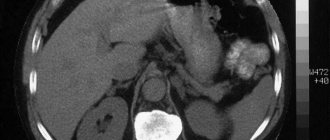
A urogram allows you to visualize a fairly large part of the human body; the image starts from the kidney area and ends with the beginning of the urethra. Due to the fact that the image is quite large (30x40 centimeters), doctors have the opportunity not only to determine the position of the kidneys and the location of the stones, but also to assess the condition of the skeletal bones and abdominal organs.
Indications, contraindications and limitations of survey urography:
A urogram is the first examination that a specialist prescribes when a patient complains of problems with the genitourinary system, however, before doing a survey urography, it is necessary to understand that the procedure has some contraindications, in the presence of which it is impossible to carry out.
Contraindications to survey urography:
Survey urography is an X-ray procedure, and therefore it is contraindicated for women who are pregnant. During pregnancy, a woman is strictly contraindicated for any examinations that include radiation, which is associated with the negative impact of the device’s rays on the fetus.
In addition to pregnancy, there is a disease in the presence of which the procedure cannot be performed - renal failure. Also, survey urography is not performed for people prone to allergic reactions to the substance used during the procedure.
Restrictions on conducting survey urography:
If a person has recently undergone examinations using barium contrast agent, survey urography is performed after the intestines are completely cleansed.
Indications for survey urography:
- suspicion of urolithiasis;
- inflammatory processes of the prostate gland;
- suspicion of neoplasms;
- cystitis, both acute and chronic types;
- pathological changes in the genitourinary system;
- dystopia, hyperplasia;
- parasitic diseases.
Preparation for survey urography:
The procedure for survey urography requires careful preparation of the patient, which, to a large extent, consists of following a special diet that allows for maximum cleansing of the intestines and stomach.
In order to obtain the most reliable readings on an x-ray image, the patient is recommended to adhere to the following diet for 2-3 days before the scheduled procedure:
- in order to avoid intestinal bloating and gas accumulation, it is necessary to refrain from eating potatoes, bread, members of the legume family, fermented milk products and sugar;
- to cleanse the intestines it is necessary to take sorbents, the most accessible and effective of which is activated carbon;
- before the appointed day in the evening and on the appointed day of the procedure in the morning, you should refuse to eat any food, the only thing you can drink is unsweetened tea;
- Before the procedure, refrain from drinking water (it can cause bloating).
If for any reason the patient experiences flatulence, the procedure is postponed until it is eliminated. Survey urography is performed in the X-ray room, immediately after checking the intestines with fluoroscopy.
Indications and contraindications for survey and excretory urography of the kidneys
| Indications | Contraindications |
The procedure without contrast is carried out:
X-rays with contrast are performed when:
| Contraindications to the procedure are due to radiation exposure, which, despite its minimal dose, can have a negative effect on:
Refusal to use contrast is necessary in the following cases:
|
How to prepare for urography?
In order for the diagnosis to obtain all the necessary data, the patient needs to properly prepare for it. The main goal is to minimize the processes of gas formation in the intestines, since gases make it difficult to visualize the areas being examined. In order to achieve the desired effect, a few days before the x-ray the patient needs to stop consuming foods that contribute to gas formation.
If necessary, the doctor may prescribe sorbents to help cleanse the intestines. No later than 12 hours before the procedure, the patient must refuse to eat food, since the procedure is performed on an empty stomach.
The use of a contrast agent, in addition to the above, will require a cleansing enema. The patient will also need to undergo an allergy test to exclude the development of an individual reaction to the contrast agent.
Preparing for the study
The quality of the picture directly depends on the condition of the intestines and bladder. To reduce diagnostic error to a minimum, ensure that the preparatory steps are carried out accurately, without neglecting the advice of your doctor. Preparation for examination using intravenous urography deserves special attention :
- For 3-4 days, eliminate foods that cause gas from your diet (dairy products, fresh fruits and vegetables, baked goods, sweets).
- In parallel with the preparatory diet, start taking enterosorbents (activated carbon and similar drugs).
- Take your last meal no later than 8-12 hours before the start of the procedure.
- On the eve of the study, use a mild laxative to thoroughly cleanse the intestines.
- On the day of your urography, limit your water intake. Immediately before the procedure, empty your bladder as much as possible.
ATTENTION! An important stage in preparing a patient for urography is an allergy test and blood biochemistry analysis to exclude renal and liver failure. In case of increased nervousness, you should take a mild sedative (it is advisable to consult a doctor).
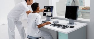
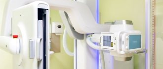
How is urography performed?
Before entering the x-ray room, the patient will need to remove all metal objects. The doctor will ask him to take a sitting or lying position, depending on the purpose of the study. So, if it is necessary to identify the mobility of the renal structures, the patient must stand; identify the location of stones or tumors - lying on your side or bending over three-quarters. It is important for the patient to follow the instructions of the radiologist - this is the only way the procedure will allow obtaining images of the desired clarity.
During survey urography without contrast, one or two pictures are taken of the area of the third and fourth vertebrae; with contrast, a contrast agent is first introduced, the time of its administration is recorded, and pictures are taken two, five, fifteen, sometimes sixty minutes after that. Thus, depending on the method of implementation, the procedure can last from 5 minutes to one hour.
Types of urography
The essence of the method comes down to the introduction of a special contrast agent into the body, which “highlights” the structure of the urinary system, helps to better examine individual structural formations and make an accurate diagnosis.
REFERENCE! Kidney urography is considered a painless procedure, but may cause some discomfort to the patient. For this reason, many people prefer to replace it with CT or MRI. However, it compares favorably with these methods due to its comparatively low cost, while being almost as effective.
There are several types of urography, which allows the attending physician to choose the optimal method of examination, taking into account the characteristics of each patient.
Survey urography
Survey urography of the kidneys is the simplest and safest diagnostic technique, as it does not require the administration of a contrast agent. It gives a general overview of the condition of the entire abdominal cavity, including the bone apparatus with adjacent organs, but at the same time allows to identify only obvious pathological changes (neoplasms, stones, extensive deformations, injuries and parasitic infections).
Taking into account modern computer technology, survey urography can be regarded as an accessible express method of examination.
Excretory urography
The classic method of contrast urography of the kidneys involves an x-ray examination after preliminary administration of an intravenous drug.
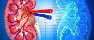
Once in the bloodstream, the “contrast” quickly reaches the filtering system of the kidneys and completely fills the entire branched “tree” of the urinary system, starting from the smallest nephrons and ending with the bladder. This significantly increases the clarity of structural formations on an x-ray. X-rays in different projections allow you to evaluate the three-dimensional structure of the urinary tract and diagnose the slightest pathological structures (tumors, cysts, stones).
By starting to take an X-ray at the moment of administration of the contrast agent, you can trace the path of its movement and assess the physiological activity of the urinary system.
Intravenous diagnostics
The method involves long-term administration of a contrast agent through a dropper, therefore it is most often used in a hospital setting to collect the most detailed information before a major intervention (for example, before surgery).
Intravenous urography of the kidneys allows not only to identify structural abnormalities, but also to accurately determine such a physiological parameter as the speed of urine passage through the urinary tract. The test is especially informative when compared with hemodynamic parameters.
What does survey urography reveal?
The procedure is highly informative, especially if it is performed using a contrast agent. Thanks to it you can identify:
- congenital structural disorders of the renal structures;
- abnormal enlargement or reduction of MPS organs;
- a pathological condition such as kidney prolapse;
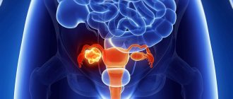
- neoplasms at different stages of development of a benign or malignant nature;
- stones, their sizes and exact location;
- any pathological changes in the structures and tissues of the MPS organs.
If the data obtained was not sufficient to make a diagnosis, the patient may be prescribed magnetic resonance imaging.
If you want to be sure that the procedure will not harm your health and will be carried out in accordance with international standards, contact the clinics of the Doctor Nearby network.