What is kidney topography
Have you been trying to cure your KIDNEYS for many years?
Head of the Institute of Nephrology: “You will be amazed at how easy it is to heal your kidneys just by taking it every day...
Read more "
Topographic anatomy is a science that studies the location of internal organs (shape and structure) and parts of the human body, and their interaction. Topography of internal organs deals with a detailed examination of the structure of the insides of the human body, the process of blood supply to each organ, the projection of organs onto the skin (used for surgeons) and their location relative to the skeleton. The subject of topography studies includes lymph circulation and changes that affect a person with age, depending on body type and gender.
The topography of the human body provides detailed theoretical information about the location of a particular organ relative to others for surgical surgery. Any practicing surgeon must imagine how his operation will proceed layer by layer, and this requires a detailed study of the discipline of topographic anatomy.
The topography of the kidneys considers the relative position of the organs themselves and those adjacent to them, and the functioning and location of the arteries, vessels and veins that carry blood, the lymphatic and nervous systems of the kidney.
The kidneys are located on opposite planes from the spine, closer to the lumbar area, behind the inner part of the peritoneum, outside its limits. Each occupies a bed, the formation of which was facilitated by a complex of muscles: on the side and behind - the large and quadratus lumborum muscles, above - the diaphragm, literally - the transverse abdominal muscle.
Protective and nourishing tissues of the kidney
The cortex connects to the fibrous capsule, from here the interlobular layers originate, invisible to the naked eye. In addition to fibers, the capsule contains a thin layer of smooth muscle. By contracting, this small muscle layer maintains the internal pressure of the kidney, which is necessary for the functioning of the renal system, exactly reproducing filtration actions.
The kidney is wrapped in a fatty capsule made of porous fibrous tissue (fat). The fatty capsule of the kidney has the thickest layer on its posterior surface. This structure helps to ensure optimal attachment of the kidney. When maintaining a diet and sudden weight loss, the fat capsule also loses weight, the dense layer melts, and there is a risk of kidney movement (a wandering kidney is diagnosed).
At the highest point, the kidney is covered by the renal fascia, characterized by a two-layer plate of leaves. The leaves located in front and behind, along the lateral edge and upper edge of the kidney, grow together, and from below, in the form of an enclosure into each other, they go along the urinary tract to the bladder. On the inner side, fascial leaves in front and behind the vessels in 65-75% of people grow together with the leaves of the other side.
Syntopy and skeletopy of the kidneys
Syntopy is the location of an organ relative to others in the human body.
Skeletotopy is the normal location of organs in the human body, relative to the human skeleton and its parts.
The kidneys have places of contact with many internal organs. But it is not the body of the kidney itself that directly concerns, but its membrane – the fascial-cellular layer.
The left kidney is located at the height of the 12th thoracic and two upper lumbar vertebrae. The right kidney is located below the left, almost 2 cm.
Syntopy of the left kidney
The upper part (upper pole) of the left kidney is united with the left adrenal gland.
The anterior region, if we consider the organ, starting from the upper pole to the bottom: 30% touches the stomach, the next 30% touches the pancreas, and the rest touches one of the loops of the small intestine.
Along the lateral edge, viewed from top to bottom: the renal membrane rests on the spleen, the flexure of the colon, and the initial part of the large intestine. Along the medial edge, the kidney rests on the pancreas. And the posterior area of the surface is attached to the muscles of the renal bed.
Syntopy of the right kidney
The right kidney is covered from above by the corresponding adrenal gland.
30% of the anterior inferior surface of the right kidney touches the right flexure of the colon. The remaining upper part is adjacent to the liver.
Along the lateral edge, the right kidney touches the terminal part of the ascending colon. Along the medial edge it touches the descending part of the duodenum. With its posterior surface, like the left kidney, the right one is attached to the muscles of the kidney bed.
Skeletotopy of the kidneys
Right kidney: from the 12th thoracic to the upper edge of the 4th lumbar vertebrae, left kidney: from the 11th thoracic to the upper edge of the 3rd lumbar vertebrae. The location of the kidneys in females is slightly different from males; the organs are located one floor of the vertebra below.
The kidneys may be located slightly higher or slightly lower, and their abnormal location is common. Most often, the right kidney is affected by such pathologies.
Vessels and nerves of the kidney
A fifth of all the blood pumped by the heart into the aorta rushes to the kidneys through the 2 renal arteries. At the renal hilum the artery branches into 4-7 interlobar arteries. The interlobar arteries pass into the arcuate arteries - between the cortex and the medulla of the kidney. Interlobular arteries branch off from the arcuate arteries, ascending into the cortex and, with a certain periodicity, creating afferent (or afferent) arterioles that deliver blood to the capillary collections of the renal glomeruli. The efferent (or efferent) arterioles distribute blood from the glomeruli to the second capillary system, which partially follows into the medulla, forming a vascular network for the renal tubules. As a result, the blood flow flows into the venules, then through the arcuate veins into the interlobar veins, from them into the renal veins and the inferior vena cava.
Lymphatic vessels from the body and fibrous capsule of the kidney come to the gates of the kidney, where they unite and travel as part of the renal pedicle to the regional lymph nodes.
The supply of organs and tissues of the kidney with nerves is accomplished by the renal nerve plexus. Branches emerge from the renal plexus to the ureter and adrenal gland.
In addition to the location of organs relative to neighboring ones and the human skeleton, topographic anatomy examines the changes that occur in the human body during certain pathologies, and the movement of organs during diseases. This helps the surgeon accurately determine the location of all important organs and systems in the human body, and intervene to introduce less imbalance. Topography is a fairly accurate science, its study is extremely important for practicing doctors. It is with the help of kidney topography, as directed by the sick patient, that the affected organ can be determined, the exception being if the pain syndrome is transmitted to the renal area.
The article is for informational (approximate) nature and cannot be used as a teaching aid
The folk way to cleanse the kidneys! Our grandmothers were treated using this recipe...
Cleaning your kidneys is easy! You need to add it during meals...
Excretory and homeostatic function
The main task of the kidneys is the formation of urine and the removal of metabolic products, excess water, vitamins, salts, hormones, drugs, etc. Therefore, laboratory urine tests are so indicative in diagnosing health in general and kidney function in particular. But the excretory function is not the only one. An equally important function is homeostatic, that is, maintaining a constant composition and properties of the internal environment - homeostasis. The kidneys maintain water and electrolyte balance; they control the level of excreted substances and their reserves in the body. Together with the lungs, the kidneys, by removing acidic and basic products, maintain the acid-base balance of the body. The kidneys secrete acids, such as phosphoric and sulfuric acids, which are formed during protein metabolism.
Blood pressure and blood secretion
It is the kidneys that play the main role in the mechanism of long-term regulation of blood pressure. And this is done by removing water and sodium chloride from the body. The kidneys synthesize prostaglandins and renin, which are involved in the mechanisms of rapid regulation of blood pressure. The kidneys also have a direct role in producing blood. This is a kind of “mini-laboratory” through which blood passes. And if there is not enough oxygen in it, then a chain of sequential reactions starts. It all starts with the production of erythropoietin, which stimulates the maturation of red blood cells. This may explain why patients who undergo hemodialysis for a long time develop severe anemia. And it is explained precisely by insufficient production of erythropoietin.
Kidney structure
Another important function of the kidneys is to cleanse the blood of products that are formed during metabolic processes. By filtering the blood in the kidneys, all waste products are removed from the body in the urine. This filter passes through two thousand liters of blood per day, that is, the entire volume of blood of an adult is filtered 35 times per day. Blood purification occurs in nephrons, and there are about two million of them in total. Each bud is bean-shaped and has a depression in it on one side. The outside of the kidneys is covered with dense fibrous tissue; there is also fatty tissue necessary for their protection and shock absorption. On the concave surface of the kidney there is an opening called the renal hilum, which includes the renal vein, the artery of the same name and the pelvis, which then passes into the ureter.
Urinary function of the kidneys
Anatomically, the structure of the kidney can be divided into medulla and cortex. If you examine the kidney in section, you will see curled and radiant areas. These rays dissect the medulla of the kidney, and pyramids are formed that look like lobules. They consist of glomeruli and tubules and the basic units of the kidney - nephrons. The glomerulus is a plexus of blood vessels that filter all incoming blood. While reading this article, the kidneys filtered the entire volume of blood, which is an average of 4-5 liters. Primary urine is formed in the glomeruli, which travels further through the system. Primary urine moves further along the nephrons and tubules and here reabsorption occurs, that is, the reabsorption of water. At the top of the renal pyramid there is a papilla that drains urine into special anatomical structures - the renal calyces. And they, connecting with each other, form the renal pelvis, which passes into the ureter.
Filtration of blood in the kidneys and their innervation
Blood is transported to the kidney by the renal artery.
As soon as it enters it, it divides into interlobar arteries, which are distinguished by a smaller caliber. And these middle arteries are divided into interlobular and arcuate, turning into capillaries. The afferent arterioles depart from the arcuate arteries; they supply blood to each glomerulus. The distribution of blood through arteries and arterioles reduces blood pressure in the organ. The main filtration occurs in the small capillaries of the cortex, and from here the blood moves into the straight vessels. This entire complex blood flow system was created with one purpose - to cleanse the blood, evaluate its condition and return it to the general bloodstream for further circulation. The innervation of the kidneys is provided by the branches of the renal plexus. In this interweaving there are branches of the vagus nerve, as well as processes that extend from the spinal nodes. This is how complex one of the most important organs in our body is - the kidneys. Take care of them! Text: Yulia Lapushkina.
General information about the kidneys
The kidneys are a paired organ of the excretory system. The kidneys are a paired organ of the excretory system. Up to 200 liters of blood are pumped through them per day, while in the human body there is only 3 liters. When pumping blood, the urinary organs cleanse its plasma of all toxic compounds, which are subsequently excreted along with urine.
The main functions of the kidneys are:
- Excretory (excretory). That is, the excretion of urine with salts, toxins and breakdown products of fats, proteins, carbohydrates.
- Metabolic. It consists of regulating the water-salt balance of the human body.
- Endocrine. It is based on the production of hormones necessary for the functioning of the body. In particular, these are erythropoietin, renin, etc.
- Synthesis function. In this case, the kidneys synthesize glucose.
Important: it is worth knowing that if the functioning of one of the functions of the urinary organs is disrupted, this is manifested by swelling of the face and limbs, increased blood pressure, and changes in the color and quality of urine. If one or more suspicious symptoms appear, you should contact a urologist immediately.
Is disability given after kidney removal and what are the conditions?
Kidney transplant
Main article: Kidney transplantation
With a decrease in the number of functioning nephrons, chronic renal failure develops, which, when progressing to end-stage renal failure, requires treatment with hemodialysis, peritoneal dialysis or kidney transplantation. Kidney transplantation is the most effective type of renal replacement therapy, including because it replaces all kidney functions, while dialysis partially compensates only for the excretory function of the kidneys, and to replace other kidney functions, the use of drugs (erythropoietin, vitamin D metabolites and etc.).
In 2011, 76 thousand kidneys were transplanted worldwide (a total of 110 thousand organs were transplanted)[4].
What is a renal pedicle and what diseases does it have?
In order to understand what a renal pedicle is and why it is needed, it is necessary to imagine the structure and location of the kidney itself in the body. These paired organs are not located at the same level; the right kidney is relatively higher than the left. The upper edge of the right is located opposite the 11th rib, and the edge of the left is opposite the 12th rib. The kidney gates are also located at different heights, on the right kidney opposite the 12th rib, on the left kidney opposite the intercostal space between the 12th and 13th ribs.
If we consider the kidneys from the back, the renal hilum is located opposite the 1st lumbar vertebra, in some cases between the 1st vertebra and the intervertebral cartilage. This is the topography of the kidneys. The hilum of the organ consists of an artery, vein, nerve fibers, vessels of the lymphatic system, as well as the pelvis, which collects urine and the ureter, which drains it into the bladder. All these vessels, nerves and branches have the common name pedicle.
The order of location of these vessels is strictly distributed. The uppermost vessel is a vein, through which blood leaves the kidney; below is an artery, through which oxygen-rich blood enters the organ. Even lower is the pelvis with the ureter. Medical students use a certain word form to remember this order, namely vein, artery, pelvis - Valya.
What is a renal pedicle?
The leg has a rather complex system of blood vessels. The artery, vein and pelvis are located in an anterior position. Based on this, we can say that the gonadal vein joins the hollow below the peduncle itself. The lumbar vein approaches the vena cava from the posterior side of the kidney, the connection occurs in the lower part of the vena cava.
Located on the right, the adrenal vein flows directly into the vena cava, entering it from behind and from the side. The vessels enter the left kidney in a slightly different order, which makes it possible to ligate the renal vein near the vena cava and at the same time the viability of the organ will be preserved. On the right side, such ligation of the vessel will lead to the death of the organ.
Intrarenal anatomy is a rather complex subject of study. Only an experienced surgeon can work with such an intricate vascular system. After all, it is so complex that it is conditionally divided into 5 segments (apical, upper anterior, middle, lower and posterior).
In iptrarenal surgery, doctors often encounter various renal anomalies, such as a horseshoe kidney, and other defects are possible. These anomalies do not allow operations to be carried out according to standard schemes. Often the doctor improvises during the operation or thinks through a unique strategy at the stage of planning the operation based on the results of the angeogram.
Pathologies
Nephropathy
Unfortunately, under unfavorable conditions, every internal human organ is susceptible to the negative effects of negative factors, which subsequently cause malfunctions.
The pelvicalyceal system of the body is no exception.
Such a complex system will work flawlessly only if it is in absolute health.
Failure of any component leads to serious disorders of the urinary system.
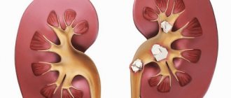
The most serious disorder, in which the calyxes of the kidneys are primarily affected, is hydrocalycosis. There are many reasons that provoke this pathology.
These include both congenital anomalies of various components of the kidneys and the occurrence of problems at any stage of life:
- kinking of the ureter;
- blocking the flow of urine;
- blockage of the ureter;
- reflux;
- infections;
- neurological pathologies.
Structure of the kidney organ
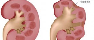
Hydrocalycosis does not manifest symptoms on its own; signals about the problem begin to arrive only after the first accompanying complications appear.
Hydrocalycosis is a disease in which the calyces of the kidneys become very stretched and dilated.
The expansion does not occur without a trace, but is accompanied by complete atrophy of the renal papilla, as a result of which the normal outflow of urine is disrupted, due to the fact that the urinary tract from the kidney to the bladder becomes practically closed.
Hydrocalycosis can affect each kidney individually or both at the same time. Medical statistics say that most often this disease affects the right kidney.
Kidney functions
Hydrocalycosis is often confused with megacalycosis, although they have nothing in common with each other.
Although with this pathology the renal calyces are significantly enlarged, the renal papillae are completely absent, but, what is very important, the outflow of urine occurs without failure.
It is the excessive increase in the size of the cups and pelvis that causes severe lumbar pain. The calyces can be enlarged up to 4 mm, and the pelvis up to 7 mm.
Evolution of the kidney
The excretory organs of vertebrates are the kidneys -
paired compact organs, the structural unit of which is represented
by the nephron.
In its most primitive form, it is a funnel that opens as a whole and is connected to the excretory canaliculus, which flows into the common excretory duct -
the ureter.
In the phylogenesis of vertebrates, the kidney went through three stages of evolution:
the pronephros - the head, or pronephros;
the primary kidney is the trunk, or mesonephros, and the secondary kidney is the pelvic, or metanephros. The forebud fully develops and functions as an independent organ in the larvae of fish and amphibians. It is located at the anterior end of the body, consists of 2-12 nephrons, the funnels of which are generally open, and the excretory tubules flow into the pronephric canal, which is connected to the cloaca. The kidney has a segmental structure. Dissimilation products are generally filtered from the blood vessels, which form glomeruli near the nephrons (Fig. 14.33, A
).
Rice. 14.33. Evolution of tefron. A-
preference;
B, C—
primary kidney;
G—
secondary kidney:
1
— collecting duct,
2—
excretory canadian,
3—
nephrostomy,
4—
coelom,
5—
capillary glomerulus,
6—
capsule, 7,
8—
tortuous canadian,
9—
nephron loop
In adult fish and amphibians, posterior to the kidneys, in the trunk segments of the body, primary kidneys are formed,
containing up to several hundred nephrons.
During ontogenesis, nephrons increase in number due to their budding from each other with subsequent differentiation. They come into contact with the circulatory system, forming glomerular capsules. The capsules have the form of double-walled cups in which vascular glomeruli are located, so that dissimilation products can flow from the blood directly into the nephron. Some nephrons of the primary kidney retain connection with the coelom through the funnels, others lose it (Fig. 14.33, B, C
).
The excretory tubules lengthen and they reabsorb water, glucose and other substances into the blood, and therefore the concentration of dissimilation products in the urine increases. However, a lot of water is lost through urine, so animals with such a kidney can only live in an aquatic or humid environment. The primary kidney retains the characteristics of a metameric structure.
In reptiles and mammals, secondary buds arise.
They are formed in the pelvic region of the body and contain hundreds of thousands of nephrons of the most perfect structure.
In a newborn child, there are about 1 million of them in the kidney. They are formed due to the repeated branching of developing nephrons. Nephrons do not have a funnel and thus lose complete connection with the coelom. loop of Henle
also appears (Fig. 14.33,
D
).
This structure of the nephron ensures not only complete filtration of blood plasma in the capsule, but also, more importantly, effective reabsorption of water, glucose, hormones, salts and other substances necessary for the body into the blood. As a result, the concentration of dissimilation products in the urine secreted by the secondary kidneys is high, but its quantity itself is small. In humans, for example, per day about 150 liters of blood plasma are filtered in the capsules of the nephrons of both kidneys, and about 2 liters of urine are excreted. This allows animals with secondary buds to be more independent of the aquatic environment and inhabit dry areas of the earth. In reptiles, secondary kidneys remain throughout their lives at the site of their original formation - in the pelvic region. They show the features of the primary metameric structure.
The kidneys of mammals are located in the lumbar region, and in most of them the external segmentation is not pronounced. In human ontogenesis, a pronounced recapitulation is revealed in the development of the kidney: the formation of first the pro-, then the meso-, and later the metanephros. The latter develops in the pelvic region, and then, due to differences in the growth rates of the spine, pelvis and abdominal organs, moves to the lumbar region. In a five-week-old embryo, the coexistence of a prebud, a primary bud, and also the rudiments of a secondary bud can be detected (Fig. 14.34).
At the initial stages of development, the human kidney is segmented. Later, its surface is smoothed out and metamerism is preserved only in the internal structure in the form of renal pyramids. Malformations of kidney development in humans, based on their phylogeny, are diverse. Preservation of mesonephros and unilateral absence of a secondary kidney have so far been described only in mice, although in principle such an anomaly is possible in humans. is relatively common ,
having one or even several ureters;
its complete doubling is also possible.
is often observed
,
associated with a violation of its movement in the 2-4th months of embryonic development (Fig. 14.35).
Rice. 14.34. A five-week human embryo with three generations of kidneys:
1
—pre-kidney,
2—
primary kidney,
3—
secondary kidney
Rice. 14.35. Ontophylogenetically determined kidney malformations:
1
—double kidney,
2—
double ureter,
3—
pelvic ectopia of the kidney,
4—
adrenal glands