| Bladder exstrophy | |
| ICD-10 | 64.1 |
| ICD-10-CM | Q64.10 |
| ICD-9 | 753.5 |
| ICD-9-CM | 753.5 |
| OMIM | 600057 |
| DiseasesDB | 33377 |
| eMedicine | ped/704 |
| MeSH | D001746 |
| Media files on Wikimedia Commons | |
Bladder exstrophy
(derived from the Greek word ekstrophe, which means displacement, eversion, eversion of the bladder) is a congenital malformation of the bladder in which the bladder is not inside, but outside. The anterior wall of the bladder is missing, as is the corresponding section of the abdominal wall, which is split, and thus the bladder is exposed. Urine flows out through the openings of the ureters. There is urethral epispadias.
With the exception of the genitourinary system, children with exstrophy are no different from other children and develop in the same way as their peers.
What abnormalities characterize exstrophy?
- Epispadias: in boys, the urethra and the head of the penis are split along the dorsal surface; in girls, the urethra is also not formed, and its outlet is located between the split clitoris and the separated labia minora.
- The corpora cavernosa are shortened due to the pubic bones diverging to the sides.
- The bladder neck and sphincter are absent. The bladder neck is the lower part of the bladder in the shape of a kind of bowl (watering can), which contains a ring of muscles that open and close the exit of urine from the bladder. It is this group of muscles (sphincter) that makes urinary control possible.
- Small bladder capacity.
- Incorrect location of the ureters (excretory tubes through which urine is discharged from the kidneys to the bladder): the ureters are located differently than necessary at the entrance to the bladder, which leads to reflux (the backflow of urine from the ureters into the kidneys).
- Divergence of the pubic bones.
- The position of the anus is higher than usual: the outlet of the rectum is located closer to the scrotum (vagina in girls), which usually does not affect bowel function.
- The navel is located lower from birth and is often poorly visible; if necessary, surgeons can form a new beautiful navel in the “right” place.
What forms of epispadias occur?
Head epispadias
With this type of pathology, the opening of the urinary canal is located under the coronal groove of the genital organ.
The urethra lies on the dorsal surface of the penis in the region of the coronal sulcus. The curvature of the penis appears. Enuresis is not observed, urination in men is accompanied by slight splashing of urine. As a rule, the development and function of the penis is not affected. This drawback is of an aesthetic nature and often does not require any changes.
Brainstem epispadias
The anal sphincter is preserved, but incontinence is often observed with an increase in intra-abdominal pressure. Often there is a splitting of the symphysis (lat. symphisis pubica). Due to the sharp curvature and contraction of the penis, prolonged sexual intercourse is impossible. The cavernous body and head are separated. The foreskin is split. From the anus to the head there is a strip of the posterior wall of the urethra.
Total epispadias
A severe form of the disease is considered total epispadias, in which the urethra looks like a funnel. It causes skin corrosion, dermatitis of the perineum and scrotum. The scrotum and penis are underdeveloped. The discrepancy of the pubic bone leads to the formation of a “duck gait”. Other anomalies of the genitourinary system include cryptorchidism (undescended testicle into the scrotum).
Education
The condition of exstrophy occurs very early in the development of the embryo, at approximately 4-5 weeks of development. The reasons for the abnormal development are not yet completely clear. It is at this time that organs and tissues of the body begin to form from separating, dividing and connecting cells. One theory suggests that it is because of some abnormal mechanism of such cell division and connection that something happens to the cloacal membrane that prevents it from closing, and this leads to the fact that the bladder ends up outside the abdominal cavity. Another theory suggests that at this stage of embryonic development, the layer of cells over the bladder is very thin, it cannot hold the bladder inside and splits, again the bladder is on the outside.
The exstrophy-epispadias complex is a spectrum of congenital malformations that includes classic bladder exstrophy, epispadias, cloacal exstrophy, and several other variants. It is believed that each of these anomalies is the result of the same embryonic error (defect).
Caudal migration of the cloacal membrane—the septum separating the cavity of the posterior/caudal intestine of the embryo—occurs in the first trimester at approximately the same time as the anterior abdominal wall matures. Mesenchyme (germinal connective tissue) does not pass from one cellular skin layer of the lower abdominal wall to another, which leads to instability of the cloacal membrane.
Premature rupture of the cloacal membrane leads to this complex of “subumbilical” abnormalities. In this case, rupture of the cloacal membrane after complete separation of the “urethro-genital” part from the gastrointestinal tract leads to classic exstrophy of the bladder, and if damage to the membrane occurs before this urorectal septum descends, then this leads to eversion of the lower urethral tract and parts of the gastrointestinal tract (cloacal exstrophy).
Anatomy
Cloacal exstrophy, also referred to as: vesico-intestinal cleft, ectopic cloaca, visceral ectopia, complicated exstrophy of the bladder, abdominal wall cleft, is the most severe form of abdominal wall anomaly. The complex of anatomical changes in the classic version of cloacal exstrophy is shown in the figures. It involves a herniated umbilical cord at the top and the intestines and bladder opening outwards at the bottom.
The bladder is split into two parts along the midline by a portion of the intestinal mucosa, with each “half-bladder” having a ureteric outlet. The intestinal mucosa, located between the halves of the bladder, is histologically an ileocecal region and can have several openings (up to four).
The most superior opening relates to the proximal intestine and can prolapse into an “elephant trunk” appearance, while the most inferior opening leads to the distal intestine. The distal colon is a caudal section (hindgut) that ends blindly and is associated with anal atresia. Between the proximal and distal openings there may be one or two more “appendicular” openings.
In all cases there are also abnormalities of the genital organs. In boys, this is undescended testicles, a split penis, each half of which also has epispadias and is closely connected to widely “spaced” pubic areas. Girls usually have a split clitoris, a double vagina, and a bicornuate uterus.
Probability
Exstrophy is considered a fairly rare defect and on average 1 child is born with exstrophy per 30,000 newborns (the frequency can range from 10,000 to 50,000). However, it can be repeated in the family. Thus, the probability of having another child with exstrophy from the same parents, according to ABC data, is 1 in 100, and the probability of having a child with exstrophy in an exstrophic person is 1 in 70. In addition, the probability of having a boy with exstrophy is 2-3 times higher than the probability of having a girl. with exstrophy.
Exstrophy can occur in twins, but if they are fraternal, both children are not known to be affected.
Cloacal exstrophy. The chance of having a baby with cloacal exstrophy is 1 in 200,000 live births and much lower among girls. This disorder is much more complex than the classic form of exstrophy and requires a very serious assessment of the associated malformations of the nervous system, upper urinary system and gastrointestinal tract. It is likely that regular catheterization may be required.
Cloacal exstrophy: symptoms, causes and treatment
Cloacal exstrophy is a rare but serious birth defect. It affects about 1 in every 250,000 births. Children with cloacal exstrophy experience problems during the development of their abdominal organs in utero.
They may be born with parts of their organs exposed on the outside of their body. They also often experience other problems with the development of abdominal organs.
For example, the bladder is often divided into two halves, like the rectum and colon.
Cloacal exstrophy must be treated surgically. Surgery is focused primarily on restoring the function of the gastrointestinal tract. With this operation, children with cloacal exstrophy can live a full life.
The term “cloacal” refers to the cloaca, the common cavity at the end of the digestive tract. Many vertebrate organisms have a cloaca. It releases both feces and genital secretions. "Exstrophy" refers to the presence of all or part of an internal organ on an external part of the body. It is most often used for abdominal organ malformations such as bladder exstrophy.
symptoms
The classic symptom of cloacal exstrophy is the presence of parts of the abdominal organs on the outside of the body. Specifically, the organs stick out through the abdominal wall in the area of the umbilical cord, where you would expect a belly button.
This is known as an omphalocele. A small omphalocele may contain only parts of the intestine. A large omphalocele may mean that many abdominal organs are protruding from the abdomen.
These organs may include the intestines, liver, and spleen.
Bladder exstrophy is also a common component of cloacal exstrophy. This is also a condition that can occur on its own. This occurs in approximately 1 in 50,000 live births. The bladder is exposed to the outer surface of the body. This is also split in half. Because of this division, the bladder cannot hold urine.
Children with cloacal exstrophy may also not have a properly formed anus. The colon may be connected to the bladder and not exit properly. They may also have spinal problems such as spina bifida.
Finally, the genitalia of children with cloacal exstrophy may also have problems forming. Male babies may have a split phallus and scrotum. The phallus may also be contained within the bladder during birth. Female babies may have a cleft clitoris or two vaginal openings. Some people have ambiguous genitalia
causes
Cloacal exstrophy is believed to be caused by problems with the development of the lower abdominal wall and abdominal organs during embryonic development. To date, doctors do not know why this happens.
Cloacal exstrophy may occur more frequently during multiple pregnancies, especially in identical twins. Both twins are not necessarily affected.
diagnostics
Cloacal exstrophy may be recognized at or before the time of birth. It is often first discovered during a prenatal ultrasound. The diagnosis will be confirmed at the time of birth.
treatment
Treatment for cloacal exstrophy usually requires the infant to undergo a series of surgeries over time. This is sometimes called a phased renovation because the renovation is done in stages.
The specific components of the reconstruction will vary depending on the specifics of your child's condition. However, there are some repair items that are more common. These include:
- Abdominal repairs are typically performed around the time of birth. This usually involves closing the bladder and creating a colostomy. Closing the bladder allows urine released by the kidneys to be retained. It can then be drained through the urethra. A colostomy is an opening from the colon to the surface of the skin. The intestines can release stool through this opening into a collection bag. This collection of bags is known as colostomy bag. At the same time, the bladder moves into the abdominal cavity and the abdominal wall closes.
- Osteotomies are surgeries that are used to realign the hip bones so that the pelvis can properly support the internal organs. Additional bladder surgeries are sometimes performed at the same time.
- Drawing procedures can be done if the baby is born with enough colon to produce hard stools. The pull-up procedure connects the colon to the rectum. This will allow the baby to pass stool through the rectum.
Gender assignment
Treatment for cloacal exstrophy may also include genital surgery. Cloacal exstrophy is, in part, a difference in sexual differentiation. Genital surgery for cloacal exstrophy is quite controversial.
Historically, all people with cloacal exstrophy have been assigned to be female due to the extensive problems seen with the structures of the phallus. However, many XY individuals with cloacal exstrophy develop a male gender identity.
Therefore, parents and clinicians should discuss gender assignment and parenting in detail before deciding how to proceed.
cope with
When your child is diagnosed with cloacal exstrophy, it can be scary. Your child will likely need several surgeries over several years. They may also need to spend some time in the hospital.
Home care for these children is also different from other children. You may need to learn how to change a colostomy bag and teach this skill to other caregivers. Your child may have other special needs.
It is important to remember that children with cloacal exstrophy can live long, healthy lives. Their lives may be different from other children's lives in some respects, but that doesn't mean they are any worse.
As a child or adult with cloacal exstrophy, you may face unique challenges with relationships and sexuality. Your genitals may be different from what you or your partners expect.
You may also be dealing with a colostomy bag or other issues that are difficult to explain to your partner. Try not to be discouraged. People with cloacal exstrophy may have romantic and sexual relationships.
People with this condition can also often have children through assisted reproduction methods.
A word from DipHealth
If you are dealing with cloacal exstrophy, it is important to seek support. If you are a parent, surgeons at children's hospitals who specialize in this condition can connect you with other parents coping with this diagnosis. There are also online support groups for parents dealing with cloacal and bladder exstrophy.
If you are a child, teenager or adult with cloacal exstrophy, support is available for you too! It can be helpful to talk to other people who are living in similar conditions.
After all, another teenager figuring out how to handle her colostomy bag at prom may be easier to talk to than even the most sympathetic adult! Online communities like Courage to Shine can also help you see how others are living and thriving with cloacal exstrophy and related conditions.
Remember that sometimes you feel overwhelmed. Don't be afraid to ask for help and information. Talk to your doctor or find a therapist who specializes in working with people with chronic illnesses. There are many people who can provide you with the information and support you need.
Source: //ru.diphealth.com/235-cloacal-exstrophy-overview-4176426-81
Exstrophy degree
The degree of exstrophy depends on how large or small the area of the exstrophied bladder is. Exstrophy is considered to include a spectrum of conditions from “simple” epispadias to cloacal exstrophy, with the classic form of exstrophy somewhere in the middle in terms of severity. In addition, exstrophy can be “partial” (without epispadias), there can be two bubbles (one “normal” and the other exstrophied), and sometimes with exstrophy the child can have so-called “combined” pathologies.
Cloaca exstrophy
Cloacal exstrophy is an extremely severe anomaly from a group of complex malformations called exstrophy-epispadias. It is extremely rare, occurring twice as often in boys as in girls. Its frequency is approximately 1:200,000-400,000 newborns.
Symptoms and diagnosis
Currently, the diagnosis of cloacal exstrophy in most cases is made by antenatal ultrasound based on the following features: lack of visualization of the bladder, a large defect or cystic formation (persistent cloacal membrane) of the anterior abdominal wall, omphalocele and abnormalities of the lumbosacral spine. 7 less frequent “minor” signs are described: anomalies of the lower extremities, kidneys, ascites, pubic discrepancy, narrowed chest, hydrocephalus, a single umbilical artery.
Causes
The embryogenesis of cloacal exstrophy is not yet clear, but it is assumed that its cause is a violation of the migration of mesoderm during the development of the lower parts of the abdominal wall, as well as the genitourinary and anorectal canals.
Associated anomalies of the urinary tract are often observed, and 1/3 of all patients with cloacal exstrophy have pelvic dystopia or renal agenesis. Spinal dysraphism is detected in 67% of children, which determines a high percentage of complications after surgery. Combined anomalies of the skeleton, in particular the vertebral bodies and lower extremities, are also common.
Girls usually have a double uterus and vagina. In boys, there is a high incidence of bilateral cryptorchidism, but in some cases the location of the testicles is usual - in the scrotum, while the testicles have a histologically normal structure.
In 85% of patients with cloacal exstrophy, an omphalocele occurs; sometimes this defect is also combined with anomalies of the gastrointestinal tract (GIT), in particular with malrotation, duplication and duodenal atresia.
Congenitally short intestine is present in 25-50% of all patients and, accordingly, developmental and growth retardation is observed in a significant number of cases initially, even before any radical reconstruction.
Treatment
Children with cloacal exstrophy should be treated in specialized centers by a team that includes a pediatric urologist, surgeon, neonatologist, neurosurgeon, orthopedist, and each of these specialists should have experience in treating this particular pathology. The role of nursing staff in urology departments and psychologists is also invaluable.
Surgical treatment of cloacal exstrophy includes reconstruction of the bladder and genitalia, preservation of all intestinal segments to minimize fluid and electrolyte losses and ensure they can be used for genitourinary tract reconstruction, soft tissue mobilization, and/or osteotomy.
Newborns with this pathology must be transferred from the maternity hospital to a specialized center immediately after birth.
The bladder area and intestines are covered with a film to minimize fluid loss and prevent damage to the mucous membrane.
The umbilical cord is tied with a ligature, but not pinched with a clamp or clamp, which can damage the bladder area and the intestine.
Provide venous access to the upper extremities. A blood test is taken before surgery, and the karyotype is also determined.
An ultrasound of the kidneys and spine is performed to determine the condition of the upper urinary tract and spinal cord and rule out (or confirm) the presence (or absence) of spinal cord tethering. Some surgeons recommend contrast examination of the outflow tract (distal irrigography) to determine the length of the colon.
Anatomically, with cloacal exstrophy, a shortened large (or cecum) intestine is visible, located between the halves of the bladder (“half-bladder”). On the surface of the cecum, openings (entrance to the lumen) of the terminal ileum, rudimentary colon and vermiform appendix are identified. The ileum may be evaginated.
The symphysis pubis is wide open, and the hips are externally rotated and separated. The penis (or clitoris) is split into right and left halves with adjacent labia or halves of a also split scrotum. At the top of this complex is the omphalocele, containing the small intestine and sometimes the liver.
There is a wide variety of variants of cloacal exstrophy and classifications of this defect.
The dimensions of the omphalocele and the platform containing the intestine determine the options for surgical correction, which in any case is performed in a certain sequence: elimination of the omphalocele - separation of the halves of the bladder from the “intestinal” platform - reconstruction of the gastrointestinal tract - formation of the bladder. Primary radical surgery for omphalocele is not always possible; in some cases it is necessary to limit ourselves to siloplasty (suturing of the Schuster pouch).
Operation
5P ureteral catheters are inserted into both ureters and secured with 5/0 absorbable sutures. The isolation begins first from above, tying with two ligatures and crossing the umbilical vessels.
The extrophied halves of the bladder are separated with a coagulator from the skin and adjacent intestine, taking great care not to damage the ureters, which, as soon as stents are inserted into them, are identified (palpated) medially.
Once isolation is complete, the length of the intestine is measured and tubularized to recreate the ileocecal valve. The terminal portion of the colon is removed as a colostomy. The vermiform appendix is preserved if possible.
The halves of the bladder are brought together and sutured according to the same principles that are used in the surgical treatment of bladder exstrophy. The urethra is tubularized over an 8P catheter, either completely (in girls) or partially (in boys).
The abdominal wall is sutured in layers with separate 3/0 absorbable sutures. The symphysis is approximated with a 0/0 suture. Sometimes this requires an osteotomy. The skin is closed with a 5/0 subepithelial absorbable suture. The stents are brought out on the sides of the suture line.
Treatment of exstrophy
Treatment of exstrophy is surgical. The treatment plan depends on the degree and type of defect. Each treatment plan is very individual, as no two children with exstrophy are completely identical. Treatment of exstrophy in children has evolved significantly and continues to improve.
Over the past 3 decades, bladder plastic surgery has become a fairly common procedure, especially as complications of urinary diversion operations have become increasingly reported. However, achieving urinary continence in the majority of patients with bladder exstrophy remains a very difficult problem that is extremely difficult to solve.
Stages of treatment
Currently, treatment may involve a series of surgeries over several years—called “staged reconstruction.” The timing, type and nature of operations that will be required depend on the specific situation and may vary from case to case.
Typically (but not always) treatment currently consists of the following steps:
- Primary closure of the bladder (first 1-10 days from birth). The pubic bones are reduced (osteotomy), the bladder is closed and shaped so that it can hold urine, the bladder is placed inside the pelvis and the anterior abdominal wall is closed.
- Genital plastic surgery can be performed at different times in girls and boys: for example, in girls, plastic surgery of the genital organs and urethra (epispadias) can be performed simultaneously with the closure of the bladder in the first stage. In boys, penile and urethroplasty (epispadias reconstruction) is usually (but not always) performed between 1 and 2 years of age. This operation is also important for urinary retention, which in turn promotes bladder growth and capacity.
- Bladder neck surgery allows you to achieve urinary control. Usually (again, not always) performed at the age of 5 years with sufficient (at least 80 ml) bladder volume. During this operation, the ureters are transplanted to prevent reflux of urine into the kidneys and a sphincter is formed.
Augmentation
If it is obvious that the quality of the bladder is unsatisfactory, that it cannot grow as a result of surgical interventions, but remains small and cannot perform its direct function, then augmentation is performed. Augmentation involves enlarging the bladder (or forming a completely new bladder) from intestinal tissue (large, small), and rarely the stomach. This may require constant catheterization, but medicine does not stand still and augmentation methods continue to be improved in order to achieve the best results in children. There are several options for augmentation and their features should be discussed with the operating surgeon.
Stoma Mitrofanova
Cystostomy
Primary bladder plastic surgery
The goals of primary bladder closure are:
- Rotation of the innominate bones to bring the pubic bones together and close the pelvic diaphragm.
- Closing the bladder and shifting it to the posterior position into the pelvic cavity.
- Ensuring free flow of urine through the urethra.
- Mobilization of the corpora cavernosa from the pelvic bones for primary lengthening of the penis.
- Closure of the anterior abdominal wall defect.
In the case of the birth of a child with bladder exstrophy, in accordance with international standards, it is advisable to correct this defect in the early stages (the first 2-3 days after birth). Such early operations are due to the possibility of bringing together the pubic bones in patients with exstrophy of the bladder without osteotomy (intersection of the iliac bones) - while the bones remain plastic. The reduction of the pubic bones provides a better effect of urinary continence, which is the most difficult task of such operations.
Surgical interventions at a later date, after 1 month and beyond, require the same operation in combination with a Chiari-type osteotomy, which is much more traumatic and is accompanied by a several times more complex and lengthy recovery period.
That is why, all over the world, patients with bladder exstrophy are concentrated in large clinics that have extensive experience in performing such operations. This approach allows us to achieve better results in the treatment of such severe pathology.
Correction of penile abnormalities in epispadias and bladder exstrophy (literature review)
Rudin Yu.E., Marukhnenko D.V., Aliev D.K.
The exstrophy-epispadias complex represents a spectrum of urogenital malformations ranging in severity from epispadias to classic bladder exstrophy and cloacal exstrophy. The exstrophy-epispadias complex is characterized by a visible defect of the lower abdominal wall, combined with either an evaginated bladder wall (classic bladder exstrophy) or a cleft urethra (epispadias). This disease occurs on average in 1 in 100,000 newborns. In the domestic literature, epispadias and exstrophy are considered as separate nosological units, however, in world practice - only as a single complex. Anatomical changes in the penis with epispadias are expressed: 1) The urethra is split right up to the external opening of the urethra, which, depending on the shape of the epispadias, can be located on the head with the capitate form, the middle and proximal part of the shaft - with the trunk and in the peno-pubical angle - with the total form of epispadias. 2) The head of the penis is flattened, the foreskin covers only its ventral surface. 3) Due to the divergence of the pubic bones and hypoplasia of the tunica albuginea along the dorsal surface, the penis is shortened and has dorsal deformation of varying degrees. In addition, most boys with total epispadias have urinary incontinence. Thus, the main goals of correcting abnormalities of the penis in the epispadias-exstrophy complex are to achieve urinary continence, create the urethra, straighten and elongate the penis to a size sufficient for normal sexual intercourse.
This paper analyzes various methods for correcting urinary incontinence and penile anomalies in the ectrophy-epispadias complex.
DEFINITION
Exstrophy-epispadias complex (EEC) is the most serious form of anterior abdominal wall malformation. With CEE, there are characteristic defects of the urinary system, musculoskeletal system, pelvis, pelvic floor, abdominal wall, genitals, and sometimes even the spine and anus [1]. There are many theories of the embryogenesis of exstrophy, however, the most significant reason for the development of bladder exstrophy (EMB) is the pressure of the tail of the embryo on the area of the urogenital diaphragm (the place where the buds of the bladder, urethra and penis are connected) during the formation of organs at 3-4 weeks intrauterine development FD Stephens, JM Hutson [2].
CEE can have varying levels of severity, ranging from a mild form of epispadias (E), with inferior and superior clefting, to the full picture of classical EMS (CEM), and cloacal exstrophy (EC) - this anomaly also occurs as part of the OEIS complex (omphalocele, exstrophy cloaca, anal atresia, sacral malformations). CEE can be subdivided into “classical” or “typical” forms of CEA (E, CEMP, and EC) and “atypical” forms of CEE (exstrophy of the accessory bladder, covered exstrophy, and pseudo-exstrophy).
STORY
The first mention of bladder exstrophy dates back to 2000 BC, and can be found on Assyrian cuneiform tablets. At that time, congenital anomalies in humans and animals were carefully recorded on tablets due to their importance as omens and were used by soothsayers for fortune telling. M. Feneley and JP Gearhart examined Assyro-Babylonian descriptions of congenital anomalies from cuneiform texts in the British Museum in London. Although references to abnormalities of the external genitalia were common (eg, hermaphroditism, absence of external genitalia, unilateral and bilateral cryptorchidism), information about abnormalities of the kidneys and bladder was sparse and difficult to interpret medically. Duplication and laterality of anomalies were described in detail due to their distinct severity, but associated malformations were not described. Based on these studies conducted with a prominent Assyriologist, the presence of an accurate description of bladder or cloacal exstrophy has not been confirmed [1].
The first recorded case of epispadias is attributed to the Byzantine Emperor Heraclius (610,641 AD). The first detailed description of exstrophy as a congenital defect by J. Schenck von Grafenberg was dated in 1595 (M. Feneley, JP Gearhart, 2000) [1]. The term “exstrophy” itself was first used by F. Chaussier in 1780 [3]. Since then, surgeons have worked to improve the results of treatment of this malformation, but progress began to emerge only from the end of the 19th century.
C. Triersch in 1869 described the closure of the exposed bladder with lateral flaps, which made it possible to achieve a bladder capacity of about 100 ml. In 1894, K. Maydl described a more successful method of urinary diversion with transplantation of Lieto's triangle into the rectum. RC Coffey, RM Nesbit and WF Leadbetter improved the technique to prevent reflux from the sigmoid colon into the ureter [4, 5, 7]. F.von Trendelenburg in 1906 reported performing a bilateral sacroiliac osteotomy and using a pelvic loop to protect the closed anterior wall of the bladder [6]. In 1942, N.N. Young reported the first case of urinary retention after closure of bladder ectrophy. In 1948, L. Michon published a case of successful treatment of a patient who underwent complete reconstruction. H. Lepor and R. D. Jeffs in 1983, H-G. Mesrobian, PP Kelalis and SA Kramer in 1988, and PGRansley in 1991. 75-80% of patients achieved urinary continence after staged reconstruction with improvements in urethroplasty and bladder augmentation [8-15].
The first successful urethroplasty for epispadias was performed by FVCantwell in 1895: complete mobilization of the urethral platform was performed, a tubularized urethra was created, which was moved under the cavernous bodies, previously rotated in the dorsal direction and connected in the middle third of the urethra [16].
EPIDEMIOLOGY
Various data have been published on the incidence of CEE, especially in relation to different subtypes, in different ethnic groups, and also regarding gender ratios. In total, when combined, the incidence of all CEA can be estimated at 1 in 10,000 births. The incidence in the ratio of male to female sex, according to various authors, varies from 1.5:1 to 6.0:1 [17-20].
The International Birth Defects Monitoring Clearinghouse estimates the average incidence of epispadias to be 2.4 per 100,000 [18]. Among the 148 cases included, only four were female patients [18]. However, it is likely that a proportion of women with urinary incontinence with epispadias remain undiagnosed [17]. Taking this into account, it has recently been reported that the male to female ratio of epispadias incidence is 1.4:1. In Europe, the incidence range of epispadias is from 0.6 per 100,000 in France to 4.7 per 100,000 in Denmark. The highest incidence of epispadias, 8.1 per 100,000 births, was observed among American Indians, while the incidence among Asian Americans was 1 per 100,000 [19].
The reported incidence of CEMP varies from 2.1 to 4.0 per 100,000 live births [17,18]. It appears that CEMP is more common among white infants, and the incidence varies depending on geographic location and socioeconomic status [20]. Although C. P. Nelson et al. [20] found an almost equal ratio of men to women in ECMP; a summary of information from numerous studies shows the gender ratio as 2.4:1 [18, 20]. Reports of high male to female incidence ratios, such as 5:1 or 6:1, are very rare. The incidence of EC ranges from 0.5 to 1 per 200,000 births [17,18,20]. In general, girls are more likely to suffer from EC. According to L. Gambhir et al. the sex ratio is 2.0:1 [19].
CLINICAL MANIFESTATIONS
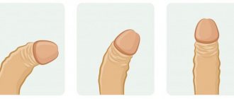
All components of CEE manifest themselves specifically and are determined during examination immediately after birth by pediatricians and obstetricians. With epispadias, the anatomical changes of the penis are pronounced: the urethra is split up to the external opening of the urethra, which, depending on the shape of the urethra, can be located on the head (Fig. 1), the middle and proximal part of the shaft of the penis (Fig. 2), in the peno-pubical corner (Fig. 3).
Rice. 1. Capitate form of epispadias
Fig.2. Stem form of epispadias
Rice. 3. Total epispadias
With total epispadias, the anterior wall of the urethra is absent along its entire length. The head of the penis is flattened, the foreskin covers only its ventral surface. Both cavernous bodies are located under the urethral platform. A careful examination allows you to identify the seminal tubercle and seminal ducts. Due to the divergence of the pubic bones and rectus abdominis muscles, the penis is shortened and has a dorsal deformation. The majority of male patients (about 70%) have total epispadias with urinary incontinence [21].
Classic exstrophy, in addition to manifestations of total epispadias, is characterized by the absence of part of the anterior abdominal wall and bladder, with the posterior wall of the bladder replacing the missing part of the anterior abdominal wall (Fig. 4). The visible lining of the bladder appears reddish at birth, and there may be mucous polyps on the surface. The navel is located lower than usual, below it there is a divergence of the rectus abdominis muscles and umbilical hernias that can be detected by palpation. The pubic bones can be felt on both sides of the evaginated bladder wall. The testicles are usually of normal size and located in the scrotum.
Often the developmental defects that make up CEE are combined with other malformations.
Rice. 4. 3-year-old boy with bladder exstrophy
Abnormalities associated with epispadias are usually limited to deformities of the external genitalia, diastasis of the pubic symphysis, and insufficiency of the bladder continence mechanism. In a review published by S. Arap et al. in 1988, described one case of renal agenesis and one case of renal ectopia in a group of 38 patients [21]. The function of the ureterovesical anastomosis in total epispadias is often insufficient; various studies describe from 30% to 40% of the incidence of vesicoureteral reflux [11, 22].
On average, one third of all patients with EMT, especially those with cloacal exstrophy, have urological abnormalities such as ureteropelvic obstruction, pelvic ectopia of the kidney, horseshoe kidney, renal hypoor agenesis, megaureter, ectopic ureter, ureterocele and duplication of the pyelocaliceal systems [1]. However, 100% of children with EMT have bilateral vesicoureteral reflux (VUR) due to impaired development of the vesicoureteral junction. In a review published by J. Ben-Chaim et al. Among boys with total epispadias, VUR is less common than in patients with EMT (82% versus 100%, respectively) [23].
TREATMENT METHODS
The goal of total epispadias correction is to achieve urinary continence while preserving the upper urinary tract and reconstruction of a cosmetically acceptable penis. Surgical treatment of urinary incontinence is almost identical to that performed for closure of bladder exstrophy.
The role of osteotomy in reconstructive operations in patients with exstrophy is reduced to bringing the bones of the symphysis closer together, which ensures a decrease in muscle tension and reliable closure of the abdominal wall, as well as moving the closed bladder deeper into the pelvis. These measures have a direct impact on improving urinary continence and reducing the risk of recurrent EMF. From the point of view of reconstruction of the penis, the reduction of the pubic bones ensures its lengthening [1]. Due to the divergence of the symphysis, the pedicles of the corpora cavernosa are stretched, which is one of the factors behind the shortening of the penis in patients with EMP up to 50% [24].
Currently, various forms of osteotomy are used in world practice. The most commonly used are posterior sagittal iliac osteotomy, transverse iliac osteotomy, diagonal mid-iliac osteotomy, combined vertical and horizontal pelvic osteotomy, and anterior pubic osteotomy (ramotomy).
HH Young reported the first effective treatment for urinary incontinence in a male patient with total epispadias in 1922 [3]. Since this time, the results of treatment of total epispadias have gradually improved [11, 21, 25, 26].
In patients with total epispadias and a large bladder capacity, reconstruction of the epispadias and bladder neck can be performed simultaneously. However, the results of treating small bladder with exstrophy and small bladder with epispadias have led to the practice of performing urethroplasty and penile lengthening before bladder neck reconstruction [22, 26].
A small, incontinent bladder in the presence of VUR is hardly an ideal case for bladder neck reconstruction and ureteral reimplantation
Initial bladder capacity is the most dominant predictor of urinary continence. In a study by S. Arap et al. There was a much higher rate of urinary continence in those patients who had adequate bladder capacity before bladder neck reconstruction than in patients with inadequate bladder capacity (71% versus 20%, respectively) [21]. Young-Dees-Leadbetter bladder neck repair, Marshall-Marchetti-Krantz urethropexy, and ureteral reimplantation are performed when bladder capacity reaches approximately 80-85 ml, which usually corresponds to the age of 4-5 years.
Achievement of urinary continence after correction of epispadias according to various authors is shown in Table 1 [11, 21, 23, 25]. Most of these patients underwent Young-Dees-Leadbetter bladder neck repair. Urinary continence was obtained in 82% of male patients (Ben-Chaim et al.) [23]. Correction of epispadias, performed before reconstruction of the bladder neck, helps to increase resistance to urine flow at the outlet of the bladder, and a possible increase in bladder capacity. Although patients with both E and EMP achieve slightly greater bladder capacity after epispadias correction, greater increases in bladder capacity are observed more often in patients with total epispadias. Increased bladder capacity may contribute to improved urinary continence in this group compared with the group of patients with classic EMF. Clinically, bladders with total epispadias are more flexible, they are easier to mobilize and reconstruct the bladder neck than with EMT.
Reconstructive operations on the penis for epispadias and exstrophy are the same.
The main objectives of penile reconstruction are:
- correction of the curvature of the cavernous bodies, their straightening;
- formation of the urethra;
- head reconstruction;
- closing a defect in the skin of the penis.
Many different methods of urethral reconstruction for total epispadias have been proposed. G. Monfort proposed using a transversely displaced flap [27]. Many authors suggested placing the reconstructed urethra between and under the corpora cavernosa (FV Cantwell; PG Ransley et al.; J.P. Gearhart et al.) [22, 31, 32]. MI Mitchell and DJ Bagley proposed a technique for complete disassembly of the penis [28]. Later, other authors reported multicenter experience with this technique - 17 patients from four institutions (M. Zaontz et al.)[29].
Table 1. Percentage of achieving urinary continence after correction of epispadias in boys
| Index | Ben-Chaim et al. (1995) [23] | Kramer, Kelalis (1982) [11] | Arap et al. (1988) [22] | Burkholder, Williams (1965) [25] |
| Number of observations | 15 | 53 | 38 | 27 |
| Reconstruction of the bladder neck was performed | 11 | 32 | 21 | 17 |
| Successful correction of urinary incontinence | 9 | 22 | 15 | 8 |
| Percentage of successful correction | 82% | 69% | 71% | 47% |
The curvature of the penis was corrected, while erectile function was preserved, and the external urethral opening was ultimately located on the glans penis. At the same time, patients were satisfied with the cosmetic results of this operation. A characteristic feature of the Mitchell operation is the complete division of the cavernous bodies and dissection of the head into two separate halves, with dissection of the urethral platform from the cavernous bodies (Fig. 5). The authors believed that complete dissection of the corpora cavernosa and head into two separate halves was necessary to facilitate medial rotation of the corpora cavernosa, with careful ventralization of the tubularized urethral platform [28]. Rotation of the corpora cavernosa leads to satisfactory correction of the dorsal curvature. However, 77% of patients subsequently require additional operations to correct hypospadias, since during “disassembly of the penis” and straightening of the corpora cavernosa, the length of the urethral platform is not enough to reach the apex of the glans penis (AT Hafez et al.) [30].
In a larger study performed by PG Ransley and I. Surer et al., positive results of treatment using the modified Cantwell Ransley technique were obtained [31,32]. To straighten a curved penis with epispadias, P. Ransley introduced the concept of excision of the dorsal chord and preservation of the dorsomedial anastomosis of the cavernous bodies above the urethra [33]. This technique involves mobilizing the urethral platform from the underlying tissues, separating the corpora cavernosa from each other. The distal part of the urethral platform, cavernous bodies and the head remain unexcised to ensure good blood supply and prevent shortening of the developing urethra (Fig. 6). The urethral platform is tubularized and placed ventrally under the corpora cavernosa. The mobilized cavernous bodies are rotated medially and fixed by applying a cavernous-cavernosostomy, ensuring effective straightening of the cavernous bodies (Fig. 7). However, in the postoperative period, in pubertal patients with severe dysplasia of the tunica albuginea along the dorsal surface of the corpora cavernosa, its significant shortening is observed in comparison with the length of the tunica albuginea on the ventral surface. In such cases, rotation and cavernous-cavernous anastomosis may not be sufficient. S-shaped curvatures of the distal part of the corpora cavernosa (cobra-type) may be observed.
The advantages of the Cantwell-Ransley and Mitchell-Bagley techniques are anatomically correct reconstruction with only a slight degree of residual deviation of the corpora cavernosa, and a low level of fistula formation due to the covering of the neourethra by the corpora cavernosa throughout its entire length. If the mobilization of the urethral platform from the cavernous bodies is sufficiently radical, then the cavernous bodies can most likely be connected without tension, without the need to perform corporotomy and complete mobilization of the neurovascular bundles. However, scarring and shortening of the neurovascular bundles can subsequently lead to serious, often irreparable curvature of the penis. One of the main requirements for these operations is the careful and thorough mobilization of the neurovascular bundle (using microsurgical optics), which helps maintain sufficient blood supply and innervation, maintain an erection and, most importantly, prevent ischemia of the glans penis.
Rice. 5. Stages of urethroplasty according to the Mitchell-Bagley method: a) isolation of the cavernous bodies; b) mobilization of the urethral platform; c) dissection of the head and separation of the cavernous bodies; d) the stage of formation of the urethra and suturing of the cavernous bodies
Rice. 6. Scheme of urethroplasty according to the Cantwell-Ransley method: a) stage of mobilization of the pedicles of the cavernous bodies; b) mobilization of the urethral platform; c) the stage of isolating neurovascular bundles
Rice. 7. Scheme of urethroplasty according to the Cantwell-Ransley method: a) stage of formation of the urethra; b) the stage of rotation and suturing of the corpora cavernosa above the urethra
CONCLUSION
The exstrophy-epispadias complex is one of the most severe diseases in the clinical practice of pediatric urologists. Every year the number of newborns with this pathology increases. The proportion of boys among such patients is 1.5-6 times higher
Despite the fact that the description and study of exstrophy and epispadias began in the 7th century. AD successful results ensuring optimal quality of life were achieved only at the beginning of the twentieth century.
Currently, the improvement of methods for surgical treatment of urinary incontinence and reconstruction of the penis for epispadias and exstrophy continues. This is a complex problem that requires the active participation of various specialists: pediatric urologists-andrologists, surgeons, traumatologists and neonatologists. Unfortunately, there are no precise guidelines for the management of such patients, and each treatment method has its positive and negative sides. The method of choice for the treatment of urinary incontinence in patients with CEA abnormalities is bladder neck plastic surgery in combination with osteotomy. The gold standard for reconstruction of the penis, in our opinion, can be considered urethroplasty with disassembly of the penis into its components, excision of the chord, mobilization of the cavernous bodies and their straightening, as well as the formation of the urethra and its immersion under the cavernous bodies. In this case, the choice of surgical technique remains with the surgeon.
LITERATURE
1.GearhartJP. The bladder exstrophy-epispadias-cloacal exstrophy complex.//InPediatricUrology.VolumeChapter 32. Edited by: Gearhart JP, Rink RC, Mouriquand PDE. Philadelphia: WB Saunders Co. 2001. P. 511–546.
2. Ashcraft K.W., Holder T.M. Pediatric surgery, Manual of St. Petersburg, Raritet-M LLC, 1999, pp. 19-20
3. Murphy LJT.The history of urology. // Charles Comas Publishers, Springfield, 1972. P.288-333.
4. ThierchC. Ueber die entstchungsweiseund operative behandlung der Epispadie, Arch Heilbundle, 1869. No. 10. P.20
5. Leadbetter WF. Considerations of the problemsincident to the performance of ureteroenterostomy:report of a technique. // J Urol, 1955. Vol.73, P 67.
6. Trendelenburg F. De la cure operatoire de l' exstrophie vesicale et de l' epispadias.//ArchKlinChir, 1892. No. 43. P.394.
7. Gambhir L, Höller T, Müller M, Schott G, Vogt H, Detlefsen B, Ebert AK, Fisch M, Beaudoin S, Stein R, Boyadjiev S, Rösch W, Utsch B, Boemers TM, Reutter H, Ludwig M. Epidemiological survey of 214 European families with Bladder Exstrophy-Epispadias Complex (BEEC). // J Urol, 2008. Vol.179. P.1539–1543.
8. Mesrobian H-GJ, Kelalis PP, Kramer SA. Long-term follow-up of 103 patients with bladder exstrophy. // J Urol, 1988. Vol.139. P.719–722.
9. Michon L. Conservative operations for exstrophy of the bladder with particular reference to urinary incontinence. // BrJ Urol, 1948. Vol 20. P.167.
10. Marshall VF, MueckeEC. // Functional closure of typical exstrophy of the bladder. // J Urol 1970. Vol. 104. P. 205.
11. Kramer SA, Kelalis P. Assessment of urinary continence in epispadias: review of 94 patients. // J Urol, 1982. Vol.128. P.290
12. Williams DI, Keaton J. Vesical exstrophy: twenty years' experience. //BrJ Surg, 1973. N 60. R. 203.
13. Lepor H, Jeffs RD: Primary bladder closure and bladder neck reconstruction in classical bladder exstrophy. // J Urol, 1983. Vol 130. P. 1142-1145.
14. Hollowell JG, Ransley PG. Surgical management of incontinence in bladder exstrophy. // BrJ U rol, 1991. Vol 68. P.543
15. James A. O' Neill, Jr, Marc I. Rowe, Jay L. Grosfeld, Eric W. Fonkalsrud, Arnold G. Coran. Pediatric Surgery, 1998. 5th edition. Vol 2. p.1709-1759.
16. Cantwell FV Operative Treatment of Epispadias by Transplantation of the Urethra. // Ann Surg, 1895. Vol. 22, N. 6. P. 689-701.
17. Boyadjiev SA, Dodson JL, RadfordCL, Ashrafi GH, Beaty TH, Mathews RI, BromanKW, Gearhart JP.Clinical and molecular characterization of the bladder exstrophy-epispadias complex: analysis of 232 families. // BJU Int, 2004. N 94. P. 1337–1343.
18. Anonymous: Epidemiology of bladder exstrophy and epispadias: a communication from the International Clearinghouse for Birth Defects Monitoring Systems. Teratology 1987. N 36. R. 221–227.
19. BennetAH. Exstrophy of the bladder treated by ureterosigmoidostomies, long term evaluation.//Urology 1973, Vol. 2. P. 165–168.
20. Nelson CP, Dunn RL, Wei JT. Contemporary epidemiology of bladder exstrophy in the United States. // J Urol. 2005. Vol.173. P.1728–1731.
21. Campbell MF. Epispadias; a report of 15 cases. // J. Urol. (Baltimore) 1952, Vol.67. P. 988
22. Arap S, Nahas WC, Giron AM. Continent epispadias: Surgical treatment of 38 cases. // J Urol 1988. Vol. 140. P. 577.
23. Ben-Chaim J, Peppas DS, Jeffs RD, Gearhart JP. Complete male epispadias: Genital reconstruction achieving continence. // J Urol 1995b; Vol.153. P. 1665.
24. Silver RI, YangA, Ben-Chaim J, Jeffs R, Gearhart JP. Penile length in adulthood aer exstrophy reconstruction. // J Urol 1997, Vol.157. N 3. P. 999–1003.
25. Burkholder GV, Williams DI. Epispadias and incontinence: Surgical treatment in 27 children. // J Urol 1965. Vol.94. P. 674.
26. Peters CA, Gearhart JP, Jeffs RD. Epispadias and incontinence: the child with a small bladder. // J Urol 1988. Vol.140. P. 1199.
27. Monfort G, Morisson-Lacombe GM, Guys JM, Coguel M. Transverse island flap and double flap procedure in the treatment of congenital epispadiasin 32 patients. // J Urol 1987. Vol.138. P. 1069.
28. Mitchell MI, Bagley DJ. Complete penile disassembly for epispadias repair: the Mitchell technique. // J Urol 1996. Vol.155. P. 300.
29. Zaontz M, Steckler RE, Shortliffe LM. Multicenter experiencewith the Mitchell technique for epispadiasrepair. // J Urol 1998; Vol.160. P. 1972
30. Hafez AT, El-Sherbiny MT, Shorrab AA. Complete primary repair of bladder exstrophy in children presenting late and those with failed initial closure:single center experience. // J Urol. 2005. Vol. 174. P. 1549-1552.
31.KajbafzadehAN,Duffy PG,Ransley PG.e evolution of penile reconstruction and epispadiasrepair:Areport of 180 cases. // J Urol 1995. Vol. 154. P. 858.
32. SurerI, Baker LA, Jeffs RD, Gearhart JP. The modified Cantwell-Ransley repairin exstrophy and epispadias: 10 year experience. // J Urol 2001. Vol.164. P.1040.
33. Ransley PG, Duffy PG, Wollin M: Bladder exstrophy closure and epispadias repair. In Paediatric Surgery. 4th edition. // Edited by: Spitz l, Nixon HH. London: Butterworths. 1988, pp. 620–632.
Magazine
Journal "Experimental and Clinical Urology" Issue No. 1 for 2016
Comments
To post comments you must log in or register