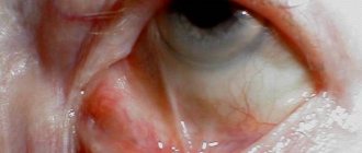
Anisometropia is an eye pathology that is associated with varying degrees of refractive power (refraction). This pathology can subsequently cause serious changes in the optical system (strabismus, amblyopia). Anisometropia is often accompanied by astigmatism, which is manifested by a defect in the cornea, lens, or eye.
In a patient with anisometry, the level of refraction of one eye may be normal, and the other eye may be impaired. In some cases, the level of refraction on both sides is the same, but the degree of decrease in visual function is different.
A person diagnosed with anisometropia may have one eye with normal refraction and the other with abnormal refraction.
If the difference in refraction level is less than 2 diopters, the patient most often does not notice it. In case of significant differences, binocular vision may be lost, since the object is alternately fixed with one eye and then with the other. This causes difficulties in orientation in space, as well as in responding to stimuli from the outside. A person perceives the environment blurry and unclear, sometimes images can merge.
Causes of anisometropia
The main causes of anisometropia:
- cataract;
- high degree of farsightedness;
- unilateral myopia;
- astigmatism;
- Iatrogenic impact.
Acquired anisometropia may result from improper intraocular lens implant power in pseudophakic patients. It develops with eye injuries, after keratoplasty of one eye.
Anisometropia in children
Congenital diseases must be taken seriously. Such conditions require mandatory treatment, as they can lead to adverse consequences.
Anisometropia in children can reach a refractive error of up to 3 diopters or more. The disease is inherited from parents. Therefore, children whose parents have a history of this condition should be screened from birth.
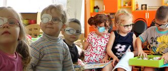
Often the disease is diagnosed in preschool and early grades, when the visual analyzer is under a lot of load. Children require correction, including the constant use of glasses.
Causes of the disease
There are congenital and acquired anisometropia. Congenital or hereditary anisometropia is common. If someone in the family has anisometropia, then the disease is likely to develop in younger generations.
Moreover, it may not appear in children at an early age, but in the future it can lead to serious consequences. In this case, it does not matter which eye sees worse in an adult: in a child, the opposite side may suffer more.
Without a hereditary predisposition, anisometropia can occur in an adult after the onset of cataracts or complications of eye surgery.
Anisometropia can be congenital or acquired. Ophthalmologists note that the physiological features of the structure of the ocular system are the main cause of diffraction disorders. Acquired pathology occurs:
- Post-existing cataract. Cloudiness of the lens in one eye changes the ability to defragate in the diseased, weak eye.
- Unilateral myopia. False myopia in a child (congenital) provokes pathological but reversible changes in the diffraction of the eyes.
- Astigmatism. Pathological deformations of the lens and cornea provoke an uncomfortable difference in the diffraction of the eyes. Postoperative period. Surgery is a jewelery process, especially on the vitreous body, lens or retina.
Treatment of the pathology is possible after consultation with an ophthalmologist. The doctor individually determines the causes of the disease and prescribes the necessary therapy. It is not recommended to wear contact lenses during treatment that cause blepharitis and other inflammatory processes of the cornea.
In moderate and severe cases of the disease, laser vision correction and correction of diffraction in the weak eye are indicated. Advanced anisometropia leads to false strabismus, myopia and general visual impairment.
Classification of anisometropia
The disease can be congenital or acquired. It develops independently or is a complication of another pathological condition.
There are 3 types of disease:
- Mixed - both organs of vision have noticeable refractive errors. The optical power, which characterizes the refractive power, and the length of the sagittal axis are different.
- The diagnosis of refractive anisometropia is made when the length of the ocular axis is equal, but the refractive power is different.
- The axial appearance of the disease means different axis lengths and the same optical power.
Depending on the severity of the pathology, three stages of development are distinguished: mild, moderate and severe.
Anisometropia
Anisometropia Source: astigmatizm.com Anisometropia is a pathology of the visual organs, which consists of different refraction of the left and right eyes.
People encounter this disease quite often. When the refraction of the eyes does not differ much, a person may not notice the disease. However, as the pathology progresses, binocular vision is lost, which leads to difficulties in orienting in space and responding to external stimuli. Let's take a closer look at what anisometropia is, what causes it, what symptoms it manifests, and how the disease is treated.
Anisometropia is an ophthalmological pathology in which the refractive power (refraction) is impaired in two eyes. This is a dangerous disease that has serious complications: strabismus or loss of vision due to inactivity of the eyes.
Often this disease develops together with astigmatism (a vision defect in which the patient sees objects unclearly due to the irregular shape of the cornea or lens).
Human eyes must have the same degree of diffraction. The lens, cornea, sclera and other elements of the eye must refract light rays equally in both the left and right eyes. In this case, the picture is clearly transmitted to the brain.
The norm is considered to be a deviation in diffraction between the two eyes of up to two diopters. The patient does not observe any discomfort, both eyes see normally, the concepts of a sick and a healthy eye are not distinguished.
When the difference in diffraction is greater than 2 diopters, a state of anisometry begins, characterized by:
- Deterioration in the quality of binocular vision. By closing your eyes one at a time, you will notice that one of them sees much better. Parents whose children suffer from anisometry notice that the child squints while reading books or studying distant objects.
- The appearance of accompanying symptoms – diplopia, myopia (false), fog in the eyes.
- Fatigue quickly when working with text or small objects. The body undergoes a process of restructuring the visual process, in which the healthy eye takes on the basic load. This causes fatigue, which is accompanied by headaches.
Basic information
It has been proven that anisometropia is caused by both different refractive powers of the optical apparatus of one and the other eye, and different lengths of the anatomical axis in both eyes. Most often it is congenital.
There are no specific figures in the ophthalmological literature regarding the frequency of anisometropia in humans. However, many authors note that mild degrees of anisometropia are more common than is commonly believed.
Anisometropia significantly affects the binocular vision apparatus. True, with a small degree of anisometropia. Despite the unequal clarity and size of the images of the object on the retinas of both eyes, binocular vision is preserved.
Anisometropia of 2.00 or more causes disruption of binocular vision (especially in children) due to difficulty merging images that are unclear in one or both eyes. In this case, the image on the retina of the worse-seeing eye begins to be suppressed, monocular vision and concomitant strabismus appear.
Anisometropes, in whom one eye has emmetropic refraction, usually use this better eye for vision. If the degree of refractive error of the second eye is small, then anisometropia is diagnosed only when checking visual acuity in an ophthalmology office.
If an anisometrope has hypermetropia in one eye and myopia in the other, binocular vision is impaired. A hypermetropic eye is used for distance vision, and a myopic eye is used for near vision.
Anisometropia can occur in different ways:
- in one eye there is normal refractive power, and in the other it is pathological;
- two eyes have the same refractive power, but different degrees of reduction in visual acuity;
- both eyes have different pathological refractive powers.
If the difference in the refractive power of the eyes is 2 diopters, then a person may simply not notice the symptoms. But if the difference is more than 2 diopters, then loss of binocular vision is possible. As a result of such complications, the image of the object is captured in turn by the right and then the left eye.
There are 3 stages of the disease:
- stage I – about 3 diopters;
- stage II – from 3 to 6 diopters;
- stage III – from 6 diopters or more.
Types of ophthalmological pathology by type of disease:
- axial - the refractive power is the same, but the length of the axis in each eye is different;
- refractive – the axes are the same, but the refractive power is different;
- combined – a combination of two types of disorders.
Symptoms of anisometropia
The clinical picture of the pathology depends on the severity. It is the same for children and adults.
Symptoms of the disease:
- At the first stage, the signs are poorly expressed. Patients complain of minor discomfort.
- The average degree of refractive error leads to a blurred image of one organ of vision, doubling of visible objects and decreased visual functions. When the eyelids of one organ of vision are closed, all signs disappear. For this reason, children and adults squint when looking at objects.
- The third degree of the disease is the most severe. It manifests itself as a sharp impairment in the ability to see a clear image with both eyes. There is a noticeable difference in the brightness and volume of the image. The visual organs quickly get tired, and cephalgia appears. The headache radiates to the brow ridges.
In the future, anisometropia leads to strabismus.
Manifestations in childhood
Source: Zrenimed.com Anisometropia in children is often diagnosed in the preschool period.
If you do not pay attention to the treatment of this disease, it can provoke the development of refractive amblyopia. When refractive index reaches two or more diopters, correction is needed, including constant wearing of glasses. A child can overcome the difference in glasses from two to five diopters without much discomfort. The younger the child, the faster it will adapt to such glasses.
However, it will take time for the little patient to learn to tolerate different glasses. Such a correction should be introduced gradually. The diopter difference in glasses increases every four months. The first corrective approach is recommended to be carried out against the background of switching off accommodation.
Anisometropia is easy to treat, especially in cases where modern methods have been used. It is better to replace glasses with the use of corrective lenses, which even a one-year-old child can successfully wear.
Considering the fact that myopic anisometropia, especially in the case of congenital myopia, can be accompanied by amblyopia and distortion of binocular vision, the doctor may recommend treatment with orthoptics and pleoptics at the same time as wearing contact lenses.
These methods have proven themselves to be an ideal correction for asymmetrical myopia.
Diagnosis of anisometropia
The disease is often discovered suddenly during a routine ophthalmological examination. In the first stages, people rarely notice abnormalities, so they do not go to the hospital.
Diagnosis is carried out based on the following studies:
- LED table for testing visual acuity. The patient sees all four colors: when all four are visible in the absence of obvious strabismus, the patient has normal binocular vision. Sees four shades also in the presence of obvious strabismus with abnormal retinal correspondence. Sees only two red colors - this indicates suppression of vision in the left eye, only three green - indicates suppression of vision in the right eye. The patient sees alternately three green and two red colors - this indicates the presence of variable suppression of vision. Three green and two red indicate the patient has diplopia.
- The patient is positioned 6 m from the Snellen chart. First, he closes one eye and reads the letters that the doctor points to, then the procedure is repeated with the second organ of vision.
- Measuring intraocular pressure. Tonometry can be applanation, dynamic contour, Goldmann, non-contact, electronic identification, rebound, impression and transpalpebral.
- The next step is to examine the fundus. This procedure is called ophthalmoscopy. It is prohibited for patients suffering from photophobia and lacrimation. These conditions will not allow the examination to be carried out normally and make examination difficult.
Additionally, skiascopy and visometry are prescribed to study the extent to which visual perception is impaired.
Correction rules and treatment
Anyone who has encountered anisometropia should know about the rules and methods of its correction. In childhood, the disease can be easily managed with surgical treatment.
Correction of the disease for complete elimination is carried out if anisometropia is in the mild and moderate stages, and in severe cases it is necessary to partially compensate for impaired vision.
It is very important to contact an experienced specialist when the first symptoms appear, and not to treat yourself, otherwise this can lead to serious consequences.
After contacting the clinic and diagnosis, treatment of anisometropia in both adults and children begins with the selection of contact lenses or glasses with correction capabilities. However, this method has one significant drawback - the patient feels discomfort.
This is due to the fact that the eyes can have a large dioptric difference, which makes wearing both lenses and glasses uncomfortable. In this regard, doctors recommend immediate correction using a laser. Nowadays, this method is considered one of the simplest and most humane in the treatment of anisometropia in children.
The main advantage of this method is that laser correction is painless and does not cause any discomfort. After the procedure, visual function is restored in just 2 weeks. With this treatment, long-term hospitalization is not required and the person soon finds himself at home.
Uncorrected anisometropia in childhood can lead to the development of anisometropic amblyopia. The difference in the strength of corrective glasses for the right and left eyes is considered acceptable within 2.0 D. For higher degrees of anisometropia, lenses are prescribed based on the better eye.
Important
An individual approach to the selection of anisometropic glasses is required, taking into account the conditions of its activity at close or far distances and, especially, the nature of the refractive error in each eye.
In some cases, it is rational to prescribe a contact lens to an eye with a higher degree of ametropia, because With contact correction, the difference in eye refraction does not matter. Implantation of intraocular lenses and refractive laser surgery can be recommended.
To reduce anisometropia, the following can be used when making glasses:
- lenses with a flatter front surface;
- reducing the thickness of the lens in the optical center;
- reduced vertex distance (the closer the lens is located to the eye, the less the size of the image on the retina changes)
The older the patient is at the time of prescription of glasses with different lenses and the greater the difference between them, the more likely the occurrence of asthenopic complaints.
If amblyopia has already occurred in the worse eye and there is no binocular vision, then any difference in correction is well tolerated.
With anisometropia, binocular intolerance often occurs. To achieve comfort you should:
- first weaken the power of the sphere on the eye with greater ametropia (and, as a rule, not the dominant one); clarification is always made with two eyes open;
- if this is not enough, proceed to manipulation of the cylinders. Cylinders located at the same angle to the horizontal (that is, 10 degrees and 170 or 20 and 160 according to Tabo) are best tolerated. The patient is put on a trial frame with a combination of lenses corresponding to the selected correction.
Using a sign projector, a cross-shaped grid is presented. The cylinder axis of the eye with a higher refraction is rotated towards the cylinder axis of the less ametropic eye until the moment of a break in the lattice and a difference in the clarity of vision of horizontal and vertical lines appears.
After a fracture appears, the axis is turned in the opposite direction until the correctness of the lattice is restored. The amount by which it is possible to rotate the cylinder axis while maintaining the correct vision of the lattice is estimated as the threshold for possible rotation of the axis and is measured in degrees on the Tabot scale.
If, after rotating the axis of one eye, the difference in the direction of the axes remains, rotate the axis of the other eye in the same way. If intolerance to the correction is associated with different amounts of astigmatism in two eyes, reduce the size of the cylinder in the eye with greater astigmatism until a feeling of comfort appears.
If there is an asymmetrical direction of the axes and a difference in the amount of astigmatism in the two eyes, first rotate the axes and then reduce the cylinder in the eye with greater astigmatism.
Astigmatic correction should be considered unbearable if there is a gross feeling of discomfort due to a distortion of the usual perception of space, “distortion” of the room, difficulties when walking up the stairs, different page sizes when reading a book.
Blurred vision, monocular and binocular double vision, pain in the eyes and brow ridges, a feeling of heaviness in the eyes, redness of the eyes, dizziness, headache, and nausea may occur.
If intolerance occurs with equal astigmatism in both eyes, the size of the cylinder is symmetrically reduced until a feeling of comfort appears.
Correction methods
Anisometropia is a dangerous disease that has serious complications. Early diagnosis guarantees complete restoration of vision without surgery.
Special corrective lenses are selected by an ophthalmologist for each patient separately. You cannot do this on your own, otherwise you will only worsen your condition.
If worn for a long time or incorrectly, microtraumas of the cornea, swelling, keratitis, and various infections may occur. Therefore, the ophthalmologist must constantly monitor the patient while wearing lenses.
The sooner vision correction for anisometropia begins, the higher the chance that vision can be restored. Correction of the disease consists mainly of wearing special corrective lenses or glasses.
There are many contraindications for wearing lenses, so the selection of lenses for each patient should be made individually by a qualified specialist. Otherwise, you can only increase the negative consequences of anisometropia.
When wearing lenses, it is better to be constantly monitored by a doctor to prevent damage to the cornea, keratitis, infections, and epithelial edema. It is also possible to use night lenses for refractive therapy.
Night lenses are good because while awake a person does not limit himself in anything that is impossible when wearing regular lenses or glasses.
Night lenses are made of special materials that are gas permeable and therefore do not cause problems associated with a lack of oxygen in the cornea. Lenses can correct the disease when the difference between the refraction of the eyes is more than 2 diopters.
Less dangerous, but also less effective, are telescopic glasses for improving vision. They represent a system of lenses - collecting and diverging. With their help, images of visible objects on the retina are enlarged, thereby eliminating problems associated with decreased vision.
Correction with glasses
Glasses can correct anisometropia when the difference in eye refraction is no more than 2 diopters. For children, there may be a big difference between the refraction of the eyes, since their visual system is still capable of changing.
In difficult cases, when correction of anisometropia does not produce results, doctors prescribe surgery to preserve vision. The operation is performed using a laser. They resort to surgical intervention in the most extreme cases, since these operations are very dangerous and are contraindicated for some.
An important criterion for prescribing or refusing surgery is the presence of corneal diseases. If any disease is present, surgery cannot be performed.
After the operation, you must follow all the doctor’s recommendations: you will have to avoid strong physical exertion and shaking, otherwise your vision may decrease.
Treatment of anisometropia
There are special contact lenses for night correction (refractive therapy). They are made of special gas-permeable materials that allow oxygen to reach the cornea. Such lenses will be effective if the difference in refractive power is about 2 diopters.
The most effective and safe methods of vision correction include glasses with telescopic lenses. Thanks to the lens system, they collect and scatter light rays. As a result, images of objects are enlarged, and vision problems are eliminated.
Telescopic glasses are effective if the difference in the refractive power of the eyes does not exceed 2 diopters. A child can wear such glasses, even if the difference is slightly greater, since the visual apparatus of younger patients has not yet changed.
There is a general rule of tolerability of different corrective glasses for anisometropia. This rule was derived based on extensive clinical experience. It states: the difference in corrective glasses prescribed to patients with anisometropia should not exceed 2.5 D.
It should be noted that some subjects (usually children) tolerate corrective glasses with a greater difference, but these cases are rare.
Correction of anisometropia, therefore, requires the ophthalmologist to carefully consider the individual tolerance of corrective glasses and a mandatory check of binocular vision with glasses.
There are many different options for choosing glasses for anisometropia. They can be reduced to the following basic principles.
- Based on tolerability, complete correction of refractive errors in both eyes should be sought.
- In the case of an average degree of anisometropia, with sufficient visual acuity, one should find some average correction that is well tolerated binocularly and gives a sufficiently high visual acuity.
- If one eye with correction has a visual acuity of 1.0, and the other eye is amblyopic and even with full correction has a visual acuity of less than 0.5, full correction should be given to the eye whose visual acuity is 1.0.
In recent years, contact lenses have been used to completely correct high-grade anisometropia, making it possible to tolerate the large difference in the refractive power of both glasses and preserving binocular vision.
One type of high anisometropia is monocular aphakia with good visual acuity in the second eye. The degree of anisometropia in this case is about 10.0 D. Unilateral aphakia is corrected with contact lenses. Correction of unilateral aphakia using conventional glasses leads only to monocular vision.
Treatment of anisometropia
It is important to start therapy immediately after diagnosis. If left untreated, the brain may choose the eye that produces the clearer image while ignoring the other. This leads to complete blindness of the weak organ of vision. In most cases it remains irreversible.
Anisometropia at one year of age must be corrected as it results in amblyopia in 76% of cases.
Conservative treatment of the disease
Anisometry is corrected with contact lenses. They are recommended for high-grade disease. Select contact lenses from an ophthalmologist or optometrist. Before choosing, it is important to measure the patient's visual acuity.
If you select the wrong contact means for vision correction, headaches and dizziness will occur. Vision does not improve, but worsens. The vision becomes double, the image becomes blurry.
Wearing glasses is possible, but contact lenses are preferred because they better correct visual perception. Glasses must be telescopic. They consist of a diverging and converging lens. Correction with glasses should be introduced gradually.
Start wearing it from 1 hour a day, gradually increasing the time of use. For children, the dioptric difference in glasses increases every four months.
Surgery
Surgery is performed at the request of the patient and when visual perception is severely impaired. There are many correction methods if there is no effect from CL.
Laser therapy is performed. Implantation of an intraocular lens is possible. Refractive surgery is performed for unilateral myopia, astigmatism and farsightedness. Photorefractive keratectomy (PRK) and laser assisted keratomileusis (LASIK) are increasingly used to correct anisometropia.
For high myopia, the lens is removed from one eye. It is replaced with an implant.
Removal of clear lenses with IOL implantation is also possible. This operation is performed if the biometry is +40 diopters and there is no lens of such power for implantation.
Clinical picture
The eye that sees worse is practically switched off from the visual act, begins to deviate to the side, strabismus and anisometropic amblyopia develop (decreased vision of the squinting eye due to inactivity). With age, the magnitude of anisometropia does not change, but the decrease in vision of the squinting eye becomes stable.
When correcting anisometropia (up to 3.0 D), a good result can be achieved: visual acuity is equalized, the image on the retina becomes the same, favorable conditions are created for binocular vision, and aniseikonia disappears. Patients with aniseikonia usually experience unpleasant sensations: pain in the eyeballs, inability to read, pain in the head.
Prevention
The congenital form of the disease cannot be prevented. To prevent the development of acquired anisometropia, the following rules should be followed:
- promptly contact an ophthalmologist if you are diagnosed with cataracts or glaucoma;
- follow postoperative rules for a speedy recovery after ophthalmic surgery;
- alternate long periods of work at the computer with short rest, during which you do eye exercises.
Patients with anisometropia should not engage in strength sports. If the degree of illness is mild, you should consult an ophthalmologist, therapist and trainer.
It is important to take vitamin complexes. The products do not contain enough useful components, so it is recommended to take dietary supplements with vitamins A and E.
Surgical method of treatment
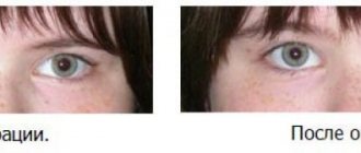
Source: ofthalm.ru If corrective glasses and lenses are ineffective, then laser surgery is performed to help preserve vision.
Surgery for anisometropia is a last resort, since any surgical intervention is very dangerous, and in some cases is even prohibited! One of these contraindications is diseases of the cornea.
In the postoperative period, it is necessary to follow the recommendations of doctors. Physical activity and shaking are strictly prohibited, as this can lead to decreased vision.
In childhood, this disease is very easy to treat surgically. Correction of anisometropia is necessary for the successful treatment of this pathology in mild and moderate stages, and in complicated and advanced cases, it will bring lost vision to a state of partial compensation.
When contacting the clinic, treatment begins with the proper selection of contact lenses or glasses with correction capabilities. But this method has one serious drawback, which is discomfort.
They arise due to the fact that the dioptric difference between both eyes can be very large, on the order of three diopters or more. This makes wearing glasses and contacts difficult.
Therefore, it is better to immediately make a correction using innovative equipment - a laser. At the moment, this method is the simplest and most humane for the treatment of anisometropia in childhood.
The biggest advantage of this treatment is that laser correction of anisometropia is absolutely painless, and the period of active restoration of visual functionality usually takes only 14 days. During this treatment, the child does not require long-term hospitalization and can undergo the procedure together with his parents.
Treatment
Correction of anisometropia depends on the degree of complexity, the age category of the patient and the type of disease. But the sooner the problem is noticed, the less effort and time it will take to solve it.
All treatment can be divided into two main methods:
- Wearing corrective lenses or telescopic glasses. Attention! Selecting lenses or glasses on your own without the advice of a specialist is very dangerous and can worsen the situation. This is fraught not only with further deterioration of vision, but also with microtraumas of the cornea, which can easily lead to infection, swelling and inflammatory processes. The design of telescopic glasses consists of two types of lenses: converging and diverging, which level vision.
- Surgical method. This is already an extreme method of treatment; doctors resort to it only in cases where non-surgical methods are ineffective. Usually these are already advanced stages. Vision correction occurs using a laser. But this can only be as prescribed by a doctor. The operation has a number of contraindications and limitations. For example, after surgery, you should not put heavy strain on your eyes, to be afraid of injuries and concussions, as this can provoke a complete regression of the disease.
Of course, recently the level of equipment of clinics has become quite high. But without consulting a specialist, taking tests and recommendations, you cannot treat anisometropia. It is also important to follow the recommendations after surgery. Then the chances of recovery will be much higher. The nature of heredity, of course, does not indicate one hundred percent that in a particular case the disease will necessarily manifest itself. However, if there is a predisposition, it is better to take preventive measures, namely: do not overstrain your eyesight, try to take vitamins for the eyes, and do relaxing exercises. But if, due to your profession, your main activity is on the computer, it is better to give your eyes an hour of rest, for example, go for a walk or do simple relaxing gymnastics.
By the way, it should be noted that contact sports, such as football, volleyball, basketball and others, are also good exercises that help strengthen the eyes. An important link in the chain of health is food. It should be rich in fiber, vitamins E, A, and group B. But cholesterol intake should be reduced.
Causes
Refractive error may be caused by structural or functional changes. In the process of development of anisometropia, structural changes, i.e., organic pathology, predominate. And dysfunctions can only slightly change the refractive power, which will not affect the quality of a person’s vision.
Causes:
- Cataract. There is a disruption in the passage of rays through the clouded lens.
- Astigmatism. Refraction is distorted due to the altered shape of the cornea or lens.
- Congenital unilateral myopia. This pathology is the most common cause of anisometropia in children. When the child grows up, the eye is finally formed, and the difference in refraction goes away on its own.
- Unilateral farsightedness. Severe hyperopia of one eye is more typical for people after 40 years of age and is associated with uneven age-related changes.
- Postoperative condition. A defect in the surgical technique or ignoring contraindications for surgery can lead to postoperative anisometropia. This happens, for example, when installing IOLs in children whose eyes have not yet fully formed.
Treatment (correction) of aniseikonia
To correct aniseikonia, special glasses are used. Such glasses are called aniseikonic. Aniseikonic glasses are discs glued together that are not capable of optically changing the refractive power of the eye. Their action is aimed only at enlarging or reducing images. Aniseiconic glasses are calculated in percentage rather than dioptre terms and are capable of increasing or decreasing visible images from 0.5 to 10%. Just like other glasses for optical correction of refractive errors, aniseiconic glasses are selected for each person individually after an ophthalmological examination.
In the medical department, everyone can undergo examination using the most modern diagnostic equipment, and based on the results, receive advice from a highly qualified specialist. The clinic is open seven days a week and operates daily from 9 a.m. to 9 p.m. Our specialists will help identify the cause of vision loss and provide competent treatment for identified pathologies.
In our clinic, appointments are carried out by the best ophthalmologists with extensive professional experience, the highest qualifications, and a huge amount of knowledge. The cost of treatment at MGK is calculated individually and will depend on the volume of treatment and diagnostic procedures performed.
Lifestyle recommendations
In order to slow down the deterioration of visual acuity with anisometropia, it is necessary to introduce some restrictions into the child’s lifestyle:
- Avoid contact sports that can lead to eye injuries. Preference is given to swimming, which is not harmful to health and at the same time strengthens the child’s muscle corset;
- nutrition adjustments. It is necessary to minimize foods rich in cholesterol in the child’s menu. The basis of the diet is fruits and vegetables. They not only have a beneficial effect on the immune system, but also have a positive effect on the state of the visual system;
- perform therapeutic exercises for the eyes. Regular exercise helps maintain clear vision and prevent eye fatigue;
- observe the work and rest schedule. When working at a computer or watching TV, you need to take regular breaks. The amount of time a child spends at the computer should be limited.
Treatment of anisometropia should begin as early as possible. With timely diagnosis and adequate therapy, complete restoration of visual function can be achieved.
Features and symptoms
Anisometropia is a type of eye disease in which both eyes have different refractive powers . The refraction of the left eye may differ from the right by more than 2 diopters. If there is a slight difference, the patient can ignore this pathology for a long time without visiting a doctor. A complication can be strabismus and even complete relaxation of the muscles of the affected visual organ, which will lead to blindness.
Features of the disease:
- Accompanied by diplopia, a blurred image in front of the eyeballs.
- Rapid eye fatigue appears during prolonged visual work.
- Visual acuity decreases.
REFERENCE: According to the International Classification of Diseases, the code for anisometropia according to IBC-10 is H52.3.
Main symptoms of the disease:
- Double image.
- Blurry outline.
- Disappearance of visual disturbances when one eye is closed.
- Alternating fixation of an object: now with the left, now with the right eye.
- Some images may appear blurry and blurred in front of your eyes.
- Amblyopia may appear. In this case, one eye may completely lose visual function.
- Aniseikonia is observed - an increase in the difference in image size and picture brightness.
- The appearance of headaches during heavy exertion, for example, while reading or working at the computer.
Symptoms are associated with impaired visual acuity of the patient, which doctors check using tables or photo scanning of the eyeball.
Complications and prognosis after treatment
The prognosis for a person is favorable. Anisometropia can be easily corrected. But if you refuse treatment, the disease can progress and become more complicated.
At a certain stage, strabismus develops. Thus, the visual apparatus seeks to compensate for the refractive error. Moreover, strabismus appears only when the direction of gaze changes.
Other possible complications of anisometropia:
- amblyopia – lazy eye, i.e. “switching off” one eye from working;
- corneal damage due to long-term wearing of contact lenses;
- the risk of infectious and inflammatory ophthalmological diseases increases;
- Anisoaccommodation is a difference in the ability of the eyes to accommodate (move the eye from one distance to another).
Why does anisometropia develop?
Anisometropia is a visual impairment in which the clinical refraction of the two eyes does not match, and the power of the optical lenses of one eye is significantly greater or less than the other.
Predisposing factors
Anisometropia can be congenital or acquired .
The most common form is the congenital form, which can be detected already during a medical examination of the child a year. But often low-grade anisometropia is ignored by doctors and patients due to the lack of complaints, until they appear in adulthood.
Acquired anisometropia can appear after an eye disease, surgical treatment or severe trauma.
Immediate causes
In childhood, the immediate cause of anisometropia is the uneven development of the two eyes relative to each other or a lag in the development of one eye against the background of the normal development of the other.
Normally, a child is born with large hypermetropic refraction (farsightedness), which decreases significantly by the end of the first year of his life (down to 1-2 diopters) and turns into normal zero refraction (emmetropia) by about 6-8 years.
If at any stage of development of the organ of vision an error occurs, and one eye begins to grow faster or slower than the other, their length and optical power cease to be the same, they refract the light beam differently, and anisometropia occurs.
If eye development occurs normally, and during routine examinations by an ophthalmologist no difference was detected in the two eyes, then in adulthood anisometropia can develop only after an injury or disease, for example, unilateral cataracts.
Description of the disease
A person diagnosed with anisometropia may have one eye with normal refraction and the other with abnormal refraction.
Sometimes the eyes have the same refraction, but a different decrease in visual acuity. Or there is a different abnormal refraction in both eyes.
If the difference in refraction between the eyes is small (less than 2 diopters), the disease may not be noticed by the person. But if the difference is significant, there is a possibility of loss of binocular vision (vision with both eyes), the object is fixed alternately with the right and left eyes, which leads to difficulties in orientation in space and reaction to external stimuli; a person perceives the surroundings unclearly, blurry, sometimes the images merge.
There are three degrees of development of anisometropia:
- weak degree of anisometropia (within 3 diopters),
- medium degree (range from 3 to 6 diopters),
- strong degree (more than 6 diopters).
There are also types of anisometropia based on the type of disease:
- axial anisometropia (when, with the same refraction of the eyes, the length of the axis of each eye differs),
- refractive anisometropia (with the same axes, the refractive powers of the eyes differ),
- mixed anisometropia (when both types of disorders are present).
Classification
Anisometropia can occur against the background of other eye diseases, or it can occur independently. There are several classifications of anisometropia based on different characteristics.
By occurrence:
- congenital;
- acquired.
Based on severity, there are 3 degrees:
- the difference in refraction from 2 to 3 diopters is weak;
- from 3 to 6 diopters – average;
- over 6 diopters – high.
According to the mechanism of occurrence:
- axial anisometropia - the same refraction, but different lengths of the eyeballs;
- refractive – the length is the same, but the refraction is different;
- mixed - a combination of the previous options.
Symptoms
With weak anisometropia, binocular vision is practically not affected; a person may not be aware of the existence of a pathology.
Impaired binocular vision is the main symptom of anisometropia.
If the difference in refractive errors between the right and left eyes exceeds 2-3 diopters, then in the visual analyzer of the brain, the pictures received from different eyes cannot merge into a single image, and binocular vision is impaired. As a result, a person sees objects indistinct and blurry, his orientation in space is disturbed, and headaches and nausea often occur.
Over time, anisometropia can lead to amblyopia (lazy eye disease) and strabismus.
Causes of visual impairment
The causes of the disease include:
- Myopia or congenital unilateral myopia. This mainly happens in children when myopia becomes more severe. Find out about progressive myopia here;
- Cataract. If the passage of sunlight through the lens is impaired, anisometropia may develop. The greatest problems come in the case of dysfunction of only one eye;
- Astigmatism is a disease that is associated with changes in the shape of the lens, cornea or eyeball, due to which the patient loses the ability to see objects clearly. Anisometropia can develop against the background of progression of this pathology;
- Postoperative complications. The difference in visual perception may be a consequence of poorly performed surgical intervention to eliminate other refractive errors (myopia, astigmatism);
- Unilateral hypermetropia. In most cases, it develops against the background of progressive presbyopia (age-related farsightedness). You can learn about hypertensive retinal angiopathy in this material.
The reasons for the development of anisometropia is the progression of other refractive pathologies. To prevent this, undergo regular ophthalmological examinations and practice good visual hygiene.
Possible consequences and complications
With a high degree of anisometropia, the visual analyzer cannot combine the images received from each eye into a single whole. The central nervous system seeks to protect itself from such discomfort, for which it ignores the received image from the eye with the greatest refractive error. As a result, the visual functions of this eye gradually fade away, amblyopia develops, or, as this condition is also called, lazy eye disease. Another complication of anisometropia can be convergent or divergent strabismus.
With anisomitropy, it becomes difficult for the patient to respond correctly to external stimuli and navigate in space.
Classification of aniseikonia
There is multiple classification of the disease:
- An optical disorder caused by different refractive powers in the eyes.
- A functional disorder that occurs when the structure of the retina or the visual perception center in the brain changes.
- Aniseikonia with accompanying anisometropia. A discrepancy in the refraction of light in the eyes, the difference of which is 2 or more diopters.
- Aniseikonia with concomitant ametropia (normal eye refraction).
- Aniseikonia with accompanying isometry (lack of refraction in the right and left eyes).
- Aniseikonia, caused by differences in the density of receptors on the retina.
Having identified the type of disorder, the doctor can determine treatment that will help completely eliminate the disease.
Diagnostics
The initial degree of anisometropia is detected by chance during a routine examination. A person with complaints about changes in visual functions comes in at a moderate or high degree. The following examinations are carried out:
- Visometry – assessment of visual acuity using Sivtsev tables.
- Perimetry is an additional study to assess the narrowing of visual fields.
- Biomicroscopy is an examination of the eye using slit lamp light.
- Ophthalmoscopy is an examination of the fundus of the eye using an ophthalmoscope.
- Skiascopy is a shadow test to assess refraction.
- Ultrasound of the eyeballs – study of the size and structure of the eyes.
- Computer refractometry is an assessment of refraction by determining the ratio of the eye axis to refractive power.
Ophthalmological tests allow you to assess the extent of preserved visual functions (how well a person sees, whether the visual fields are narrowed, etc.). And more detailed studies, such as ultrasound and refractometry, are designed to detect the cause of changes in the eye organ.
To watch a practical video about the study of visual fields using perimetry:
Prognosis and prevention
The prognosis for life and work ability is favorable. Specific methods of prevention have not been developed. Nonspecific preventive measures are limited to monitoring visual acuity and clinical refraction. Persons who have undergone eye surgery over the past 2 years are advised to consult an ophthalmologist once every 6 months with mandatory refractometry and visometry. When diagnosing clinical refractive errors in children over 1 year of age, it is necessary to carry out timely correction of visual dysfunction in order to prevent the development of strabismus. In patients younger than 12 months, the use of special treatment methods is not indicated.
Maintaining the correct visual load regime
To maintain proper visual and physical activity, it is highly recommended to alternate eye strain (for example, reading or watching television) with active rest.
But if with myopia, which does not exceed 3 diopters, there are no restrictions on physical activity, then with 3 diopters and above, jumping, heavy lifting and a number of other activities are limited. It is also recommended to follow the tips below:
- Always maintain good lighting when reading or working on a computer.
- Do not read in moving vehicles, this increases eye strain.
- Do not sit closer than 40 cm from the computer.
- Every 55 minutes of working at the computer, take at least a 5-minute break.
Wearing glasses/lenses
If your doctor has prescribed you glasses or contacts (the selection principle is indicated above), it is important to take into account the specifics of the patient - where he spends more time, at the computer and reading - or in activities that involve looking into the distance.
Two pairs of glasses/lenses can be prescribed - for near and for distance, they need to be alternated depending on what type of activity is currently prevailing.
Visual gymnastics
There are many different eye exercises that help improve blood circulation to the eyes and train the ciliary muscle. Let's give a few examples.
- "Mark on the window." Place a two-centimeter mark on the window glass. Stand at a distance of 30 cm from her and look from her to some object at least 5 meters away - and back. Repeat 10 times.
- Sit down, close your eyes tightly for 5 seconds, then open them sharply for 5 seconds. Repeat 8 times.
- Move your eyes vertically and horizontally with maximum amplitude, slowly, evenly. Repeat 8 times.
- Move your eyes in a circle, first 4 times in one direction, then 5 times in the other. Repeat 2-3 times.
- Sit down, close your eyes and massage your eyelids with light circular movements for about a minute.
Complications of anisometropia
If left untreated, especially in early childhood, anisometropia can lead to serious consequences. Since the part of the brain responsible for vision receives a sufficient amount of information from one eye, and a blurred picture comes from the second, the brain gradually “turns off” the poorly functioning eye, the nerve connections between them are functionally lost, and vision continues to deteriorate.
Because of this, strabismus and amblyopia occur, known to us as “lazy eye,” and this is a more serious disease. The sooner treatment is started, the greater the chance that vision will be restored, and strabismus will not have to be corrected surgically.
In adult patients, anisometry can lead to diplopia - double vision of surrounding objects, which can be accompanied by dizziness, headache and eye pain. The presence of complaints will be affected by the degree of ametropia, but even with a slight blurring of the incoming information, discomfort in the eyes is possible. After prescribed optical vision correction, in the absence of concomitant pathology, the complaints cease to be a concern.
How to stop the progression of the disease
Before carrying out treatment, you need to make sure that you are dealing with myopathy, and not with its false variety, which is also called spasm of accommodation. Read about the treatment of progressive myopia in children and adults at this link.
Fighting accommodation spasm
Treatment tactics for the latter depend on the etiology of the disease.
Drug treatment
If the doctor chooses a medicinal approach, the patient takes a number of drugs that dilate his pupil.
Self-selection of medications is prohibited; you must follow only the instructions of the attending physician.
Optical correction
This method is used only in extreme cases and only for a short period of time. It consists in the fact that the patient wears minus glasses, as well as polydiaphragms or perforation glasses. The latter are a “screen” with small holes placed in front of the patient’s eyes. Due to this, the depth of focus increases and visual acuity improves.
Surgery
Accommodation spasms are often characterized by fairly low effectiveness of conservative therapy, so theoretically surgical treatment can also be prescribed, but at the moment this is not a common practice, but isolated exceptions.
Functional treatment
There are other treatment options for spasm of accommodation:
- optical-reflex training;
- physiotherapy;
- home training (for example, exercises using a mark on glass);
- hardware treatment (for example, local barotherapy).
In general, the prognosis for spasm of accommodation is not very positive - treatment may remain ineffective, and the likelihood of relapse is high. The severity of relapses may vary. Often the spasms resolve spontaneously even without major intervention.
Fighting myopia
Now let's look at the approaches to therapy used when a patient is diagnosed with progressive myopia.
Non-drug treatment
This treatment option includes:
- Nutrition balanced in vitamins and proteins.
- Physiotherapeutic procedures, such as electrical stimulation through the skin, which helps improve blood circulation in the retina.
- Eye exercises specially selected by your doctor.
- Targeted reduction of eye strain (for example, reducing time spent in front of the TV, computer, etc.).
- Frequent walks and exercise, including swimming.
- Eye exercises specially selected by your doctor.
Drug treatment
This method cannot guarantee that vision will return to previous levels, but it can stabilize the condition of the eyes.
Also, drug treatment stops the active progression of the disease.
The following categories of drugs are used:
- Drugs that have a positive effect on strengthening the sclera.
- Drugs that lower pressure inside the eye.
- Drugs that help activate metabolic processes in the retina and blood vessels of the eyes.
- Drugs that help restore the ciliary muscle.
Self-medication in this case is unacceptable; you can only take medications prescribed by your doctor. Their selection takes into account the individual characteristics of a person.
Non-surgical treatment
This is the name for therapy using glasses or contact lenses.
For myopia
Children use glasses in any case.
In adults, everything depends on the specific situation and stage at which the disease is located.
- Glasses. They are used for myopia of both stronger and weaker degrees. But with a weak degree, it is necessary to minimize the period of wearing them, since with prolonged use they can weaken the eye muscles. It is strongly recommended to use glasses when vision ranges from 3 to 6 diopters. It is recommended to remove glasses when reading or working with small objects.
- Contact lenses. This is a more useful option for the muscles - contact lenses, as it were, form a common optical system with the eye - and the muscles begin to work as healthy. As a result, in the early stages of myopia, a better effect is achieved than with glasses, but with a high degree of myopia, the effect will be just the opposite, since the lenses are very close to the eye.
There is a special type of lenses - orthokeratological. They are able to change the shape of the cornea - they are put on at night, taken off in the morning, after which a person sees better all day. The best option is in the range from 1.5 to 5 diopters. This article will tell you about vitamins for eyes for myopia.
Surgical
If myopia has reached six diopters, you can proceed to more drastic measures - surgical sclera-strengthening operations. There are, among other things, the following:
- Scleral strengthening injection. A special polymer substance is injected into the posterior part of the eye, where it enters the sclera and becomes a kind of framework that promotes the growth of connective tissue.
- Posterior scleroplasty. Small fragments of special scleroplastic tissue are inserted into the incisions in the back wall of the eye. It is used for myopia, which is caused by an increase in the size of the eye. A common indication is during pregnancy.
- Keratoplasty. An operation that involves replacing the cornea.
- Refractive lens replacement. It is performed for advanced myopia.
- Lensectomy. Replacing the lens with an implant.
Causes of myopia progression
Unlike congenital myopia, progressive myopia is a pathology, the development of which is caused not by any one, but by a whole set of different factors. It is based on genetic inheritance - if parents suffered from this disease, then their child also increases the likelihood of developing the problem. Read about lens replacement surgery for astigmatism here.
The following provoking factors are genetically determined:
- weakness of the eye muscles;
- scleral weakness;
- thickening of the cornea;
- thickening of the lens.
External factors that additionally influence the development of pathology include:
- some infectious diseases;
- prolonged sitting in front of a computer/TV;
- severe strain on vision;
- hormonal and immune system disorders;
- lack of a number of microelements;
- incorrect organization of work processes, including prolonged work at close range.
Myopia also directly depends on age. At a young age, before primary school, the chance of its occurrence is minimal, no more than 6%. By high school, this probability increases to 16%, and after 20 years, the risk of its development gradually decreases, again reaching minimum levels at a more mature age.
Correction of anisometropia
This pathology of the visual system is very severe. It requires early diagnosis and timely treatment through surgery.
Initially, corrective lenses are selected for the patient. You cannot choose glasses or lenses on your own; a deviation of even one diopter can lead to severe deterioration of vision. With prolonged use of incorrect lenses, swelling develops, the cornea is injured, keratitis and infections occur. For the same reason, while wearing even correctly selected lenses, the patient should regularly visit an ophthalmologist.
To correct night vision, other lenses are required. They are made from gas-permeable materials that facilitate the free penetration of oxygen into the cornea of the eye. However, such lenses are effective only with a deformation of 2-3 diopters.
The safest method of vision correction for anisometropia is considered to be glasses with telescopic lenses. They help to collect and disperse light rays, which allows you to magnify the image of an object and eliminate vision problems.
Glasses with telescopic lenses help when the difference in refractive power is no more than 2 diopters. Children are allowed to use such glasses with a slightly larger difference. This is due to the fact that the child’s visual apparatus does not change completely.
Meniscal glasses (concave-convex, convex-concave lenses that allow the collection and scattering of light rays) help patients tolerate strong vision correction more easily than aspheric lenses (one or both surfaces are not spherical).
Today, doctors successfully correct defects with a difference of up to 5 diopters. However, in this case, much depends on the patient’s willingness to do exercises and lifestyle changes.
The goal of correction for anisometropia is to achieve binocular fixation for all distances. Even if spectacle correction fails, the patient will be able to capture the distance image with two eyes and the near image with one (or vice versa).