The physiology of sensory systems is based on reflex activity. The pupillary reflex is the friendly reaction of both pupils to light. Its adequacy is determined by the coordinated activity of all components of the neural arch, consisting of 4 neurons and the brain center. The eyes do not immediately react to flash or darkness. It takes a fraction of a second for the impulse to reach the brain areas. Too sluggish a reaction indicates pathology at some stage of the reflex chain.
Normally, the direct reaction of constriction and dilation of the pupillary fissure in response to fluctuations in lighting around the human head depends on the adequate activity of afferent and efferent nerve fibers. It is also influenced by the functioning of the center in the optical cortex of the occipital hemispheres of the brain.
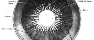
Anatomy of the eyeball and nerves
The arc of the pupillary reflex begins on the retina and passes through several nerve regions. In order to better navigate the sequence of movement of impulses to the target point, below is a diagram of the anatomical structure of the eye analyzer:
- Cornea. It is the first obstacle in the path of the light beam. This transparent structure consists of a dense row of cells, the structure of which is dominated by cytoplasm.
- Front camera. It does not contain liquid. This cavity limits the pupillary opening in front.
- Pupil. It is a hole surrounded on all sides by the iris. It is the pigmentation of the latter that gives the eyes their color.
- Lens. It is considered the second refractive structure after the cornea. According to its anatomy, the lens is a biconvex lens, capable of changing curvature due to the contraction and relaxation of the accommodative muscles and the ciliary body.
- Rear camera. It is filled with vitreous humor, which is a gel-like mass that conducts light rays.
- Retina. This is a collection of nerve cells - rods and cones. The former capture light, the latter determine the color of objects around.
- Optic nerve. It conducts light impulses accumulated by rods and cones to the optic tract.
- Two-humped bodies. They are structures of the central nervous system.
- Axons heading to the Jakubovich or Edinger-Westphal nuclei. These fibers represent the afferent site of the unconditioned reflex.
- Axons of parasympathetic oculomotor nerves to the ciliary ganglion.
- Short fibers of neurons of the ciliary ganglion to the muscles that constrict the pupil. They close the reflex arc.
Pupillary reflex pathway
The muscle fibers of the iris, arranged in a ring shape, narrow the pupil, while the radial fibers dilate it.
M. sphincter pupillae et m. ciliaris are excited by postganglionic cholinergic fibers of the oculomotor nerve (nn ciliares breves). Consequently, this nerve simultaneously constricts the pupil and increases the curvature of the lens. Relaxation of the ciliary muscle and contraction of the radial muscle of the iris are caused by irritation of the cervical sympathetic trunk. When the oculomotor nerve is irritated and the tone of the sympathetic nerve is reduced, the pupil narrows, and when the sympathetic nerve is irritated and the tone of the oculomotor nerve is turned off, it dilates. The pupillary reflex is caused by receptors in the retina, eye muscles and pain. The motor reactions of both eyes are of a friendly nature.
What is he?
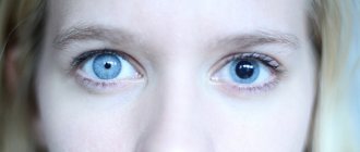
Depending on the lighting, a person’s pupil changes: in weak light it expands, in strong light it narrows.
The normal reaction of the pupil to light or photoreaction is a narrowing of the pupillary fissure when there is an abundant supply of light photons and its widening in low light. The pupillary reflex pathways begin on the light-refracting structures of the eyeball. Captured by the light-sensitive cells of the retina - rods and cones - photons of light are recorded by specific pigments and arrive in the form of nerve impulses to the optic nerve. From there, through neurotransmitters along myelinated fibers, the impulse passes into the afferent part of the nerve pathway. Afferentation ends at the level of the midbrain nuclei of Yakubovich or Edinger-Westphal. They are also called accessory nuclear structures of the oculomotor nerve. From the tegmentum of the brain stem, impulses through the conductive area enter the muscle fibers, causing the pupillary fissure to expand and contract.
14.1.2. Pupil. Norm and pathology of pupillary reactions
In children of the first year of life, the pupil is narrow (2 mm), reacts poorly to light, and dilates poorly. In the sighted eye, the size of the pupil constantly changes from 2 to 8 mm under the influence of changes in illumination. In room conditions with moderate lighting, the pupil diameter is about 3 mm, and in young people the pupils are wider, and with age they become narrower.
Under the influence of the tone of the two muscles of the iris, the size of the pupil changes: the sphincter contracts the pupil (miosis), and the dilator ensures its dilation (mydriasis). Constant movements of the pupil - excursions - dose the flow of light into the eye.
The change in the diameter of the pupillary opening occurs reflexively:
- in response to irritation of the retina by light;
- when setting to clearly see an object at different distances (accommodation);
- with convergence (convergence) and divergence (divergence) of the visual axes;
- as a reaction to other irritations.
Reflex dilation of the pupil can occur in response to a sharp sound signal, irritation of the vestibular apparatus during rotation, or with unpleasant sensations in the nasopharynx. Observations are described confirming the dilation of the pupil under great physical stress, even with a strong handshake, when pressing on individual areas in the neck, as well as in response to a painful stimulus in any part of the body. Maximum mydriasis (up to 7-9 mm) can be observed during painful shock, as well as during mental stress (fear, anger, orgasm). The reaction of pupil dilation or constriction can be developed as a conditioned reflex to the words dark or light.
The reflex from the trigeminal nerve (trigeminopupillary reflex) explains the rapidly alternating dilation and contraction of the pupil when touching the conjunctiva, cornea, eyelid skin and periorbital region.
The reflex arc of the pupillary reaction to bright light is represented by four links. It starts from the photoreceptors of the retina (I), which received light stimulation. The signal is transmitted along the optic nerve and optic tract to the anterior colliculus of the brain (II). The efferent part of the arc of the pupillary reflex ends here. From here, the impulse to constrict the pupil will go through the ciliary node (III), located in the ciliary body of the eye, to the nerve endings of the sphincter of the pupil (IV). After 0.7-0.8 s the pupil will contract. The entire reflex path takes about 1 s. The impulse to dilate the pupil comes from the spinal center through the superior cervical sympathetic ganglion to the pupillary dilator (see Fig. 3.4).
Drug dilation of the pupil occurs under the influence of drugs belonging to the mydriatic group (adrenaline, phenylephrine, atropine, etc.). The most persistent dilation of the pupil is a 1% atropine sulfate solution. After a single instillation in a healthy eye, mydriasis can persist for up to 1 week. Short-acting mydriatics (tropicamide, midriacil) dilate the pupil for 1-2 hours. Constriction of the pupil occurs when miotics are instilled (pilocarpine, carbachol, acetylcholine, etc.). The severity of the reaction to miotics and mydriatics varies from person to person and depends on the ratio of the tone of the sympathetic and parasympathetic nervous systems, as well as the state of the muscular apparatus of the iris.
Changes in the reactions of the pupil and its shape can be caused by an eye disease (iridocyclitis, trauma, glaucoma), and also occurs with various lesions of the peripheral, intermediate and central parts of the innervation of the muscles of the iris, with injuries, tumors, vascular diseases of the brain, upper cervical ganglion, nerve trunks .
After a contusion of the eyeball, post-traumatic mydriasis may occur as a consequence of sphincter paralysis or dilator spasm. Pathological mydriasis develops in various diseases of the thoracic and abdominal organs (cardiopulmonary pathology, cholecystitis, appendicitis, etc.) due to irritation of the peripheral sympathetic pupillomotor pathway.
Paralysis and paresis of the peripheral parts of the sympathetic nervous system cause miosis in combination with narrowing of the palpebral fissure and enophthalmos (Horner's triad).
For hysteria, epilepsy, thyrotoxicosis, and sometimes in healthy people. The width of the pupils changes independently of the influence of any visible factors at uncertain intervals and inconsistently in the two eyes. In this case, other eye pathology may be absent.
Changes in pupillary reactions are one of the symptoms of many general somatic syndromes.
If the reaction of the pupils to light, accommodation and convergence is absent, then this is paralytic immobility of the pupil due to pathology of the parasympathetic nerves.
Methods for studying pupillary reactions are described in Chapter 6.
- < Back
- Forward >
How does the verification take place?
Pupillary response to light is studied in ophthalmology clinics or physical therapy offices. Its demonstration is made possible with the help of a special lamp that supplies pulsating light with different frequencies and strengths. Under the influence of light rays, the nerves that conduct impulses are excited and the doctor registers reflex movements. Using the same technique, convergence and divergence are studied. Their adequacy indicates full binocular vision. Before starting the study, it is necessary to take into account the concomitant medical history. If the diagnosis is carried out in a person who abuses psychoactive substances, is intoxicated, or has a complicated neurological history, adjustments should be made in advance for these characteristics. Physiology studies the mechanics of testing and the boundaries of normal and pathological results.
Normal limits
The change in diameter with normal vision occurs synchronously; in another case, pathology is diagnosed.
The reaction of the pupils to an increase or decrease in the intensity of the glow should be bilateral and synchronous. A slight difference in diameter is allowed if a person has previously been diagnosed with unilateral myopia or hypermetropia. These medical terms refer to nearsightedness or farsightedness in one eye. In such patients, the affected eyeball must capture slightly less or more light, thereby regulating the number of photons reaching the retina. In healthy people, the pupillary diameter varies between 1.2-7.8 mm. In a brown-eyed person, this value will always be higher, since the dark pigment melanin additionally protects the retina from excessive insolation.
Detection of pathology
In a scientific publication, Dr. med. Fomenko V.N. “Mathematical models of pupillary reactions of the human eye” is about a special science - pupillometry. It also discusses the possibility of constructing graphs and models with the help of which it will be possible to study the dynamics of the reaction in individual individuals.
Eye reflexes should be checked at every routine visit to the ophthalmologist. This is important for the prevention of serious diseases and the complications they entail. During a diagnostic search, when the muscular apparatus that moves the iris around the pupillary fissure responds to neurotransmitters at the neuromuscular junction, the expected reaction occurs. If it is insufficient, this indicates a violation at some stage of the arc. Pathologies include neuromuscular dysfunction, multiple sclerosis, benign or malignant neoplasms. A complete lack of reaction indicates a severe stage of the disease. People suffering from drug addiction or using medications containing alkaloids or atropine may have an inadequate response to external lighting.
Causes of weakened or absent corneal response
A weak or zero reaction of the cornea to external influences can be caused by many diseases and abnormal conditions of the body. It is not possible to make a diagnosis from this test alone, but it is an important basis for additional neurological examination. As a rule, after the study, depending on the results, the following pathologies can be assumed:
- If there is no reaction, the specialist can make a preliminary diagnosis - deformation of the pons . The pons is one of the parts of the brain that belongs (together with the cerebellum) to the hindbrain. Its function is to transmit information from the spinal cord to the brain. It controls the facial and trigeminal nerves.
- If the reflex towards the hearing organs decreases, the presence of an acoustic neuroma . This is a benign tumor, which is manifested by decreased hearing in one half of the face, painful sensations and impaired swallowing function. The tumor is removed surgically or using radiotherapy. Sometimes a neuroma requires only observation.
- A disruption in the cornea's response to touch is not necessarily the result of internal pathologies. The corneal reflex may be reduced or even disappear if the patient takes large quantities of medications that have very powerful sedative properties . These drugs include benzodiazepines, barbiturates, some antipsychotics and antidepressants, as well as alcohol. Antiemetics and analgesics can also affect the reflex.
- In case of traumatic brain injury, the doctor can detect both revival and suppression of the reaction. In the first case, improvements are observed and a quick exit from the coma is possible, the second case is more severe: depression is usually observed with a fracture of the temporal bone.
Conducting pathways of the pupillary and corneal reflexes
Any change in the normal reaction of the cornea to external irritation requires immediate medical attention, as this may be a symptom of a serious illness. Do not hesitate to get help and carry out all the necessary procedures as quickly as possible.
Treatment of abnormalities
The importance of the pupillary reflex is difficult to overestimate, since it signals many severe pathologies. If this function is impaired due to the progression of a benign or malignant tumor, the patient is indicated for neurosurgery. If the cause is a vessel aneurysm, it is necessary to perform angiography, after which plastic surgery of the affected artery is performed. In milder cases, when dysfunction is caused by neurotransmitter deficiency or synaptic pathology, drug treatment is undertaken. For myasthenia gravis, anticholinesterase drugs are used, which reduce the amount of the enzyme cholinesterase, which is the main link in the pathological chain. Physiotherapy and psychotherapy are recommended for patients with neurological disorders.