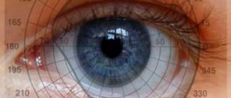
Visual field
is “ a spatial array of visual sensations available for observation in introspection psychological experiments
» [1]
The visual field is sometimes confused with the visual field. The field of view is everything that at a given moment in time causes light to fall on the retina. This input is processed by the visual system, which computes the visual field as an output.
In optometry and ophthalmology, the visual field test is used to determine whether the visual field is affected by medical conditions such as scotoma or more extensive vision loss or even decreased visual threshold.
Loss of visual fields
Normally, a person can perceive the fingers of a hand that is moved to the side by 85 degrees. If this angle is smaller, then the patient experiences a narrowing of the field of vision.
If the subject can perceive only half of the space, then there is a loss of half of the visual field. This symptom often accompanies serious diseases of the central nervous system, including the brain.
In order to more accurately diagnose the pathology in a patient with loss of visual fields, it is necessary to consult a doctor. Various techniques are used to examine these patients.
When half of the visual fields or even quarters are lost, we are talking about hemianopsia. Usually this pathology is bilateral, that is, the visual field is damaged on both sides.
Sometimes the loss of visual fields is concentric in nature. In this case, the condition may worsen to the point of tube vision. A similar symptom occurs with optic nerve atrophy or severe glaucoma. Sometimes this narrowing of the visual field is temporary and is associated with psychopathy.
With focal loss of the visual field, we are talking about a scotoma, which is characterized by the appearance of shadows or islands of absent or decreased vision. In some cases, scotoma can be detected only during a special examination of the patient, that is, he himself does not notice the visual impairment.
If the scotoma is located in the central zone, then most likely it is associated with macular degeneration and age-related changes in the area of the macula. Due to the fact that very effective methods of treating these serious diseases have recently appeared, you should follow all the instructions of your doctor.
Diagnostic methods
Diagnosis of changes in visual fields consists of measuring them, as well as identifying the possible cause of this condition. Previously, labor-intensive and time-consuming methods were used (manual perimetry).
Currently, this process is automated thanks to the use of computer technology in ophthalmology (automated perimetry), and therefore takes only a few minutes. Ophthalmologists also evaluate the nature of central vision.
In addition to the listed research methods, those that are aimed at diagnosing the cause of this condition are recommended. These include:
- measurement of intraocular pressure;
- ophthalmoscopy – examination of the fundus of the eye;
- ultrasound examination of the eyeball;
- computed tomography of the brain;
- 24-hour blood pressure monitoring.
It may also be necessary to consult a neurologist to exclude the central mechanisms of this pathological condition.
Examination helps to clarify hemianopsia.
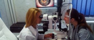
The simplest method for diagnosing narrowing of the visual fields is to compare it between the physician and the patient using the Donders method. The technique is used when a person is in serious condition (paralyzed, bedridden), a small child, or in the absence of the necessary digital devices in a medical institution.
To perform the diagnosis, the specialist and the person being examined must, being at a distance of 1 m, turn to face each other. Everyone covers one eye.
The patient looks into the doctor's open eye. And the specialist begins to slowly move his hand or a small table to the center of the field of view.
The patient tells the doctor when he sees her.
Visual field loss can be diagnosed even at home.
As a rule, a healthy person sees his arm extended to the side by at least 85 degrees. If this angle is smaller in a person, it means that there are deviations from the norm, which means that the brain or nervous system is not in order. The doctor finds out which organ is not in order after a thorough examination.
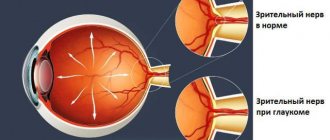
It is quite difficult to diagnose open-angle glaucoma in the early stages of development (this is only possible by chance, during a routine examination). At the same time, with progressive narrowing of the visual fields, all necessary diagnostic tests should be carried out as soon as possible, the true cause of the disease should be identified and appropriate treatment should be started to prevent further damage to the optic nerve.
Which doctor should I contact for glaucoma?
A doctor of any specialty should be able to recognize an acute attack of glaucoma, who in this case should immediately provide first aid to the patient. After stopping an acute attack, as well as if any signs of slowly progressing glaucoma appear, you should consult an ophthalmologist (a doctor who diagnoses and treats eye diseases).
Only he will be able to adequately assess all the signs of the disease and make an accurate diagnosis. In addition, only an ophthalmologist’s office has all the tools necessary for a full examination of the eye and identifying the cause of the disease.
It is worth noting that, if necessary, the ophthalmologist can refer the patient for consultation to other specialists (for example, to an endocrinologist in the presence of diabetes mellitus, to an oncologist if a tumor is suspected in the eye area), but only after measuring intraocular pressure and excluding (or stopping) an acute attack glaucoma.
Measuring intraocular pressure in glaucoma
Measuring intraocular pressure is the first and most informative test that is prescribed if glaucoma is suspected. However, it is worth remembering that sometimes a patient can develop glaucoma with normal intraocular pressure, so it is unacceptable to exclude this disease based on IOP testing alone.
Diagnosis of such a disease is carried out taking into account the study of visual fields using computer perimetry. The presence of clinical symptoms of the disease is usually confirmed by additional laboratory tests.
Often, the symptoms of hemianopsia indicate the presence of serious brain damage. In order to clarify the diagnosis, computed tomography, as well as magnetic resonance imaging and radiography of the skull are performed. How is visual field loss treated?
Reasons for violations
Depending on the cause of visual field loss, the nature of the pathology may be different. Usually there is a malfunction of the receiving apparatus of the optical system. If the pathology manifests itself as a so-called curtain on one side, then most likely the cause of the disease lies in disruption of the conduction pathways or retinal detachment. In the latter case, the disturbance of the visual field is accompanied by distortion of the shape of objects and a break in straight lines. The size of the visual field defect may also differ between the morning and evening hours. In some cases, the patient perceives surrounding objects in the form of floating figures. Retinal detachment often develops against the background of severe myopia, traumatic injury to the eye, or degeneration of the cells of this layer.
If there is bilateral loss of visual fields from the side of the temples, then we are probably talking about a pituitary adenoma.
If the field of vision is disturbed in the form of a translucent or dense curtain, which is located on the nasal side, then this indicates high intraocular pressure. Also, with glaucoma, rainbow circles appear when looking at point sources of light or fog in front of the eyes.
A translucent curtain on one side may appear when the transparency of the optical media of the eye decreases. These include cataract, cataract, pterygium, vitreous opacification.
When the central part of the visual field is lost, the cause of the disease is most often caused by a malnutrition of this area due to macular degeneration or pathology of the optic nerve and its atrophy. With macular degeneration, there is also a violation of the perception of the shape of objects, uneven changes in image size, and curvature of lines.
With concentric (up to tubular) narrowing of the visual field, we are usually talking about pigmentary degeneration of the retinal substance. At the same time, central visual acuity remains normal for quite a long time. Also, a concentric narrowing of the visual field is observed in glaucoma, but in this case the acuity of central vision is also reduced.
Typically, a concentric narrowing of the visual field is manifested by the fact that a person spends a very long time looking for the keyhole in the door, cannot navigate in an unfamiliar environment, etc.
With sclerotic changes in the arteries of the brain, the nutrition of nerve cells in the cortical visual centers is disrupted. This condition can also cause a concentric narrowing of the visual field, but the acuity of central vision also decreases, and there are other symptoms of brain malnutrition (forgetfulness, dizziness).
Causes
The causes of scotomas and narrowing of visual fields are different. They can be associated with damage to the nervous system, its central part, and the organ of vision itself. The main causative factors are the following:
- glaucoma is a disease in which intraocular pressure increases;
- swollen optic disc;
- chorioretinitis - inflammation of the retina and choroid;
- inflammation of the optic nerve;
- optic nerve atrophy;
- retinitis pigmentosa;
- arterial hypertension with all its negative consequences;
- neuroses and neurosis-like conditions;
- strokes;
- cerebral hemorrhages;
- brain injuries;
- brain tumors.
The disease in the initial stages is practically not felt due to the peculiarities of pathophysiology. Often the disorder is diagnosed during a routine medical examination, causing shock in the patient.
As the disease progresses, it becomes difficult for a person to read, watch TV, work at a computer, or navigate in space. The eyes begin to hurt more often, vision becomes less sharp, objects “float”.
The main causes of pathophysiological changes in peripheral vision are considered:
- mechanical damage to the retina (due to physical, sports activities, stressful situations, head injuries);
- glaucoma;
- cataract;
- stroke;
- atherosclerosis;
- vegetative-vascular dystonia;
- benign or malignant neoplasms;
- circulatory disorders;
- hypertension;
- osteochondrosis;
- diabetes;
- degenerative processes in the retina (detachment, thinning);
- vascular disorders;
- age (after 60 years).
Normal visual field values are considered to be: 55° from the inner and inner upper sides, 90° from the outer and outer lower sides, 70° from the upper outer side, 50° from the inner lower side, 65° from the lower side. Violation of indicators indicates diseases of the brain or eyes.
A decrease in the boundaries of lateral vision to 5-10° is diagnosed as a concentric narrowing of the visual field. Without treatment, the disorder progresses to tunnel vision, a pathologically limited ability to see.
A change in a certain area of the visual field is considered local loss. The disorder can be unilateral (homonymous hemianopsia) - loss of left or right zones, and bilateral (heteronymous hemiapsia) - loss of opposite regions.
There is symmetrical and asymmetrical impairment of peripheral vision. Symmetrical loss of the temporal halves of the visual fields is classified as bitemporal hemianopsia, and symmetrical loss of the nasal halves is binasal hemianopsia.
There is a loss of only a quarter of the visual field on both sides - homonymous square hemianopsia.
Scotomas
Scotomas, localized areas lacking visual function, are periodically diagnosed. Violations vary in shape (arc, circle, oval) and location (sectoral, pericentral, central, paracentral, peripheral).
Scotomas are divided into negative and positive. In the first option, the pathology is not felt by the person and is detected during special examinations. In the second case, the disorder is described by the patient as a cloudy spot or shadow in the field of vision.
With an absolute scotoma, the ability to see in the affected area completely disappears. If the patient notes that objects become unclear, “blurry,” then the pathology is diagnosed as relative.
There is a physiological scotoma. The disorder has the appearance of an oval-shaped blind spot located in the temporal region of the visual field.
The nature of visual field loss depends on the cause that causes it. The most common cause is diseases of the light-receiving apparatus of the eye. If the loss of the visual field looks like a curtain on any side, the cause is either retinal detachment or a disease of the visual system pathways.
With glaucoma, the patient experiences a deterioration in the visual field - a gradual narrowing of the visual field, first from the side of the nose, then from the central part (concentric narrowing of the visual field). Despite the fact that the visual field is reduced in many neurological diseases, this symptom is quite typical in glaucoma.
In medicine, the visual field is considered to be the space that a person sees in front of him without turning his head and focusing his eyes as much as possible on one point. What is directly in the focus of vision, a person sees with so-called central vision. What is on the sides is perceived by peripheral vision and is seen unclearly.
Among the reasons that cause distortion of visual fields, doctors name:
- stroke;
- brain tumors;
- inflammation in the brain;
- optic nerve injuries;
- deep sclerosis of the optic nerves.
If half or a quarter of the visual fields are poorly visible or not visible at all, then this phenomenon is called hemianopia. It is observed in both eyes. There is another type of prolapse when the concentric picture is not visible. This symptom accompanies the last stage of glaucoma.
There may be various reasons why peripheral vision is impaired. The most common are the following:
- Damage to the retina of the eye. Damage may vary. In this case, minimal damage to the retinal nerve cells contributes to dysfunction. This entails impaired peripheral vision. Such injuries include vascular eye disease, retinal detachment, or retinal dissection. Often, degeneration (deterioration of biological characteristics) and dystrophy (systemic disorders) of the retina of the eye lead to the destruction of retinal nerve cells.
- Glaucoma. This is a disease that is associated with increased intraocular pressure. The patient suffers damage to the optic nerve. And one of the characteristic symptoms of this disease is disturbances in the field of vision.
- Damage to the optic nerve may result from poor circulation in the vessels that supply the nerve. Another cause may be injury, tumor, or inflammation of the optic nerve.
- Increased intracranial pressure, which causes damage to the optic nerve.
- Brain damage. May be caused by injury, brain tumor, or vascular circulatory problems and hemorrhage.
Lateral vision is a necessary component of human vision. Deterioration or loss of peripheral vision is a symptom of many serious diseases. It is necessary to have your vision checked periodically. At the first sign of eye problems, you should seek help from a specialist. This is the only way to prevent the development of a serious illness.
Some diseases lead to a narrowing of the visual field, in which the patient loses, to some extent, peripheral or central visibility. There are two types of this symptom and reasons for the narrowing of the visual field:
- concentric narrowing, characterized by the global extent of the lesion;
- local narrowing that occurs in a specific area.
- increased IOP (ophthalmohypertension)
- age over 50 years
- ethnicity (glaucoma is more common among the Negroid race)
— chronic eye diseases (iridocyclitis, chorioretinitis, cataracts)
- history of eye injuries
– general diseases (atherosclerosis, hypertension, obesity, diabetes mellitus)
- stress
- long-term use of certain medications (antidepressants, psychotropic substances, antihistamines, etc.)
The relevance of considering this issue is that, according to statistics, about 30% of patients with stroke do not notice or do not attach importance to the manifestations of the disease.
Vascular pathology of the retina or disturbances of the blood supply in the area of the visual center, optic chiasm due to transient disturbances in the nutrition of neurons or small foci of stroke - the disease can proceed unnoticed and not cause concern in patients.
There are many reasons that can cause loss of the visual field. They affect not only the organs of vision, but also serve as a consequence of a serious disorder in the brain.
The most common causes of visual field impairment include cataracts, along with glaucoma, optic nerve pathology, eye injuries, retinal detachment, neurological diseases, high blood pressure, atherosclerosis and diabetes.
If some part of the image is observed as if through a translucent curtain, then we are most likely talking about cataracts. At the initial stage of glaucoma, the center of vision is usually affected, and only then the pathology can affect the peripheral areas. The causes of visual field loss should be identified by a doctor.
Hemianopsia is congenital or acquired. The most common causes of visual field loss are:
- The presence of vascular lesions of the brain in the form of hemorrhagic or ischemic strokes.
- Development of brain injuries.
- The presence of a brain tumor of any course (it does not matter whether it is benign or malignant).
- The presence of transient or transient cerebral circulatory disorders.
- The presence of hysterical reactions, hydrocephalus, migraines and epileptic seizures.
Hemianopsia may be transient in nature with a transient vascular disorder or against the background of migraine. The nature of such a transient disease is explained by short-term swelling of certain areas of the brain.
If the swelling of this area of the nervous system subsides, then blindness regresses and restoration of visual function is achieved. The appearance of such a symptom in a neurological disease as hemianopsia makes it possible to diagnose and clearly establish the area of brain damage.
Torsion fields and their nature
What are torsion fields? Does this concept have a real basis or is it an unproven theory from the section of esotericism and pseudoscience? Torsion fields are reciprocating movements of an electromagnetic field that turn into a spiral. As mentioned above, the theory of torsion fields excited the minds of scientists at the beginning of the last century, but there was no real evidence of their existence, or they were indirect and subjective. The first breakthrough in this matter occurred in the 80s, when physicist Oleg Gritskevich created a water engine based on the concept of torsion fields. Oleg Gritskevich combined the torsion of water with a magnetic field, using as a basis the “Ranke tube”, developed by the French physicist back in 1932. The device created by Gritskevich resembled a “donut” in appearance, inside which water circulated, heating up to a high temperature. Gitskevich’s invention was not just an interesting exhibit; the installation generated energy and supplied it to a small scientific town.
Then Gritskevich went to the USA with his colleagues and there he produced a more advanced example of his invention - a powerful hydromagnetic dynamo. But, apparently, oil tycoons intervened, for whom mass production of such an invention would have meant a complete collapse of the business, and research was soon curtailed.
Gritskevich explains the principle of his invention as follows. The water molecule has the shape of a pyramid. There are about a million such molecules in one cubic centimeter of water. At a pressure in the pipe of 10 atmospheres, the vortex spinning the water breaks the “pyramids” of water molecules, the hydrogen and oxygen atoms are separated, and when they unite again into molecules, a powerful release of energy occurs.
So, according to the Shipov-Akimov theory, the energy of swirling water is extracted from the physical vacuum. According to their research, the torsion field is generated by special geometric shapes. For example, a pyramid generates a powerful torsion field. Thus, architectural forms can be energy generators or portals to other layers of reality. It has long been suggested that the Egyptian pyramids are not tombs at all, but ancient energy generators (well, something like modern nuclear power plants) or portals for moving to other dimensions. Of course, modern science (both physics and history) denies such assumptions, because we will have to reconsider not only our view of energy and space, but also ask the question that previous generations of earthlings were in many ways smarter and more developed than us. And this means casting doubt on the generally accepted theory that past generations with stone axes ran after mammoths and communicated with each other by inarticulate mooing. Can modern science take such a radical step? The question is rhetorical.
Any geometric figure changes the property of ether - the element of space. This subtle matter “twists” and a torsion field is formed. As we know, theory without practice is dead. You can endlessly read about torsion fields in the works of talented scientists, but it’s easier to check everything through personal experience.
Torsion fields. Practical use
Anyone, even without special education, can create the simplest torsion generator at home. To do this, you need to take four neodymium magnets and tighten them, for example, placing them on the fan blades. The faster the rotation, the more powerful the formation of the torsion field from the vacuum will be. How can this invention be used? According to Akimov, various negative energy, which can exist in the room or directly cause illness in the body, leaves the area of formation of a powerful torsion field. Akimov even described examples of patients recovering after using such devices in an apartment.
The use of such a device for the formation of a torsion field will allow you to feel this very field on a purely physiological level - a metallic taste in the mouth and other symptoms. However, Akimov warned that it is not enough to form a torsion field using such a device. For a torsion field to benefit a person, you need to be able to structure it, and not everyone can do this. Otherwise, the unstructured torsion field will damage the human aura, and instead of a positive impact there will be a process of destruction.
Thus, anyone can generate a torsion field, but not everyone can structure it to use it for its intended purpose - to heal diseases or increase personal energy or the energy of space. Forming a torsion field without the proper ability to handle it is like giving a child a grenade.
How is the verification done?
To determine whether a patient has visual field defects, a complete examination is necessary. In this case, the doctor will be able to determine the area of the lesion, as well as the level of change in the structure of the optical system. This will help establish the diagnosis of the disease or lead to the need to conduct a number of additional examinations.
To evaluate visual fields, you can use one of the generally accepted techniques.
An experiment that is easy to carry out will allow you to approximately assess the state of your vision. In this case, you need to look into the distance and extend your arms to the sides (at shoulder level). After this you need to move your fingers. With normal peripheral vision, a person can easily notice the movement of the fingers. If the patient cannot notice the movement of the fingers, then he has lost peripheral vision.
Some people think that only central vision is important, but this is not true. For example, in the absence of peripheral vision, it is impossible to navigate in space, drive a car, etc.
The quality of vision can be affected by various diseases, including glaucoma. In this case, the field of view gradually decreases, that is, its concentric narrowing. This symptom is a reason to immediately seek medical help.
When carrying out diagnostic manipulations, the doctor can accurately determine the localization of damage in the optical system (before or after the optic chiasm, directly in the chiasm zone).
If the ophthalmologist identified a scotoma on only one side, then the damage is located up to the chiasm, that is, it affects either the retinal receptors or the optic nerve fibers.
Visual disorders can be present independently or combined with other pathologies of the central structures of the nervous system, which include disorders of consciousness, motor activity, speech, etc. Sometimes they are a consequence of impaired blood flow in the arteries that supply blood to the visual centers of the brain. Most often, young or middle-aged patients are susceptible to this condition.
With vegetative-vascular disorders, the first thing that appears is loss of the visual field. After a few minutes, these defects move left and right. They can also be felt when the eyelids are closed. This leads to a significant decrease in visual acuity, and then to severe headaches.
You can help a patient in this condition if you let him rest in his own bed, after unbuttoning the tight clothes. In addition, you can use receptor drugs, for example, give the patient a validol tablet to dissolve. If this condition recurs, then in addition to the ophthalmologist, you should definitely visit a neurologist.
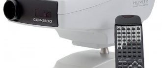
To assess the patient's condition, you need to use special computerized installations. In them, against a dark background, light points flash unevenly, which can have the same or different brightness and size. After this, the installation registers those areas that did not fall into the field of view.
Computer perimetry
Computer perimetry has become a modern method for assessing the narrowing of visual fields (and not only)
as a fairly simple method used in most medical institutions in ophthalmology offices. For glaucoma, its use is mandatory.
Under the field of view
refers to the gap that can be seen by one or two eyes at the same time.
To carry out diagnostics, special equipment is used - in a concave sphere with a stand. The subject needs to fix his chin on this stand and focus his gaze on a point in the center of the sphere. A point moves towards the center of the sphere, which at a certain moment the patient’s gaze must fixate.
The essence of the study is to register the indicator when the patient’s eye recorded (noticed) an object moving in the periphery. The moment when this object is seen by the eye is called the boundary of the visual field. This examination is performed monocularly (for one eye).
The internal fields located on the side of the nose and the external ones (on the side of the temple) are recorded for each eye. As a result of the diagnosis, a map of the visual fields is drawn, and then it is deciphered. Normally, the indicators will be close to the following.
Today, standard instrumental examination using a concave sphere can be replaced by more accurate and faster examination using a computer.
This diagnosis is recommended for suspected simulation of visual impairment or aggravation (tendency to exaggerate symptoms).
There are several assessment tests within this study. They make it possible to assess the condition of the retina from different angles.
- Kinetic perimetry
. Judging by the name, it is clear that the movement of a certain object (usually a black dot) is tracked across the field of a darkened screen along a certain trajectory. The patient monitors its movement and at the moment when the point ceases to be visible, gives a signal through the remote control, which he holds in his hand. The boundaries of the visual field outlined in this way provide reliable information not only about the condition of the eyes, but also about the functioning of the brain. - Static perimetry
. The patient observes a stationary object. The object appears at a variety of points on the boundaries of the visual field. In this case, the brightness of the object constantly changes. This approach allows you to assess the sensitivity threshold of the eye, which has preventive value. Including static perimetry, it is possible to identify the early stages of glaucoma. Since this is a computer diagnostic, it is the machine that determines exactly when a person’s eye has focused on a stationary object. This way he can testify to the sensitivity threshold of the eye. - Amsper's test
. This is a very simple diagnosis to identify pathology of the macula, that is, disorders that may be within the spot called macula. The patient is asked to focus on an object that is located in the center of the grid. In the absence of pathologies, the drawing will be viewed without distortion. If certain areas of the image are not clearly visible, fall out entirely, or are visible in the form of spots, then this indicates that there is a pathology in the center of the retina. Thanks to the Amsper test, you can determine what condition the central part of the retina has, as well as what the field of vision is. The main thing is to be able to fix your vision on one object in the center of the picture, which shows a grating. - Campimetry
determines the state of visual function. The patient must look at a white object moving inside a black square along a given path. The device must record all the places where the dot disappears, as well as those where it later appears. The dimensions of a square are meter by meter. It is located one meter from the patient’s eyes. A special table is used to track the readings. This way you can determine at what stage of the disease the retina is. The results are displayed on a special zone map, which shows how the retina (its photoreceptors) works. The state of vision is judged by the configuration of the contours of vision. By examining and analyzing the map, one judges the presence of defects and blind spots that exceed the physiological norm of human vision. For example, a person normally has a blind spot (scotoma) in the area of the optic nerve. There can be several cattle (they are called “positive”, “negative”, “absolute”, “relative”). The diagnosis is made based on how much the number and shape of scotomas exceed the norm.
Peripheral vision
Description
Peripheral vision
is a function of the rod and cone apparatus of the entire optically active retina and is determined by the field of view. The visual field is the space visible to the eyes (the eye), which a person sees with a motionless, fixed gaze. Peripheral vision helps to navigate in space.
The field of view of each eye has specific parameters. They are determined by the border of the optically active retina and may be limited to the upper edge of the orbit or the dorsum of the nose. The normal boundaries of the field of vision for white color are as follows: outward - 90°, upward outward - 70°, upward inward - 55°, inward - 55°, downward inward - 50°, downward - 65°, downward outward - 90°. The field of vision changes with retinal diseases, glaucoma, and pathology in the visual pathway. These changes consist of a concentric or local narrowing of the boundaries and the appearance of prolapses (scotomas) in the field of vision
. In the normal field of vision there are physiological scotomas: a blind spot in the temporal half of the visual field at 15° from the point of fixation and angioscotomas. The blind spot contributes to the projection of the optic disc, which does not contain photoreceptors. Angioscotomas are located around it. These band-like lesions in the visual field are associated with large retinal vessels that cover the photoreceptor cells.
Concentric narrowing of the visual field
on all sides is characteristic of retinal pigmentary dystrophy and damage to the optic nerve. The field of view may decrease down to a tube, when only a section of 5-10° remains in the center. The patient can still read, but cannot independently navigate in space.
Symmetrical loss in the visual fields of the right and left eyes
- a symptom indicating the presence of a tumor, hemorrhage or inflammation at the base of the brain, pituitary gland or optic tracts.
Heteronymous bitemporal hemianopsia
- this is a symmetrical half loss of the temporal fields of vision of both eyes. It occurs when there are lesions within the chiasm of intersecting nerve fibers coming from the nasal halves of the retina of the right and left eyes.
Heteronymous binasal symmetrical hemianopsia
It is rare, for example, with severe sclerosis of the carotid arteries, which equally compress the chiasm on both sides. Homonymous hemianopsia
- this is a half-like (right- or left-sided) loss of visual fields in both eyes. It occurs in the presence of pathology affecting one of the visual tracts. If the right optic tract is affected, then left-sided homonymous hemianopia occurs, i.e., the left halves of the visual fields of both eyes fall out. When the left optic tract is damaged, right-sided hemianopsia develops.
In the initial stage of a tumor or inflammatory process, only part of the optic tract may be compressed. In this case, symmetrical homonymous square hemianopias
, i.e., a quarter of the field of vision is lost in both the right and left eyes. When a brain tumor affects the cortical parts of the visual pathways, the vertical line of homonymous loss of visual fields does not involve the central parts, it bypasses the point of fixation, i.e., the projection zone of the macula. This is explained by the fact that fibers from the neuroelements of the central part of the retina go to both hemispheres of the brain.
Pathological processes in the retina and optic nerve can cause changes in the boundaries of the visual field of various shapes. Glaucoma, for example, is characterized by a narrowing of the field of vision on the nasal side.
Local loss of internal nodes of the visual field, not related to its boundaries, are called scotomas
. Scotomas can be absolute (complete loss of visual function) and relative (decreased perception of an object in the studied area of the visual field). The presence of scotomas indicates focal lesions of the retina and visual pathways. Scotoma can be positive or negative. A positive scotoma is seen by the patient himself as a dark or gray spot in front of the eye. This loss of vision occurs when there is damage to the retina and optic nerve. The patient himself does not detect a negative scotoma; it is detected during examination. Typically, the presence of such a scotoma indicates damage to the pathways.
Atrial scotomas
- These are suddenly appearing short-term moving deposits in the field of view. Even when the patient closes his eyes, he sees bright, flickering zigzag lines extending to the periphery. This symptom is a sign of cerebral vascular spasm. Atrial scotomas may appear with indefinite frequency. When they appear, the patient should immediately take an antispasmodic drug.
Based on the location of scotomas in the field of view, they are distinguished
- peripheral,
- central
- and paracentral scotomas.
At a distance of 12-18° from the center in the temporal half there is a blind spot. This is a physiological absolute scotoma. It corresponds to the projection of the optic nerve head. Enlargement of the blind spot has important diagnostic value. Central and paracentral scotomas
appear when the papillomacular bundle of the optic nerve, retina and choroid are damaged. Central scotoma may be the first manifestation of multiple sclerosis.
The visual field can be roughly assessed using a simple and publicly available control method. In this test, the health care worker's normal field of vision is compared with the patient's field of vision. The patient is seated opposite him with his back to the light at a distance of 0.5-1 m. The visual field of each eye is examined separately
. To do this, cover opposite eyes with the palm of your hand, for example, the left eye of the patient and the right eye of the researcher, then, conversely, the right eye of the patient and the left eye of the medical worker. The patient looks into the open eye of the researcher, who smoothly moves his hand from the periphery to the center from different sides, moving his fingers slightly. The hand is placed in the middle of the distance between the patient and the doctor. The patient must indicate the moment when he notices the appearance of the doctor’s hand in his field of vision. This method reveals significant narrowing of the boundaries and gross defects in the field of view. This method is considered indicative, since it does not allow obtaining a numerical expression of the degree of narrowing of the boundaries of the field of view. The method can be used in cases where it is impossible to conduct research using instruments, including in bedridden patients.
Accurate determination of the boundaries of the field of view is carried out using instrumental methods. These include campimetry
— study of the field of view on a concave spherical surface. Campimetry has limited use; it is used to study the central parts of the visual field within 30-40° from the center. The perimeters have the form of an arc or hemisphere. The simplest device for studying the visual field is the Förster perimeter, which is a 180° black arc (on a stand) that can be shifted in different directions. The outer surface of the arc is divided into degrees from 0 in the center to 90° at the periphery. For research, white or colored paper objects are used, attached to the ends of long rods. Paper circles have different diameters. To determine the outer boundaries of the visual field, a white object with a diameter of 3 mm is used, to measure defects inside the visual field, a white object with a diameter of 1 mm is used, and colored objects have a diameter of 5 mm.
During the examination, the patient's head is placed on a stand so that the eye being examined is in the center of the arc (hemisphere), and the other eye is covered with a bandage. In addition, throughout the entire study, the subject must fix the mark in the center of the device. It is also necessary for the patient to adapt to the conditions of the study within 5-10 minutes. The doctor moves white or colored marks along the arc of the Förster perimeter in various research meridians from the periphery to the center, thus determining the boundaries of their detection, i.e. boundaries of the field of view.
In projection perimeters
a light object is projected onto the arc or inner surface of the hemispherical perimeter (spheroperimeter). You can use objects of different sizes, brightness and colors. This allows for quantitative perimetry. In this case, two objects of different sizes are used, but the amount of reflected light from them is the same. This technique allows for early diagnosis of diseases in which the field of vision changes.
Dynamic (kinetic) perimetry is the most widely used
, in which an object moves in space from the periphery to the center along the radii of a circle. Nowadays, static perimetry is being increasingly introduced - the study of the visual field using stationary objects, the size and brightness of which change. Automatic, computer-controlled static perimeters are used. The researcher selects a program for presenting test objects to the patient. On a hemispherical or some other screen, white or colored marks move or flash in various meridians. The corresponding sensor records the test subject's indicators, indicating the boundaries of the visual field and areas of loss in it on a special form or in the form of a computer printout. When determining the boundaries of the visual field for white, a round mark with a diameter of 3 mm is usually used. If vision is poor, you can increase the brightness of the tag illumination or use a tag with a larger diameter. Perimetry for different colors is carried out with a 5 mm mark. Due to the fact that the peripheral part of the visual field is achromatic, the color mark is initially perceived as white or gray of varying brightness, and only upon entering the chromatic zone of the visual field does it acquire the appropriate color (blue, green, red), and only after that the subject should register a luminous object. The field of vision has the widest boundaries for blue and yellow colors, and the narrowest for green.
The information content of perimetry increases when using marks of different diameters and brightness - the so-called quantitative, or quantitative, perimetry
. It allows you to determine the initial changes in glaucoma, degenerative lesions of the retina and other eye diseases. To study twilight and night (scotopic) visual fields, the weakest background brightness and low illumination of the mark are used to assess the function of the rod apparatus of the retina.
, visual contrast perimetry has come into practice.
, which is a method of evaluating spatial vision using black-and-white or color stripes of different space and different frequencies, presented in the form of tables or on a computer display. Impaired perception of different spatial frequencies (gratings) indicates the presence of changes in the corresponding areas of the retina or visual field.
Regardless of the perimeter model, when studying the friction field, you must adhere to the following rules
:
- the field of view in each eye is examined in turn, the second eye is securely closed using a bandage that does not limit the field of view of the eye being examined;
- the eye being examined must be located exactly opposite the fixation mark in the center of the arc (hemisphere) of the perimeter, and at the entrance of perimetry the central mark must be constantly fixed;
- before starting the study, you need to instruct the patient, show fixation and moving marks, explain what answers are expected from him; the study must be carried out along at least eight, and preferably twelve radii of the circle;
- if the field of vision for colors is examined, then its peripheral border is marked not when the patient first noticed the mark, but at the moment when he confidently distinguishes its color. The results of the visual field study are applied to standard forms. They indicate the normal boundaries of the visual field for each eye. Narrowing of the visual fields or scotomas identified in the patient are shaded.
By the nature of the visual field limitation, one can determine the localization of the lesion in certain parts of the visual pathway, the stage of glaucoma, the degree of degenerative lesion, etc.
—-
Article from the book: Eye diseases. Complete Guide | Perederiy V.A.
https://glaza.by/, Moscow 01/22/14 06:15 0
29683
Central and peripheral vision
In this article we will focus on central and peripheral vision.
What are their differences? How is their quality determined? What are the differences between peripheral and central vision in humans and animals, and how do animals see in general? And how to improve peripheral vision...
This and much, much more will be discussed in this article.
Central and peripheral vision. Interesting information.
First about central vision.
This is the most important element of human visual function.
It got its name because... provided by the central portion of the retina and the central fovea. Gives a person the opportunity to distinguish shapes and small details of objects, therefore its second name is shaped vision.
Even if it decreases slightly, a person will immediately feel it.
The main characteristic of central vision is visual acuity. Her research is of great importance in assessing the entire human visual apparatus, for tracking various pathological processes in the organs of vision.
Visual acuity refers to the ability of the human eye to distinguish between two points in space located close to each other, at a certain distance from the person.
Let us also pay attention to such a concept as the visual angle, which is the angle formed between the two extreme points of the object in question and the nodal point of the eye.
It turns out that the larger the visual angle, the lower its acuity.
Now about peripheral vision.
It provides a person’s orientation in space and makes it possible to see in darkness and semi-darkness.
How to understand what is central and what is peripheral vision? Turn your head to the right, catch an object with your eyes, for example, a picture on the wall, and fix your gaze on any individual element of it. You see him well, clearly, don’t you?
This is thanks to central vision. But besides this object, which you see so well, a large number of different things also come into your field of vision. This is, for example, a door to another room, a closet that stands next to the painting you have chosen, a dog sitting on the floor a little further away. You see all these objects unclearly, but you still see, you have the ability to capture their movement and react to it.
This is peripheral vision.
Both human eyes, without moving, are capable of covering 180 degrees along the horizontal meridian and a little less - about 130 degrees along the vertical.
As we have already noticed, the acuity of peripheral vision is less than central. This is explained by the fact that the number of cones, from the center to the peripheral parts of the retina, decreases significantly.
Peripheral vision is characterized by the so-called visual field. This is the space that is perceived by a fixed gaze.
Peripheral vision is invaluable to humans.
It is thanks to it that free, habitual movement in the space surrounding a person and orientation in the environment around us is possible.
If peripheral vision is lost for some reason, then even with full preservation of central vision, the individual cannot move independently, he will bump into every object on his way, and the ability to see large objects with his gaze will be lost.
What kind of vision is considered good?
Now consider the following questions: how the quality of central and peripheral vision is measured, as well as what indicators are considered normal.
First about central vision. We are accustomed to the fact that if a person sees well, they say about him “one in both eyes.”
What does it mean? That each eye individually can distinguish two closely spaced points in space, which give an image on the retina at an angle of one minute. So it turns out to be one for both eyes.
By the way, this is only the lower norm. There are people who have vision of 1,2, 2 or more.
We most often use the Golovin-Sivtsev table to determine visual acuity, the same one with the well-known letters Ш B in the upper part. A person sits in front of the table at a distance of 5 meters and closes alternately his right and left eyes. The doctor points to the letters in the table, and the patient says them out loud.
The vision of a person who can see the tenth line with one eye is considered normal. Peripheral vision.
It is characterized by a field of view. Its change is an early and sometimes the only sign of some eye ailments.
The dynamics of changes in the visual field makes it possible to assess the course of the disease, as well as the effectiveness of its treatment. In addition, through the study of this parameter, atypical processes in the brain are revealed.
Studying the visual field is determining its boundaries, identifying defects in visual function within them. To achieve these goals, various methods are used.
The simplest of them is the control one. Allows you to quickly, literally in a few minutes, without using any instruments, determine a person’s field of vision.
The essence of this method is to compare the physician’s peripheral vision (which should be normal) with the patient’s peripheral vision.
It looks like this. The doctor and the patient sit opposite each other at a distance of one meter, each of them closes one eye (opposite eyes close), and the open eyes act as a point of fixation. Then the doctor begins to slowly move his hand, which is located on the side, out of the field of view, and gradually bring it closer to the center of the field of view. The patient must indicate the moment when he sees her. The study is repeated from all sides.
Using this method, a person's peripheral vision is only roughly assessed.
There are also more complex methods that give deeper results, such as campimetry and perimetry.
The boundaries of the visual field may vary from person to person and depend, among other things, on the level of intelligence and the structural features of the patient’s face.
Normal indicators for white skin color are as follows: upwards - 50⁰, outwards - 90⁰, upwards outwards - 70⁰, upwards inwards - 60⁰, downwards outwards - 90⁰, downwards - 60⁰, downwards inwards - 50⁰, inwards - 50⁰.
Color perception in central and peripheral vision. It has been experimentally established that human eyes can distinguish up to 150,000 shades and color tones.
This ability has an impact on various aspects of a person’s life.
Color vision enriches the picture of the world, gives the individual more useful information, and influences his psychophysical state.
Colors are actively used everywhere - in painting, industry, scientific research...
The so-called cones, light-sensitive cells that are found in the human eye, are responsible for color vision. But the rods are responsible for night vision. There are three types of cones in the retina, each of which is most sensitive to the blue, green and red parts of the spectrum.
Of course, the picture we get thanks to central vision is better saturated with colors compared to the result of peripheral vision. Peripheral vision is better at picking up brighter colors, such as red or black.
Women and men, it turns out, see differently!
Interestingly, women and men see things somewhat differently.
Due to certain differences in the structure of the eyes, representatives of the fair sex are able to distinguish more colors and shades than the majority of humanity.
In addition, scientists have proven that men have better developed central vision, while women have better peripheral vision.
This is explained by the nature of the activities of people of different sexes in ancient times.
Men went hunting, where it was important to clearly concentrate on one object and not see anything else. And the women looked after the housing and had to quickly notice the slightest changes, disturbances in the usual flow of everyday life (for example, quickly notice a snake crawling into a cave).
There is statistical evidence to support this statement. For example, in 1997, in the UK, 4,132 children were injured in road accidents, of which 60% were boys and 40% were girls.
In addition, insurance companies note that women are much less likely than men to be involved in car accidents involving side impacts at intersections. But parallel parking is more difficult for beautiful ladies.
Women also see better in the dark and notice more small details in a wide field compared to men. At the same time, the eyes of the latter are well adapted to tracking an object at a long distance.
If we take into account other physiological characteristics of women and men, the following advice will be formed - during a long trip it is best to alternate as follows - give the woman the day, and the man the night.
And a few more interesting facts.
Beautiful ladies' eyes get tired more slowly than men's.
In addition, women's eyes are better suited for observing objects at close range, so they can, for example, thread a needle much faster and more dexterously than men.
People, animals and their vision. Since childhood, people have been fascinated by the question: how do animals, our beloved cats and dogs, soaring birds, creatures swimming in the sea, see?
Scientists have been studying the structure of the eyes of birds, animals and fish for a long time so that we can finally find out the answers that interest us.
Let's start with our favorite pets - dogs and cats.
The way they see the world is significantly different from the way a person sees the world. This happens for several reasons. First.
Visual acuity in these animals is significantly lower than in humans. A dog, for example, has vision of approximately 0.3, and cats generally have 0.1. At the same time, these animals have an incredibly wide field of vision, much wider than that of humans.
The conclusion can be drawn as follows: the eyes of animals are maximally adapted for panoramic vision.
This is due to both the structure of the retina and the anatomical location of the organs.
Second.
Animals see much better than humans in the dark.
It is also interesting that dogs and cats see even better at night than during the day. All thanks to the special structure of the retina and the presence of a special reflective layer.
Third.
Our pets, unlike humans, distinguish moving objects better than static objects.
Moreover, animals have a unique ability to determine the distance at which an object is located.
Fourth. There are differences in the perception of colors. And this despite the fact that the structure of the cornea and lens in animals and humans is practically no different.
Humans can distinguish many more colors than dogs and cats.
And this is due to the structural features of the eyes. For example, a dog’s eyes have fewer “cones” responsible for color perception than a human’s. Therefore, they distinguish less colors.
Previously, there was a general theory that the vision of animals, cats and dogs, is black and white.
Now about other animals and birds.
Monkeys, for example, see three times better than humans.
Eagles, vultures, and falcons have extraordinary visual acuity. The latter can clearly see a target up to 10 cm in size at a distance of about 1.5 km. And the vulture is able to distinguish small rodents that are located 5 km away from it.
The record holder in panoramic vision is the woodcock. It's almost circular!
But the pigeon we are all familiar with has a viewing angle of approximately 340 degrees.
Deep-sea fish see well in absolute darkness, seahorses and chameleons can generally look in different directions at the same time, and all because their eyes move independently of each other.
How does our vision change throughout our lives?
How does our vision, both central and peripheral, change during life? What kind of vision are we born with, and what kind of vision do we come to old age with? Let's pay attention to these issues.
At different periods of life, people have different visual acuity.
When a person is born, he has low visual acuity. At four months of age this figure is approximately 0.06, by one year it grows to 0.1–0.3, and only by five years (in some cases it takes up to 15 years) vision becomes normal.
Over time, the situation changes. This is due to the fact that the eyes, like any other organs, undergo certain age-related changes; their activity gradually decreases.
It is believed that deterioration of visual acuity is an inevitable or almost inevitable phenomenon in old age. Let us highlight the following points.
| * | With age, the size of the pupils decreases due to weakening of the muscles that are responsible for their regulation. As a result, the reaction of the pupils to the light flux worsens. This means that the older a person gets, the more light he needs for reading and other activities. In addition, in old age, changes in lighting brightness are very painful. |
| * | Also, with age, the eyes recognize colors worse, the contrast and brightness of the image decreases. This is a consequence of a decrease in the number of retinal cells that are responsible for the perception of colors, shades, contrast and brightness. The world around an elderly person seems to fade and become dull. |
What happens to peripheral vision? It also gets worse with age - lateral vision worsens, visual fields narrow.
This is very important to know and take into account, especially for people who continue to lead an active lifestyle, drive a car, etc.
A significant deterioration in peripheral vision occurs after 65 years.
The following conclusion can be drawn.
A decrease in central and peripheral vision with age is normal, because the eyes, like any other organ of the human body, are susceptible to aging.
I can’t be with poor eyesight... Many of us already knew from childhood what we wanted to be in adulthood.
Some dreamed of becoming a pilot, some a car mechanic, some a photographer.
Everyone would like to do exactly what they like in life – no more, no less. And what a surprise and disappointment it can be when, upon receiving a medical certificate for admission to a particular educational institution, it turns out that the long-awaited profession will not become yours, and all because of poor eyesight.
Some people don’t even think that it can become a real obstacle to the implementation of plans for the future.
So, let's figure out which professions require good vision. It turns out there are not so few of them.
For example, visual acuity is necessary for jewelers, watchmakers, people involved in precision small instrument making in the electrical and radio engineering industries, in optical-mechanical production, as well as those who have a typographic profession (this could be a typesetter, proofreader, etc.).
Undoubtedly, the vision of a photographer, seamstress, or shoemaker must be sharp.
In all of the above cases, the quality of central vision is more important, but there are professions where peripheral vision also plays a role.
For example, an aircraft pilot. No one will argue that his peripheral vision should be as good as his central vision.
The profession of a driver is similar. Well-developed peripheral vision will allow you to avoid many dangerous and unpleasant situations, including emergency situations on the road.
In addition, auto mechanics must have excellent vision (both central and peripheral). This is one of the important requirements for candidates when hiring for this position.
Don't forget about the athletes either. For example, football players, hockey players, and handball players have peripheral vision that approaches ideal.
There are also professions where it is very important to correctly distinguish colors (preservation of color vision).
These are, for example, designers, seamstresses, shoemakers, and workers in the radio engineering industry.
We train peripheral vision. A couple of exercises. You've probably heard about speed reading courses.
The organizers undertake to teach you, in a couple of months and for not such a large amount of money, to swallow books one by one, and remember their content perfectly. So, the lion’s share of time in the courses is devoted to the development of peripheral vision. Subsequently, a person will not need to move his eyes along the lines of a book; he will immediately be able to see the entire page.
Therefore, if you set yourself the goal of developing excellent peripheral vision in a short time, you can enroll in speed reading courses, and in the near future you will notice significant changes and improvements.
But not everyone wants to spend time on such events.
For those who want to improve their peripheral vision at home in a quiet environment, here are a few exercises.
Exercise No. 1. Stand near the window and fix your gaze on some object on the street. This could be a satellite dish on a neighboring house, someone's balcony, or a slide on the playground.
Recorded? Now, without moving your eyes and head, name the objects that are near your chosen object.
Exercise No. 2.
Open the book you are currently reading.
Choose a word on one of the pages and fix your gaze on it. Now, without moving your pupils, try to read the words around the one on which you fixed your gaze.
Exercise No. 3.
For this you will need a newspaper.
In it you need to find the narrowest column, and then take a red pen and draw a straight thin line in the center of the column, from top to bottom. Now, glancing only along the red line, without turning your pupils to the right and left, try to read the contents of the column.
Don't worry if you can't do it the first time.
When you are successful with a narrow column, choose a wider one, etc.
Soon you will be able to look at entire pages of books and magazines.
Source: https://glaza.by/fakty/620/Tsentralnoe_i_perifericheskoe_zrenie.html
tags: patients, vision, central, peripheral
| Tweet |
Focal defects (scotomas)
If vision is reduced or absent in a certain area, the boundaries of which are not adjacent to the outer contour of the visual field, then we are talking about a scotoma. In this case, visual defects may not be perceived by the patient, because the image is completed by the second eye. Such scotomas are called negative. With positive scotomas, the patient perceives the defect as a spot or shadow located in the field of view.
The shape of scotomas can be different (sector, arc, oval, circle, irregular polygon). Depending on the location of scotomas relative to the central point of fixation, they also have different names (peripheral, sectoral, pericentral, paracentral, central). If vision is completely absent in the area of the defect, then the scotoma is called absolute; otherwise, it is called relative (only the clarity of perception is impaired).
An interesting fact is that in one patient the scotoma can be both relative and absolute (when examining the visual field using markers of different colors).
In addition to various pathological scotomas, each patient also has so-called physiological scotomas. These include the blind spot and vascular pattern.
In the first case, we are talking about an oval-shaped absolute scotoma, which is located in the temporal zone of the visual field. This scotoma corresponds to the projection of the optic nerve head. There is no light-receiving apparatus in the blind spot area. A physiological scotoma has clear dimensions and location. If a change in these parameters occurs, for example, an increase in size, then the scotoma becomes pathological. In particular, an increase in the size of the blind spot is observed with papilledema, glaucoma, and hypertension.
To identify scotomas, doctors previously resorted to rather labor-intensive visual field studies. Recently, mainly automatic perimeters are used, as well as testers for central vision, which greatly simplifies the procedure and reduces its execution time to several minutes.
Island of Good Vision
The visual field is uneven. It is not uniform and has maximum resolution in its central part.
Harry Moss Traquair described our visual field in 1927 as “an island of vision or a hill of vision surrounded by a sea of blindness.”[4] We have a corresponding modified definition of “Island of Vision.” Its empirical elliptical constraints on the length of the (horizontal) axis, our blind spots.
Kim Lloveras i Montserrat argue[5] that in the Roman era, people were aware of the characteristics of our blind spots in the horizontal limits of central good vision. In addition, they know that a greater change in clarity leads to a greater change in the perception of space. The observer feels inside the central space and understands its boundaries, which "envelop it", making them a very useful principle of architectural design. Personal exhibition exhibition La Experiencia del Espacio
[6] the Barcelona school of architecture accompanied this phenomenon by using a large ellipse (TK), which is 3.10 meters (twice the height of a person) and has a major axis of 3.94, located 6.38 m from the observer (these dimensions are very similar to those proposed by Traquair for the limits of the "visual island").
Changing the boundaries of the field of view
The narrowing of the boundaries of the visual field can be concentric, that is, global, or local. In the latter case, the formation of a defect occurs in a certain area, while on the rest of the perimeter the boundaries of the visual field are not broken.
Concentric narrowing
With concentric narrowing, much depends on the degree of this process. Thus, in severe cases, so-called tube vision is formed, in which peripheral perception is almost completely lost.
Concentric narrowing of vision can be associated with various pathologies, including neuroses, neurasthenia, and hysteria. In such conditions of the nervous system, the narrowing of the visual field is functional in nature.
However, concentric narrowing of the visual field is more often associated with organic pathology, for example, peripheral chorioretinitis, atrophy or neuritis of the optic nerve fibers, retinitis pigmentosa, and glaucoma.
To accurately determine the nature of the narrowing of the visual field (functional or organic), it is necessary to conduct a series of studies. They use objects of different sizes, colors, and brightness. In case of functional deviations, the size of the object and its other characteristics do not affect the result of the study. In addition, the patient’s ability to navigate in space is used as a distinguishing feature. If this property is violated, then most likely we are talking about an organic lesion.
With local narrowing of the visual field, the process can be bilateral or unilateral. With bilateral lesions, the defects can be located symmetrically or in different areas of the visual field.
In this case, some characteristic areas of vision loss, for example, hemianopia (half loss of visual fields), are of important diagnostic importance. In this condition, we are talking about damage to the visual pathway in the chiasm zone or closer to the central structures.
Hemianopsia can be diagnosed independently, but more often such visual impairments are identified during examination of the patient.
Hemianopsia can be homonymous (loss of the temporal half on one side and the nasal half on the other) or heteronymous (simultaneous loss of the nasal or temporal halves on both sides). There is also quadrant hemianopsia, when the beginning of the defect coincides with the fixation point.
Diagnostics of the visual field condition
When diagnosing pathologies of the visual field, it is necessary to identify the cause of vision anomalies. This will allow you to prescribe treatment in a timely manner and prevent the development of pathology. The professionalism of an ophthalmologist plays an important role when making a diagnosis.
The specialist may prescribe one of the following studies:
- Kinetic and static perimetry.
- Visometry.
- Biomicroscopy of the eye.
In some cases, a visual assessment allows you to determine the state of the visual field. The doctor and patient sit opposite each other and cover one eye with their hand. Then the ophthalmologist begins to move the other hand, which is holding an object. As soon as an object appears in the patient’s field of vision, he must inform the specialist about it. If both the doctor and the patient saw the object at the same point, it means that the patient’s visual field is normal.
How fov works in CS:GO
Let's say right away that the viewing angle directly depends on the aspect ratio of the monitor. Those. The wider the viewing angle, the more you see. Let's look at everything in practice so that you can better understand what works and how. Just pay attention to the right wall. This is what the picture looks like with an aspect ratio of 16:9 and a viewing angle of 90.
And like this - with a ratio of 16:10 and a viewing angle of 84.
And this is what 4:3 looks like with an angle of 74.
Well, so – 5:4 and 70.
And this is what, for example, a 16:3 ratio looks like with a viewing angle of 143.
Hemianopsia
Homonymous hemianopsia most often occurs as a result of pathological space-occupying formations in the brain (tumor, abscess, hematoma) or with retrochiasmatic lesions of the optic pathway (opposite side). In such patients, hemianoptic scotomas may be detected, which are located in symmetrical areas of the visual field.
In heteronymous hemianopsia, the defects can be located on the outer side (bitemporal hemianopsia) or on the inner side (binasal hemianopsia). In the first case, the visual pathway in the chiasm zone is affected, which is characteristic of a tumor process in the pituitary tissue. With binasal hemianopsia, there is damage to the uncrossed fibers of the optic pathway in the chiasm zone. This can occur when the internal carotid artery aneurysm puts pressure on the external nerve fibers in the decussation area.