LABORATORY TESTS IN NEUROLOGY ARE AVAILABLE IN THE BRANCHES:
Laboratory examinations in neurology in the Primorsky region
Address: St. Petersburg , Primorsky district, st. Repisheva, 13
Laboratory examinations in neurology in the Petrograd region
Address: St. Petersburg , Petrogradsky district, st. Lenina, 5
Laboratory examinations in neurology in Vsevolozhsk
Address: Vsevolozhsk , Oktyabrsky Prospekt, 96 A
The most commonly performed tests are erythrocyte sedimentation rate, morphological studies, peripheral blood smear, urine and cerebrospinal fluid analysis.
Let's look at some of the basic laboratory techniques used in neurology.
Examination of cerebrospinal fluid (spinal cord puncture)
The examination involves collecting cerebrospinal fluid by puncturing it with a special needle, which is inserted into the spinal canal (subarachnoid space). Analysis of cerebrospinal fluid is mainly aimed at studying its physicochemical properties, the presence of immunoglobulin G and identifying pathogenic microorganisms. Changes in the composition of the cerebrospinal fluid make it possible to diagnose certain diseases of the central nervous system (purulent bacterial meningitis, traumatic brain injury, hemorrhagic stroke, cysts) and spinal nerves. The procedure is performed on patients of any age and pregnant women.
Methods for studying the nervous system.
Methods for studying the nervous system
The main methods for studying the central nervous system and neuromuscular system are electroencephalography (EEG), rheoencephalography (REG), electromyography (EMG), which determine static stability, muscle tone, tendon reflexes, etc.
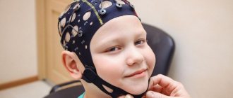
Electroencephalography (EEG)
— a method for recording electrical activity (biocurrents) of brain tissue for the purpose of objectively assessing the functional state of the brain. It is of great importance for diagnosing brain injury, vascular and inflammatory diseases of the brain, as well as for monitoring the functional state of an athlete, identifying early forms of neuroses, for treatment and for selection into sports sections (especially boxing, karate and other sports related with blows to the head). When analyzing data obtained both at rest and under functional loads, various external influences in the form of light, sound, etc.), the amplitude of the waves, their frequency and rhythm are taken into account. In a healthy person, alpha waves predominate (oscillation frequency 8-12 per 1 s), recorded only when the subject’s eyes are closed. In the presence of afferent light impulses with open eyes, the alpha rhythm completely disappears and is restored again when the eyes are closed. This phenomenon is called the fundamental rhythm activation reaction. Normally it should be registered. In 35-40% of people in the right hemisphere, the amplitude of alpha waves is slightly higher than in the left, and there is also some difference in the frequency of oscillations - by 0.5-1 oscillations per second. With head injuries, the alpha rhythm is absent, but oscillations of high frequency and amplitude and slow waves appear. In addition, the EEG method can diagnose early signs of neuroses (overwork, overtraining) in athletes.
Rheoencephalography (REG)
- a method for studying cerebral blood flow, based on recording rhythmic changes in the electrical resistance of brain tissue due to pulse fluctuations in the blood supply of blood vessels. The rheoencephalogram consists of repeating waves and teeth. When assessing it, the characteristics of the teeth, the amplitude of the rheographic (systolic) waves, etc. are taken into account. The state of vascular tone can also be judged by the steepness of the ascending phase. Pathological indicators are deepening of the incisura and an increase in the dicrotic tooth with a shift downward along the descending part of the curve, which characterizes a decrease in the tone of the vessel wall. The REG method is used in the diagnosis of chronic disorders of cerebral circulation, vegetative-vascular dystonia, headaches and other changes in the blood vessels of the brain, as well as in the diagnosis of pathological processes resulting from injuries, concussions and diseases that secondary affect blood circulation in the cerebral vessels (cervical osteochondrosis , aneurysms, etc.).
Electromyography (EMG)
- a method for studying the functioning of skeletal muscles by recording their electrical activity - biocurrents, biopotentials. Electromyographs are used to record EMG. The removal of muscle biopotentials is carried out using surface (overhead) or needle-shaped (injected) electrodes. When studying the muscles of the limbs, electromyograms are most often recorded from the muscles of the same name on both sides. First, the resting EM is recorded in a maximally relaxed state of the entire muscle, and then in its tonic tension. Using EMG, it is possible to determine at an early stage (and prevent the occurrence of muscle and tendon injuries) changes in muscle biopotentials, to judge the functional capacity of the neuromuscular system, especially the muscles most loaded in training. Using EMG, in combination with biochemical studies (determination of histamine, urea in the blood), early signs of neuroses (overfatigue, overtraining) can be determined. In addition, multiple myography determines the work of muscles in the motor cycle (for example, in rowers, boxers during testing). EMG characterizes muscle activity, the state of the peripheral and central motor neuron. EMG analysis is given by amplitude, shape, rhythm, frequency of potential oscillations and other parameters. In addition, when analyzing EMG, the latent period between the signal for muscle contraction and the appearance of the first oscillations on the EMG and the latent period for the disappearance of oscillations after the command to stop contractions are determined.
Chronaximetry
- a method for studying the excitability of nerves depending on the time of action of the stimulus. First, the rheobase is determined—the current strength that causes the threshold contraction—and then the chronaxy. Chronancy is the minimum time for a current of two rheobases to pass, which gives the minimum reduction. Chronaxy is calculated in sigmas (thousandths of a second). Normally, the chronaxy of various muscles is 0.0001-0.001 s. It has been established that proximal muscles have less chronaxy than distal ones. The muscle and the nerve that innervates it have the same chronaxy (isochronism). Muscles that are synergists also have the same chronaxy. On the upper limbs, the chronaxy of the flexor muscles is two times less than the chronaxy of the extensor muscles; on the lower limbs, the opposite ratio is observed. In athletes, muscle chronaxy sharply decreases and the difference in chronaxy (anisochronaxy) of flexors and extensors may increase due to overtraining (overfatigue), myositis, paratenonitis of the gastrocnemius muscle, etc.
Stability in static position
can be studied using stabilography, tremorography, Romberg's test, etc.
Romberg's test
reveals imbalance in a standing position. Maintaining normal coordination of movements occurs due to the joint activity of several parts of the central nervous system. These include the cerebellum, vestibular apparatus, conductors of deep muscle sensitivity, and the cortex of the frontal and temporal regions. The central organ for coordinating movements is the cerebellum. The Romberg test is carried out in four modes with a gradual decrease in the support area. In all cases, the subject's hands are raised forward, fingers spread and eyes closed. “Very good” if in each pose the athlete maintains balance for 15 seconds and there is no body swaying, trembling of the hands or eyelids (tremor). For tremor, a “satisfactory” rating is given. If the balance is disturbed within 15 s, the test is assessed as “unsatisfactory”. This test is of practical use in acrobatics, gymnastics, trampolining, figure skating and other sports where coordination is important.
Determination of balance in static poses
Regular training helps improve coordination of movements.
In a number of sports (acrobatics, artistic gymnastics, diving, figure skating, etc.) this method is an informative indicator in assessing the functional state of the central nervous system and neuromuscular system. With overwork, head injury and other conditions, these indicators change significantly. The Yarotsky test
allows you to determine the sensitivity threshold of the vestibular analyzer.
The test is performed in the initial standing position with eyes closed, while the athlete, on command, begins rotational movements of the head at a fast pace. The time of head rotation until the athlete loses balance is recorded. In healthy individuals, the time to maintain balance is on average 28 s, in trained athletes - 90 s or more. The sensitivity level threshold of the vestibular analyzer mainly depends on heredity, but under the influence of training it can be increased. Finger-nose test.
The subject is asked to touch the tip of his nose with his index finger with his eyes open and then with his eyes closed.
Normally, there is a hit, touching the tip of the nose. In case of brain injuries, neuroses (overwork, overtraining) and other functional conditions, there is a miss (miss), trembling (tremor) of the index finger or hand. The tapping test
determines the maximum frequency of hand movements. To carry out the test, you must have a stopwatch, a pencil and a sheet of paper, which is divided into four equal parts by two lines. Dots are placed in the first square for 10 seconds at maximum speed, then a 10-second rest period and the procedure is repeated again from the second square to the third and fourth. The total duration of the test is 40 s. To evaluate the test, count the number of dots in each square. Trained athletes have a maximum frequency of wrist movements of more than 70 in 10 seconds. A decrease in the number of points from square to square indicates insufficient stability of the motor sphere and nervous system. The decrease in the lability of nervous processes occurs in steps (with an increase in the frequency of movements in the 2nd or 3rd squares) - indicating a slowdown in the processes of processing. This test is used in acrobatics, fencing, gaming and other sports.
Nervous system research, analyzers.
Kinesthetic sensitivity is examined with a hand dynamometer. First, the maximum force is determined. Then the athlete, looking at the dynamometer, squeezes it 3-4 times with a force equal to, for example, 50% of the maximum. Then this effort is repeated 3-5 times (pauses between repetitions are 30 s), without visual control. Kinesthetic sensitivity is measured by the deviation from the obtained value (in percent). If the difference between the given and actual effort does not exceed 20%, kinesthetic sensitivity is assessed as normal.
Muscle tone study.
Muscle tone is a certain degree of normally observed muscle tension, which is maintained reflexively. The afferent part of the reflex arc is formed by conductors of muscle-articular sensitivity, carrying impulses from proprioceptors of muscles, joints and tendons to the spinal cord. The efferent part is the peripheral motor neuron. In addition, the cerebellum and extrapyramidal system are involved in the regulation of muscle tone. Muscle tone is determined by V.I. tonometer. Dubrovsky and E.I. Deryabina (1973) in a calm state (plastic tone) and tension (contractile tone). An increase in muscle tone is called muscle hypertension (hypertonicity), no change is called atony, a decrease is called hypotension. An increase in muscle tone is observed with fatigue (especially chronic), with injuries and diseases of the musculoskeletal system (MSA) and other functional disorders. A decrease in tone is observed with prolonged rest, lack of training in athletes, after removal of plaster casts, etc.
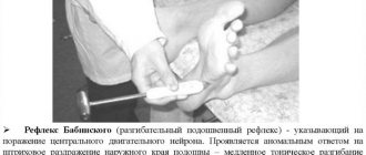
Reflex Research
. Reflex is the basis of the activity of the entire nervous system. Reflexes are divided into unconditioned (innate reactions of the body to various exteroceptive and interoceptive stimuli) and conditioned (new temporary connections developed on the basis of unconditioned reflexes as a result of the individual experience of each person). Depending on the site of evocation of the reflex (reflexogenic zone), all unconditioned reflexes can be divided into superficial, deep, distant and reflexes of internal organs. In turn, superficial reflexes are divided into cutaneous and mucous membranes; deep - tendon, periosteal and articular; distant - for light, auditory and olfactory. When examining abdominal reflexes, to completely relax the abdominal wall, the athlete needs to bend his legs at the knee joints. Using a blunt needle or quill pen, the doctor makes a line irritation 3-4 fingers above the navel parallel to the costal arch. Normally, contraction of the abdominal muscles on the corresponding side is observed. When examining the plantar reflex, the doctor stimulates along the inner or outer edge of the sole. Normally, there is flexion of the toes. Deep reflexes (knee, Achilles tendon, biceps, triceps) are among the most constant. The knee reflex is caused by striking the quadriceps tendon below the kneecap with a hammer; Achilles reflex - hitting the Achilles tendon with a hammer; the triceps reflex is caused by a blow to the triceps tendon above the olecranon; biceps reflex - with a blow to the tendon in the elbow bend. The blow with a hammer is applied abruptly, evenly, precisely on a given tendon. With chronic fatigue, athletes experience a decrease in tendon reflexes, and with neuroses - an increase. With osteochondrosis, lumbosacral radiculitis, neuritis and other diseases, a decrease or disappearance of reflexes is observed.
Studies of visual acuity, color perception, visual field. Visual acuity
is examined using tables located at a distance of 5 m from the subject. If he distinguishes 10 rows of letters on the table, then visual acuity is equal to one, but if only large letters, the 1st row, are distinguished, then visual acuity is 0.1, etc. d.
Visual acuity is of great importance when selecting for sports. So, for example, for divers, weightlifters, boxers, wrestlers with vision of -5 and below, sports are contraindicated! Color perception is studied using a set of colored strips of paper. With injuries (lesions) to the subcortical visual centers and partially or completely to the cortical zone, color recognition is impaired, most often red and green. If color vision is impaired, auto and cycling and many other sports are contraindicated. The field of view is determined by the perimeter. This is a metal arc attached to a stand and rotating around a horizontal axis. The inner surface of the arc is divided into degrees (from zero at the center to 90°). The number of degrees marked on the arc shows the boundary of the field of view. The boundaries of the normal field of vision for white color: internal - 60°; lower - 70°; upper - 60°. 90° indicates deviations from the norm. Evaluation of the visual analyzer is important in team sports, acrobatics, gymnastics, trampolining, fencing, etc. Hearing examination.
Hearing acuity is examined at a distance of 5 m. The doctor pronounces the words in a whisper and offers to repeat them.
In case of injury or illness, hearing loss is observed (auditory neuritis). Most often observed in boxers, water polo players, shooters, etc. Analyzer research.
A complex functional system consisting of a receptor, an afferent pathway and a zone of the cerebral cortex where this type of sensitivity is projected is referred to as an analyzer. The central nervous system (CNS) receives information about the external world and the internal state of the body from reception organs specialized in the perception of irritations. Many reception organs are called sense organs, because as a result of their irritation and the receipt of impulses from them in the cerebral hemispheres, sensations, perceptions, ideas arise, that is, various forms of sensory reflection of the external world. As a result of information from receptors entering the central nervous system, various acts of behavior arise and general mental activity is built.
The topic is vast
Neuroendocrine research
Neuroendocrine studies are carried out by administering substances that increase or block the synaptic transmission of nerve impulses in certain structures of the brain, making it possible to study the effect of the secretion of a specific hormone.
An example of such an examination is the determination of the amount of somatotropin (growth hormone that is secreted by the anterior pituitary gland) after the administration of the appropriate drug. In patients with depressive syndrome, the secretion of somatotropin is impaired; sometimes, to clarify the diagnosis, it may be necessary to record a video EEG or undergo an MRI of the brain.
High-intensity pulsed magnetic therapy (HIMT)
High-intensity pulsed magnetic therapy (HIMT) is a therapeutic effect on the human body of a pulsed electromagnetic field that is several times higher than the intensity of traditional magnetotherapy.
The VIMT method is painless (at an induction of 0.35 T and above, only a sensation of “mechanical push” appears), since the induction current is directly proportional to the electrical conductivity of the tissues; weak currents are induced in the skin, adipose and bone tissues, not reaching the threshold for excitation of pain receptors. Moreover, by activating weakly myelinated A - and C - afferent nerve fibers, VIMT is able to block afferent impulses from the pain focus through the mechanism of the so-called “peripheral gate block” (at the level of the dorsal horns of the spinal cord), which leads to persistent pain relief.
There are indications that VIMT is especially effective for chronic pain (Ponomarenko G.N., 1998). Along with this, the excitation of thick myelinated, A - and, A - efferents restores impaired muscle tone, and starting from an induction of 0.5 T causes distinct muscle contractions. In this case, VIMT evenly penetrates the human body, its effect is noticeable at a distance of up to 10 centimeters from the inductor, therefore the induced currents affect all fibers of the nerve trunk.
Finally, the frequency of induced current pulses coincides with the maximum impulse of vegetative B-fibers, which determines the possibility of trophic effects of VIMT.
Exposure to VIMT causes a significant increase in local blood flow, which helps remove cell autolysis products from the lesion and, as a result, reduce the inflammatory response. The charge of cells, the dispersion of colloids and the permeability of cell membranes change, which leads to a pronounced anti-edematous effect. VIMT stimulates the processes of reparative tissue regeneration and their metabolism. The regulatory effect of VIMT on antioxidant systems was noted.
Thus, the main therapeutic effects of VIMT are:
- neuromyostimulating,
- analgesic,
- vasoactive,
- decongestant,
- anti-inflammatory,
- trophic,
- regenerative.
The nervous system is most sensitive to VIMT, followed by the endocrine, sensory organs, cardiovascular, blood, muscular, digestive, excretory, respiratory and skeletal systems.
INDICATIONS FOR HIGH INTENSITY PULSE MAGNETOTHERAPY
- Neurology: diseases and injuries of the peripheral nervous system, including after reconstructive operations on the nerves, vertebrogenic diseases of the nervous system, vascular diseases of the brain and spinal cord with focal neurological deficits, craniocerebral and spinal trauma with motor and sensory disorders, children's cerebral paralysis (hemiparetic form, spastic diplegia), consequences of neuroinfections, hereditary neuromuscular diseases, Raynaud's disease (syndrome).
- Traumatology and orthopedics: damage to the musculoskeletal system (bruises, ligament damage, bone fractures), soft tissue injuries, muscle wasting as a result of physical inactivity caused by trauma, scoliotic disease in children.
- Arthrology: deforming osteoarthritis, ankylosing spondylitis (ankylosing spondylitis), periarthrosis, epicondyloses, arthritis, periarthritis, epicondylitis, heel spurs.
- Surgery: slow-healing and infected wounds, trophic ulcers, boils, carbuncles, abscesses, hidradenitis, phlegmon - after surgery, mastitis, chronic osteomyelitis, thermal burns, occlusive diseases of peripheral arteries.
- Pulmonology: chronic bronchitis in remission, bronchial asthma, pleural adhesions after dry and exudative pleurisy, bronchiectasis.
- Gastroenterology: hypomotor-evacuation disorders of the stomach (including after gastrectomy and vagotomy), hypomotor dysfunction of the colon and gallbladder, peptic ulcer of the stomach and duodenum.
- Cardiology: hypertension stage I–II, vegetative - vascular dystonia.
- Gynecology: inflammatory diseases of the uterus and appendages, ovarian hypofunction, inflammatory changes after surgical delivery, menstrual dysfunction, algomenorrhea.
- Urology: atony of the bladder, weakness of the sphincter and detrusor, prostatitis, sexual disorders in men, condition after lithotripsy, ureteral stone.
- Ophthalmology: partial atrophy of the optic nerve.
- Occupational diseases: vibration disease.
- Dentistry: arthrosis of the temporomandibular joint, periodontal disease, filling pain.
For diagnostic purposes, VIMT is used to assess the functional state of the motor pathway (including the first motor neuron) through transcranial magnetic pulse stimulation (TMS) with determination of the amplitude of the evoked motor potential and measurement of central motor conduction time, for mapping the functions of the cerebral cortex, and intraoperative monitoring. This, in particular, has important prognostic significance in so-called “motor neuron diseases” and allows one to quickly assess the effectiveness of the therapy.
CONTRAINDICATIONS FOR VIMT
General contraindications for use: severe arterial hypotension, systemic blood diseases, bleeding tendency, thrombophlebitis, thromboembolic disease, bone fractures before immobilization, grade III diffuse toxic goiter, acute purulent inflammatory processes, abscesses and phlegmons before opening and draining cavities, cholelithiasis , epilepsy, presence of an implanted pacemaker, febrile conditions, pregnancy.
If you want to learn more about magnetic stimulation, we recommend that you read the following literature:
Nikitin S.S., Kurenkov A.L. Magnetic stimulation in the diagnosis and treatment of diseases of the nervous system. Guide for doctors.
Marcolin MA, Padberg F. Transcranial Brain Stimulation for Treatment of Psychiatric Disorders
Hallett M., Chokroverty S. Magnetic Stimulation in Clinical Neurophysiology