Each person is a microcosm. This is not a pretentious saying of another poet, but the real everyday life of anatomy and histology. In the body of each of us there is a huge number of complex microstructures, a violation in any of them will lead to loss of health or even death. A good example is the loop of Henle; the average person doesn’t even know what it is, and any failure is a guaranteed visit to the doctor.
Our body fluid
We are not only what we eat, but also what we drink:
- The human body should receive at least 2-3 liters of fluid daily;
- None of us can survive a week without water;
- ~4 liters of blood constantly moves through the veins and arteries of a person;
- More than 200 liters of primary urine are produced per day;
- The circulatory network of the kidney is much more developed compared to other organs.
Dehydration is the worst torture. Remember yourself the morning after the holiday, if you can somehow cope with a headache and general “worship”, then dehydration causes real inconvenience.
That's why:
- Drink only what will not harm your kidneys;
- Don't deny yourself fluids;
- Make sure that swelling does not appear;
- At the first problem with your kidneys, go to the doctor.
You can really live with one kidney if the second one can take on all functions in full. Otherwise, you will have to get acquainted with hemodialysis and find out what organ transplantation is. Life expectancy after such manipulations leaves much to be desired.
How does a nephron work?
The human kidneys pump all the blood through themselves, separating fluid and small elements and gradually forming urine from this. They also regulate the amount and composition of fluid in the body.
Therefore, edema is so often associated with impaired renal function and especially glomerulonephritis, and the diseases themselves are detected by the composition of urine. A urine test shows whether nephron filters allow something unnecessary to pass through and whether they return what is needed into the bloodstream.
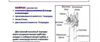
Each nephron functions in several stages, so that urine is formed as a result of four processes:
- Filtration (purification of fluid from the blood that needs to be removed).
- Reabsorption (return of residual nutrients into the blood).
- Secretion (removal of necessary substances from cells).
- Removing excess and unhealthy substances with urine.
In this way, through the kidneys, the composition of the blood and the volume of fluid in the tissues are normalized, and blood pressure returns to its normal values. Since the structure of the nephron is related to its functions, the problem in any of these processes is physical destruction or deformation of the nephron.
Where is the loop of Henle located?
The loop of Henle is located in the kidneys
human body:
- Part of the nephron;
- Connects the proximal and distal tubules;
- Found in both the cortex and medulla.
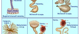
This is not a single structure, like the same nose. The count goes into millions of nephrons and the same number of loops. For a more complete understanding, you should study the structure of the nephron under a microscope (pictured at the end of the article):
- The loop is located between the proximal and distal tubules;
- It is an integral part of the Shumlyansky-Bowman capsule;
- Originates from the proximal tubule located in the cortex;
- Descends into the renal medulla;
- The ascending part returns to the cortex, ending in the distal tubule.
Again, these are all microstructures and a rough attempt to explain the location of the loop in 3D space. Schematically, it is sketched as a tapering loop going down from a thick tubule, and then, rising, up to the same thick tubule.
Types of nephrons
The types of nephrons functioning in the kidney differ in their morphological features and the work they perform:
- Superficial and intracortical cortices - 80-85% of all nephrons.
- Juxtamedullary - make up 15-20% of their total number.
Cortical
There are superficial and intracortical cortical types of nephrons. The renal corpuscle of the superficial cortical renal units is located at a distance of 1 mm from the glomerular capsule in the outer part of the renal cortex, and the corpuscle of the intracortical units is located in the middle part of the cortex.
A feature of the cortical variety is the presence of a short loop of Henle, reaching only the outer part of the medullary renal substance. The cortical nephron is a structural unit of the kidney, the main function of which is the formation of primary urine.
Loop of Henle: functions
The entire range of functions comes down to one result:
- Na, Cl and K are reabsorbed in the loop;
- Up to 20% of the initial amount of Na ions is reabsorbed; this element “pulls” the liquid with it;
- In the ascending part, magnesium and calcium are absorbed back into the body;
- In exchange for the ions and fluid received, the body releases urea, which enters the fluid traveling along the distal tubule;
- All the described processes occur in the renal medulla.
200 liters of emerging primary urine must somehow be brought to a reasonable 1.5-2 liters. And the described actions are one of the body’s mechanisms for regulating water-salt balance.
Ultimately, the secreted fluid will lose:
- Glucose;
- Proteins;
- Most of the volume;
- Part of mineral elements;
- Everything that can be useful to the body.
Passing through the loop of Henle, primary urine becomes more and more similar in its characteristics to secondary urine. It is this that will be removed from the body along with everything that has been deemed unsuitable for further use.
Thousands of studies have been devoted to this topic. The structure of the nephron and all the processes occurring in it during filtration and reabsorption of fluid have been studied in detail. For more detailed information, it is better to refer to scientific literature. It is very difficult to read it without prior preparation due to the abundance of scientific terms and the deepest immersion in the topic.
How does a nephron work?
Parts of the nephron are responsible for several stages of filtration and formation of urine:
- After primary filtration in the glomerulus, the liquid enters the capsule.
- There are still useful substances in it. They are reabsorbed (reabsorbed) through the wall of the tubule into the blood.
- Then the substances are secreted (removed from the cells) in the wall of the canal itself, entering partly into the bloodstream and partly into the canal. The tubule passes secreted substances from the blood into the nephron.
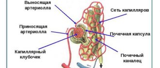
The anatomy of the nephron includes a glomerulus of capillaries, a capsule where the initial urine enters after sifting out the proteins, two tubules with “windows” in which urine is formed in its final composition (secondary urine). The tubules are connected to each other by the loop of Henle, and by the connecting canal to the collecting duct.
Malhypian corpuscle of the kidney
The nephron begins with the renal glomerulus, which is saturated with capillaries and “connected” to the arteriole, which is responsible for blood circulation in the kidney. The glomerulus is also called the glomerulus.
After initial filtration by the glomerulus, the primary urine is received and filtered by the nephron capsule. The round and wide part of the nephron, where the glomerulus and capsule are located, is called the Malpighian corpuscle of the kidney.
In the nephron capsule of the kidneys, primary filtered urine accumulates, which so far differs little from blood plasma in composition.
The state of the glomerulus and capsule is determined by analyzing the glomerular filtration rate. The state of the glomerular filtering capacity is indicated by an increased level of creatinine in a blood test. This is due to the fact that creatinine is normally actively filtered.
These two analyzes indicate the condition of the kidneys and their building units.
Proximal tubule
Primary urine is a liquid purified from proteins, which from the capsule enters the proximal, that is, descending tubule. This liquid contains a lot of sodium, potassium, calcium, magnesium, glucose, phosphates, sulfates and other substances beneficial to the blood. These elements should almost completely return to the blood through the windows of the tubules. The walls of the tubule are responsible for transporting and retaining toxic substances.
Loop of Henle
The loop of Henle is where the descending renal tubule meets the ascending tubule. Where it is adjacent to the descending part, water is excreted more intensively and osmolality increases (that is, the saturation of the remaining liquid with the necessary substances). In the ascending part, salts continue to be absorbed into the blood.
The loop of Henle is compared to a hairpin due to its shape.
Distal tubule
In the distal (ascending) tubule, the process of reabsorption of substances needed by the blood continues. If the proximal section has been damaged and reabsorption in it is impaired, then the distal tubule takes over its job of returning substances to the blood.
The sections of a healthy tubule function accurately: it accurately recognizes all substances necessary for the blood and ensures their return to the blood.
Connecting tubule
The connecting section belongs to the collecting duct. It connects it to the distal tubule.
collecting duct
The renal collecting system is not always classified as a nephron and is sometimes excluded from its scheme. The collecting ducts “exit” into the cortex and medulla of the kidney. They remove water, but almost no sodium and other substances that require reabsorption.
The collecting duct is sensitive to the hormone vasopressin (ADH), which itself depends on the amount of water in the tissues.
The hormone vasopressin is responsible for fluid exchange in the body.
If there is excess fluid, then there is no vasopressin in the bloodstream. In this case, the tube stops removing water and sucks in sodium. If dehydration occurs in the body, then a state of increased blood osmolality occurs, that is, the blood is saturated with salts. In this case, there is a lot of ADH, and the tube “receives the command” to concentrate salts in the urine.
Thus, a change in the osmoregulatory function, which is detected by sediment in the urine, indicates the condition of the kidneys and the body, and affects the diagnosis.
Blood supply
90% of renal blood flow occurs in the renal cortex, and in general the kidney is supplied with blood 100 times more intensely than the muscle at rest.
The organ has two different circles of blood circulation: the large cortical and the small pericerebral.
The kidney and nephron respond to the blood supply. With anemia, ischemia, and vascular sclerosis, functions decrease. With long-term kidney diseases, the capillaries in the glomeruli and other living parts of the organ are replaced by fibrous tissue and lose all functions.
Kidney failure - consequences
Kidney problems can occur for various reasons:
- Acute and chronic infections;
- Intoxication;
- Operations and injuries;
- Sepsis;
- Shock;
- Burn disease;
- Obstructions and tumors of the genitourinary tract.
Acute failure is dangerous, as the name implies, due to rapid pathological changes. But don’t discount chronic disease – it can also cause death. Today, hemodialysis is available to almost everyone in need in our country, which cannot be said about transplantation. Therefore, people are forced to regularly carry out this procedure for years, when the glomerular filtration rate decreases to a critical level.
Urologists advise everyone to perform their first kidney ultrasound in childhood or adolescence, because even now they come across patients who live up to 30-40 years and only when they see a doctor they find out that all this time they had only one kidney.
After serving in the army, starting a family, working in a hazardous industry, playing sports for 10 years, a person learns that he is almost disabled.
Recommendations
- Elsevier, Dorland's Illustrated Medical Dictionary
, Elsevier. - "THh306".
- ^ a b c d f f gram
Dunn R. B.;
Kudrath W.; Passo SS.; Wilson L.B. (2011). "8". Kaplan USMLE Step 1 Physiology Lecture Notes
. pp. 209–223. - Human Anatomy, 7th edition (p.705)
Microscopic structure of a nephron
The loop of Henle is an integral part of the nephron:
- It is located between the proximal and distal tubules;
- Located in both the medulla and cortex;
- From its lumen Na and water from primary urine are absorbed back into the blood;
- Instead of the “taken” water, urea comes in.
There are more than one or even two loops of Henle in our body. Estimated – up to 2.5 million. And problems immediately arise in hundreds of thousands of structures during the development of the pathological process. Despite its miniature size, it is in this area that 20% of Na ions and water are reabsorbed, which has a significant impact on the volume of secondary urine formed.
The process of “taking” ions and reabsorbing water occurs in the medulla, due to the rich blood supply to this area of the kidney. Regulation of the amount of calcium and magnesium excreted in the urine is also regulated at the level of the ascending part of the loop.
Despite the importance of the structure, the loop of Henle is known only to histologists and doctors. Few people try to understand how the human body works at the microscopic level, to understand the principle of functioning of systems. But what is the brain alone worth, any galaxy is inferior to it in terms of the complexity of the device.
Types and functions of nephrons
Location in the cortex, outer medulla, or inner medulla influences what function the nephron performs.
These structural units are of three types:
- superficial, or superficial, which are located closer to the upper edge of the renal cortex;
- located in the inner part of the cortex, or intracortical;
- located in the cortex, but close to the medulla - juxtamedullary.
Sometimes they are divided into the same types based on length, which is also responsible for functional features.
The largest number of nephrons is located in the inner part of the renal cortex. In total, each kidney has about 1 million working units, a third of them work simultaneously.
Super official
Cortical, or cortical types of nephrons are divided into superficial and intracortical. They make up the majority of nephrons. Their structure is distinguished by a short loop of Henle. The prefix “super” indicates proximity to the outer, upper part.
Intracortical
Both superficial and intracortical nephrons are located in the outer renal structure, the cortex. The prefix “super” means “on top”, and “intra” means “inside”. Both of these types are similar in structure and differ from the next one.
Juxtamedullary
In the juxtamedullary glomeruli, the exit vessels are wider than the entry vessels. Due to this, they form the juxtamedullary, a shorter method of blood circulation in the kidneys. They have the function of not only filtration, but also drainage.
Their proximal tubule and loop of Henle extend deeper into the medulla, that is, into the medulla of the kidney. Due to this, the juxtamedullary type is sensitive to processes of salt concentration (osmolality) in the medulla.
Juxta means "near" in Latin, so the name refers to the proximity to the inner medulla.
Although these nephrons make up about 20% of the total number, they are often depicted on diagrams due to the length of the loop.
Structure of the Nephron
The picture below shows a common nephron. This nephron contains the loop of Henle, so it is a mammalian nephron. While the nephron loop is special to mammals, the rest of the structure is visible in all vertebrates. The glomerulus is a network of capillaries within the glomerular capsule (also known as Bowman's capsule). Although the figure shows that the glomerular capsule and the rest of the renal tubules look similar in the graph below, they are actually composed of a wide variety of cell types designed to extract and retain certain chemicals in the tubules.
Each nephron consists of one main interlobular artery supplying one renal tubule. Each vertebrate kidney contains hundreds to millions of nephrons, each of which produces urine and sends it to the bladder. The cells in each nephron are arranged so that the most concentrated cells are at the bottom of the nephron, and the cells at the top are less concentrated. The cells at the exit of the nephron are the most concentrated and therefore extract as much water as possible from the ultrafiltrate before sending it to the bladder.
- kidney - An organ containing hundreds or millions of individual nephrons used to concentrate urine from the blood.
- Liver – An organ in the body that metabolizes certain molecules in the blood and controls the levels of various important blood components.
- renal – related to the kidneys.