Aniridia is a congenital absence of the iris, which is accompanied by photophobia, decreased quality of vision and irreversible health consequences. Patients with this rare pathology are prone to the development of cataracts, glaucoma and opacification of the cornea. Treatment is predominantly surgical. Conservative therapy is also used, but is symptomatic in nature, and can also be used to prevent common complications.
Causes
Depending on the origin, ophthalmologists distinguish two forms of aniridia - congenital and acquired. The second type of pathology develops due to the separation of the iris from the root during traumatic exposure.
Aniridia
characterized by the absence of the iris and is a genetic abnormality. The main reason for the development of the disease is a mutation of the PAX6 gene on chromosome 11, which is responsible for the formation of the eyeball and nervous system organs. The disease is often combined with cataracts and glaucoma. Aniridia is inherited as an autosomal dominant trait.
Causes
Most often, the risk zone includes people who have inherited this disease (relatives also suffered from it). This gene is inherited in approximately 65% of cases.
Irideremia is equally common among men and women, and also does not depend on place of residence.
Important! There are cases when cataracts develop along with this pathology. This occurs when the expression of the disease is low. If the anterior part of the eye is also included in the process, then congenital glaucoma often develops. Synechia occurs in a mature person. We recommend reading individual articles about these diseases on our website.
The main cause of aniridia is a mutation in the PAX6 gene, which is located on chromosome 11.
Using this gene, the body encodes transcription, which occurs during the development of the embryo and is responsible for the formation of:
- organ of vision;
- nervous system;
- nose;
- pancreas.
When this gene is damaged, these processes are disrupted and a different anatomical structure is formed. Such changes in the body occur at 13-14 weeks of gestation.
Also, the cause of aniridia can be a head injury, which leads to separation of the eye membrane from the root.
Heredity
As we have already said, aniridia is most often inherited. But the genetic disorders themselves can be different, these include:
- autosomal dominant form is the most common; bilateral lesions (both eyes) of varying expression are observed;
- autosomal recessive - aniridia is rare, Gillespie syndrome is usually detected (irideremia, impaired coordination and decreased intelligence are detected);
- sporadic - occurs in one third of cases, it is typically accompanied by other pathologies (Williams tumor, mental retardation, diseases of the genitourinary system).
Symptoms
The clinical manifestations of the disease depend on the degree of damage to the iris. The frequent combination of aniridia with glaucoma and cataracts worsens the overall prognosis of the disease, which greatly reduces the patient’s quality of life and leads to disability due to progressive blindness.
Additional signs of the disease:
- decreased visual acuity;
- involuntary oscillatory movements of the eyeballs (nystagmus);
- strabismus;
- increased photosensitivity.
Aniridia can be partial, complete, or combined with Gillespie syndrome and WARG syndrome. In the full form of the disease, remnants of the iris root are preserved, but due to underdevelopment of the eyeball, the quality of vision decreases. The partial form of the pathology has less pronounced manifestations of the disease and can proceed for a long time with minor manifestations.
WARG syndrome in combination with aniridia is manifested by the formation of nephroblastoma, genitourinary disorders and obvious mental retardation.
Some patients additionally develop pancreatic diseases and serious abnormalities in kidney function.
Gillespie syndrome occurs in combination with the main symptoms of aniridia and manifestations of mental retardation with cerebellar ataxia.
In some patients, not only vision but also hearing decreases. In severe forms of the disease, children are born with serious developmental defects, brain damage, underdevelopment of the bones of the body and skull, and absence of eyes. In most patients, against the background of photophobia, characteristic changes in appearance occur: the habit of looking from under the brows appears, the palpebral fissure narrows, and pronounced wrinkles appear around the eyes.
Classification and symptoms
Doctors divide the disease into congenital and acquired (heredity and injury) and into partial and complete. Next we will take a closer look at the latter types.
Important! Signs differ depending on the severity of the disease and the form of damage. Therefore, we will analyze the symptoms separately by type of disease.
Full
The full form is characterized by:
- the presence of remnants of the iris root (sometimes);
- polar cataract as a concomitant pathology;
- lens dislocation;
- peripheral vascularized corneal dystrophy;
- macular hypoplasia;
- glaucoma of varying severity;
- nystagmus;
- optic nerve hypoplasia;
- deterioration of visual acuity (ranging from 0.05 to 0.16; associated with insufficient development of the eye and depends on medical history);
- eye sensitivity to light;
- presence of strabismus.
Partial
Partial aniridia is characterized by the same symptoms as complete aniridia. But since this disease is mild, its manifestations will be mild. Unlike the full form, the manifestations of this form cannot always be noticed; only hypoplasia (thinning) of the iris stroma is visible to the naked eye, while other signs can only be detected during diagnosis.
The patient's appearance will be hemihypertrophic. At the initial manifestation and at an early stage, the disease may not have any pronounced symptoms at all. This happens due to underdiagnosis.
Diagnostics
Diagnostics includes the following procedures:
- ophthalmoscopy;
- ultrasound biomicroscopy;
- tonometry;
- clinical refraction studies;
- genetic studies (for congenital aniridia).
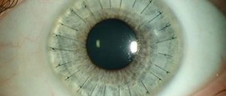
During a classic examination, the ophthalmologist does not detect the iris. With partial aniridia, fragments of the iris may remain. The doctor must measure intraocular pressure during tonometry. When it increases, the risk of developing glaucoma increases sharply. With aniridia, blood vessels do not penetrate the cornea, but pass along its surface, forming characteristic growths. Because of this, the outflow of tears is disrupted, intraocular pressure increases and glaucoma progresses.
The examination must be comprehensive so that specialists can assess not only the condition of the organ of vision, but also other body systems, including the kidneys and liver. The treatment tactics for the disease are selected taking into account the results of diagnosis and examination of the patient.
Symptoms of aniridia
Ophthalmic symptoms of aniridia may include:
- increased photosensitivity of the eyes;
- uncontrolled eye movements (nystagmus);
- decreased visual acuity;
- strabismus;
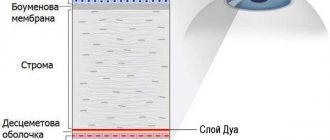
- decreased transparency of the cornea;
- cataract and glaucoma;
- causeless loss of vision (amblyopia);
- structural defects of the lens, retina, optic nerve.
Some patients develop specific facial expressions - wrinkles on the forehead, narrowing of the palpebral fissure, which appears against the background of a pathological reaction of the eye to light. Many patients with aniridia always look from under their brows.
As a rule, with the disease, the cornea of the eye does not have vessels, or their number is pathologically reduced. Most of the vessels do not penetrate the cornea, but are present on its surface in the form of growths (pannus).
Normally, the angle of the anterior chamber of the eye (the place located between the iris and cornea) is responsible for maintaining the required intraocular pressure and draining the fluid in the eye. With aniridia, the absence of the iris and the proliferation of altered vessels leads to disruption of the outflow of tear fluid, which leads to an increase in blood pressure and the development of glaucoma.
Pathology can also lead to irreversible changes in the lens of the eye. The lens is a biconvex lens located behind the iris, responsible for refraction of light rays entering the retina and sending converted signals to the brain with the participation of the optic nerves.
Aniridia causes displacement of the lens (subluxation), so over time the patient may experience opacity of light in this part of the eye and the appearance of cataracts.
With aniridia, the area of the retina that controls clarity of vision - the fovea - is also underdeveloped, so foveal hypoplasia joins the pathology, reducing the quality of vision. The optic nerve also undergoes abnormalities, which further aggravates the situation.
Taken together, all these deviations and developmental defects lead to severe disturbances in the functioning of the patient’s visual organs.
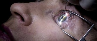
Treatment
The main method of treating aniridia is the implantation of an artificial iris of a suitable shade. The operation is used mainly for congenital forms of the disease. Acquired aniridia is accompanied by a high risk of corneal damage during surgical interventions. Therefore, surgery in this case may be contraindicated.
Due to increased photosensitivity, patients are recommended to use sunglasses when outdoors.
Conservative therapy includes the use of symptomatic remedies that maintain the health of the visual organ and prevent the occurrence of complications. Ophthalmologists prescribe drops based on beta blockers, prostaglandins and other active agents to control intraocular pressure and prevent glaucoma.
Read in a separate article: Iritis: causes, symptoms, diagnosis and therapy
If there are contraindications to surgical treatment, patients are recommended to use contact lenses that hide the absence of the iris. With the help of special moisturizing local products, it is possible to prevent the development of keratopathy, which is accompanied by destruction and clouding of the surface layers of the cornea.
The main task of specialists who treat people with aniridia is to socialize the patient and maintain a decent quality of life. Not so long ago, specialists did not have the opportunity to perform surgical correction of the disease and could only offer the use of contact lenses with light filters. But every year more and more treatment methods appear, including surgery.
Reconstructive surgeries for implantation of the irido-lens diaphragm have become available to patients. Such surgical interventions are still traumatic and can be dangerous for the patient. But in the future, experts plan to improve surgical treatment techniques. Implantation can be carried out through micro-incisions, thereby reducing the degree of trauma and reducing the risk of developing undesirable health consequences.
Useful video
Aniridia is a rather complex disease that develops against the background of an acquired mutation or spontaneous disorders in the patient’s genes. There are no measures to prevent eye diseases. The only method of preventing the development of pathology of the visual apparatus is gene monitoring of the fetus in parents with such a pathology.
Author's rating
Author of the article
Alexandrova O.M.
Articles written
2031
about the author
Was the article helpful?
Rate the material on a five-point scale!
If you have any questions or want to share your opinion or experience, write a comment below.
Diagnostic measures
The main measures that allow a thorough examination of a patient with suspected aniridia:
- Visual examination of the anterior parts of the eyes;
- Examination of the patient's fundus using special ophthalmic instruments;
- Procedure for local measurement of intraocular pressure;
- Visual examination of the anterior chamber of the ocular apparatus;
- Study of clinical refraction;
- Biomicroscopy.
Upon direct examination of the anterior part of the eye, the ophthalmologist notices a partial or complete absence of the iris.
Traumatic aniridia is characterized by the presence of “fragments” of the iris. They can be located in the organ in any order. Through ophthalmoscopy, the doctor can identify hypoplastic changes in the central segment of the retina and optic nerve. This is a typical sign that a person has total aniridia.
Intraocular pressure is measured using an ophthalmic tonometer. High rates indicate the presence of glaucoma or a high risk of this pathological condition. In this case, gonioscopy plays the role of an auxiliary technique - the doctor can examine the anterior chamber of the ocular apparatus in more detail.
Symptoms
Symptoms of aniridia
The full form of this eye disease is characterized by the absence of the iris, which will be clearly visible even without diagnosis.
The general clinical picture will be characterized as follows:
- decreased visual acuity;
- nystagmus;
- photophobia;
- corneal clouding;
- increased intraocular pressure;
- amblyopia;
- abnormalities of the lens, optic nerve and retina.
If we talk about the partial form of eye disease, then the clinical picture will be characterized in the same way, but less pronounced. If aniridia is associated with WAGR syndrome, then the symptoms will occur in a much more complex form and be characterized as follows:
- impaired mental function;
- disturbances in the functioning of the genitourinary system and gastrointestinal tract;
- development of oncological processes;
- pathological enlargement of the muscles of one half of the body.
If there is a complication with Gillespie syndrome, the clinical picture will be supplemented by hearing loss, ptosis, and pulmonary artery stenosis.
Symptoms and signs
The iris does not develop correctly in one or both eyes before the baby is born, but vision is usually preserved in some mild cases of aniridia . There are believed to be at least four types of aniridia . First type . Some people with this type of aniridia may not be aware of the eye problem because the pupils appear normal and usually only one eye is affected by iris thinning.
The second type of aniridia. Problems with the iris can occur independently, or in parallel with other diseases and abnormalities. Associated disorders may include cataracts (clouding of the lens of the eye), glaucoma (gradual loss of vision due to increased pressure inside the eyeball, which can be accompanied by varying degrees of pain), or superficial clouding of the cornea. Rapid involuntary movement of the eyeball is also possible.
The third type of aniridia is associated with mental retardation and is called Gillespie syndrome. The fourth type develops along with Wilms tumor (kidney cancer), genitourinary abnormalities and possible mental retardation.