Epidemic hemorrhagic conjunctivitis belongs to the category of acute infectious pathologies caused by enterovirus type 70. The virus enters the eye shell through contact, through dirty hands, hygiene products, and eye drops. People of any age and gender are susceptible to the disease. What are the symptoms and treatment of enteroviral conjunctivitis?
Symptoms of the disease
The disease has a rapid course. The incubation period after entering an enterovirus infection is 1–2 days. First, one eye is affected, then symptoms appear in the other. The disease manifests itself acutely.
The main symptoms of hemorrhagic conjunctivitis:
- it hurts the eyes, painful sensations arise;
- increased sensitivity to bright light appears;
- the corners of the eyes burn and tingle;
- tear fluid is intensively produced;
- eyelids swell;
- there is a feeling that there is a foreign body in the eye;
- discharge of a mucous and purulent-mucous nature appears;
- hemorrhage under the conjunctiva;
- the sensitivity of the cornea decreases;
- follicles appear on the membrane of the eye.
In addition to eye symptoms, general signs occur:
- tracheitis, bronchitis;
- inflammation of the lymph nodes;
- increased body temperature;
- headache.
The period during which the patient feels unwell ranges from 10 days to a month.
In the absence of adequate treatment, epidemic hemorrhagic conjunctivitis becomes chronic. The patient feels discomfort in the eye area, irritation. The outer side of the eyelid swells and turns red.
The severity of symptoms varies depending on the type of disease. Thus, acute bacterial conjunctivitis is characterized by severe photosensitivity and lacrimation. With adenoviral conjunctivitis, the respiratory system is affected. The epidemic type is manifested by severe hemorrhages in the eyelid and sclera.
Symptoms
According to the principles of clinical medicine, the symptoms of hemorrhagic conjunctivitis directly depend on the etiology and concomitant causes of the disease. Common symptoms of all forms are swelling and redness of the conjunctiva of the eyelids, accompanied by the presence of copious mucous discharge or a viscous purulent yellow discharge. The causative agent of adenoviral or chlamydial pathology causes noticeable lesions. An acute form of follicular conjunctivitis is diagnosed. The appearance of the conjunctiva is quite characteristic. Even with a visual examination, small translucent pink formations are visible. There are two stages of manifestation of a viral infection:
- The acute phase is characterized by a rapid onset and rapid development. Pain and pain appear in one eye, after which the process moves to the other. General malaise, headache and temperature fluctuations are observed for one to three weeks. Externally, there is active redness, accompanied by pinpoint hemorrhages. Discomfort manifests itself in the discharge of mucous, mucopurulent or purulent discharge.
- The chronic phase begins imperceptibly, gradually, without sudden activation. The course is persistent and long-lasting. Discomfort consists of unpleasant sensations, a feeling of irritation from the presence of a foreign body in the eye. The outer side of the eyelid becomes slightly red and swollen.
Symptoms depend on different types of disease. Acute bacterial conjunctivitis is characterized by severe photophobia, accompanied by active lacrimation. The conjunctivae are red, swollen, with numerous pinpoint hemorrhages. Symptoms of adenoviral conjunctivitis are concomitant signs of respiratory disease. It is characterized by an acute onset, lacrimation, redness, swelling of the conjunctiva, and rare pinpoint hemorrhages. The discharge is scanty, mainly of the mucous type. The lower transitional fold of the conjunctiva is sprinkled with follicular formations. The epidemic type of hemorrhagic conjunctivitis is hemorrhages on the eyelid and eyeball, which resolve depending on physiological changes and size. People who have close contact with the environment, with soil, some herbs, vegetables, fruits, and sick animals, are at risk of developing fungal conjunctivitis under certain conditions. The causative agent is various types of fungi - varieties of actinomycetes, molds, yeasts and others.
Experts call allergic lesions of the eye mucosa a separate subtype of inflammation of the conjunctiva. There are as many allergens as there are manifestations of a complex of symptoms. The most common is drug-induced hemorrhagic conjunctivitis. It is a consequence of independent untested use of the drug. Signs with a clear clinical picture are visually determined after six hours. This is a rapid increase in conjunctival edema, itching, burning, and copious mucous discharge. Along with this type of inflammation, conjunctivitis with hay fever is actively diagnosed. They are characterized by precise seasonality, accompanied by rhinitis, exacerbation during the flowering period of certain plants. Burning sensation, sharp pain, itching, active lacrimation and severe photophobia, redness, swelling of the conjunctiva, copious mucous discharge, all signs are characterized by synchronous damage to the organs of vision.
Symptoms of hemorrhagic conjunctivitis
Hemorrhagic conjunctivitis tends to develop rapidly; it has virtually no incubation period; symptoms appear 12-48 hours after infection. At first, as a rule, only one eye is affected, then the infection spreads to the other. Epidemic hemorrhagic conjunctivitis manifests itself very acutely and is usually accompanied by:
- pain and pain in the eyes;
- photophobia;
- a burning sensation and tingling sensation in the corners of the eyes;
- lacrimation;
- swelling of the eyelids;
- sensation of a foreign object in the eye;
- the presence of mucous or mucopurulent discharge;
- subconjunctival hemorrhage;
- decreased sensitivity of the cornea;
- small follicular rashes.
All of the above symptoms develop against a background of general malaise, headache, fever, tracheitis and bronchitis. Sometimes there is an enlargement of the lymph nodes. Regardless of the cause that caused hemorrhagic conjunctivitis, symptoms can appear from 10 days to a month.
Epidemic hemorrhagic conjunctivitis
Epidemic hemorrhagic conjunctivitis is a highly contagious acute ophthalmic infection caused by picornaviruses, affecting the conjunctiva of the eyes and accompanied by massive subconjunctival hemorrhages. Epidemic hemorrhagic conjunctivitis occurs with severe hyperemia, swelling and chemosis of the mucous membrane, pain in the eyes, photophobia, serous-purulent discharge, subconjunctival hemorrhages that occur against the background of general symptoms (adenopathy of the pre-auricular lymph nodes, headache, fever, tracheobronchitis, etc.). For the purpose of differential diagnosis, eye biomicroscopy, fluorescein instillation test, serological, virological and cytological studies are performed. Treatment of epidemic hemorrhagic conjunctivitis is carried out using instillations of antiviral, antiallergic and antibacterial drugs. Symptoms of epidemic hemorrhagic conjunctivitis
Epidemic hemorrhagic conjunctivitis usually begins acutely, first affecting one eye, and after 8-24 hours the second. Due to severe pain and photophobia, the patient seeks help on the first day. The conjunctiva is sharply hyperemic, chemosis and follicular conjunctivitis are noted.
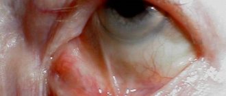
Small and large subconjunctival hemorrhages appear on the conjunctiva of the eyelids and eyeball. Discharge from the conjunctiva is mucous or mucopurulent. Extensive hemorrhages can involve almost the entire conjunctiva of the sclera. Changes in the cornea are minor - pinpoint epithelial infiltrates that disappear without a trace.
Enlarged preauricular lymph nodes are palpated.
The clinical manifestations of conjunctivitis are very peculiar. This is primarily an acute onset. Incubation takes 1-2 days (sometimes 8-12 hours).
The first symptom of conjunctivitis is a feeling of pain in the eyes, the inability to look at the light. In this condition, the patient consults a doctor. On examination, swelling of the eyelids, chemosis of the conjunctiva, its infiltration, and individual follicles on the lower transitional fold are noted. The discharge is usually not very copious, mucous or mucopurulent in nature.
Hemorrhages into the tissue of the conjunctiva and under the conjunctiva are typical, appearing in the first hours of the disease and disappearing after a few days, and in some cases after 2 weeks. They have different sizes and different shapes. Sometimes this is a continuous hemorrhage located over the entire area of the conjunctiva of the sclera, sometimes it is a hemorrhage in the form of a smear. In some cases, microbleeds in the form of petechiae are observed. It is impossible to see them with the naked eye. When examining such patients, it is necessary to use the biomicroscopy method.
Barely noticeable hemorrhages should be looked for in the upper half of the conjunctiva of the sclera, where they are often concentrated.
The second clinical sign, pathognomonic for this conjunctivitis, is the appearance in the conjunctiva of small, point-shaped spots of white or white-yellow color. They resemble meibomian gland infarctions, which are well known to ophthalmologists.
This symptom does not occur in other clinical forms of viral conjunctivitis. It owes its origin to the cytopathic action of the virus that causes hemorrhagic conjunctivitis. Invading the excretory ducts of the mucous membranes and accessory lacrimal glands of the conjunctiva, the virus causes their blockage with necrotic cells lining the duct. The clinical picture of conjunctivitis is generally complemented by adenopathy of the preauricular lymph glands, expressed in their soreness and obvious enlargement. In some cases, keratitis develops. Its originality lies in the superficial epithelial localization of the process.
Small infiltrates usually appear on the cornea, stained with a 2% fluorescein solution. After a few days, the symptoms of keratitis disappear almost without a trace. As for the symptoms of conjunctivitis, they last on average 10 days, sometimes up to 2 weeks. Trace reactions may remain for some time, which leads to complaints of discomfort during work, the sensation of a foreign body in the eye. The clinical picture of conjunctivitis can be combined with general phenomena such as weakness, malaise, and increased body temperature.
In such cases, the diagnosis of influenza or catarrh of the upper respiratory tract is erroneously made, against the background of which the therapist may not take into account or falsely interpret eye symptoms. Differential diagnosis of epidemic hemorrhagic conjunctivitis should also be carried out with such seemingly unrelated conditions as occupational conjunctivitis, electric ophthalmia, snow ophthalmia.
They are related to hemorrhagic conjunctivitis by the commonality of subjective sensations in the form of acute pain, photophobia, lacrimation, with which a person who has been exposed to iodine vapor at work or ultraviolet irradiation may come to the appointment. A thorough examination carried out after instillation of a 0.5% dicaine solution into the conjunctival cavity allows us to diagnose hemorrhagic conjunctivitis based on the pathognomonic symptoms described above.
Treatment
Antiviral agents are used. To suppress a secondary bacterial infection, broad-spectrum antibiotics are prescribed in the form of instillations 3-6 times a day, sulfonamides (10% sulfapyridazine sodium solution, 30% sulfacyl sodium solution, etc.) 3-4 times a day.
To resolve subepithelial infiltrates, a 0.1% solution of dexazone, a 6% solution of polyglucin, a 3% solution of potassium iodide, a 0.1% solution of lidase and ronidase are instilled. Treatment should be carried out for a long time.
Diagnostics
The disease can be diagnosed based on clinical manifestations and patient complaints. To confirm the diagnosis, laboratory tests of mucus are performed to detect viruses and serological tests. The biomicroscopy technique is used.
When examining the patient, the doctor detects swelling, hemorrhage under the conjunctiva of varying severity, and small pinpoint follicles. Infiltrative areas on the cornea are determined by a fluorescein staining test. Sometimes cytological analysis of scrapings from the conjunctiva may be necessary.
Epidemic hemorrhagic conjunctivitis in children and adults requires differential diagnosis with influenza, ARVI, electroophthalmia, foreign body in the eye, allergic conjunctivitis, snow blindness.
Symptoms
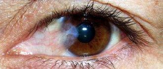
Distinctive signs of hemorrhagic conjunctivitis are infiltration and hyperemia of the mucous membrane of the eye, numerous hemorrhages (hemorrhages) under the conjunctiva, follicular rashes on the folds of the eyelids.
A few days are enough for the infection to spread to the other eye.
The incubation period lasts from 12 hours to 2 days. Initially, the lesion is unilateral, but after 1-3 days the pathological process affects the second organ of vision. Symptoms appear within 10-14 days. The feeling of the presence of a foreign body and discomfort in the eyes during work that requires visual strain usually bothers you for quite a long time. The pathology has an abrupt onset and is accompanied by symptoms such as:
- burning in the eyes;
- sensitivity to light;
- copious mucous or purulent discharge from the eyes;
- swelling and redness of the mucous membrane of the eyelids;
- sensation of a foreign object in the eye;
- lacrimation;
- weakness;
- bronchitis;
- elevated temperature;
- tracheitis;
- lymphadenitis;
- headache.
Treatment and prevention
The first manifestations of this disease require the patient to contact a specialist. Accurate, comprehensive diagnosis allows you to prescribe effective treatment, avoid further complications and not get sick again in the future. The most effective technique is biological microscopy. It shows extensive hyperemia with subconjunctival hemorrhages of various sizes and shapes, with small pinpoint follicles. The diagnosis is clarified using virological, serological and cytological studies of scrapings from the affected conjunctiva. Therapy consists of the following methods and techniques:
- Bacterial conjunctivitis is cured by instilling a 0.25% solution of chloramphenicol or sodium sulfacyl. The area of the conjunctival sac is washed with a solution of furatsilin, potassium permanganate, and 1% oletethrin ointment is placed under it.
- For therapy of viral etiology, the doctor prescribes the use of human leukocyte interferon, pyrogenal, and poludan. A high therapeutic effect can be obtained from 0.5% florenal, 0.05% bonaftone and analogues from a series of eye ointments.
- Chlamydia conjunctivitis responds quickly to antiviral drugs and tetracycline antibiotics.
- Fungal conjunctivitis is eliminated by prescribing nystatin, levorin, amphotericin B, local treatment.
- Therapy for allergic conjunctivitis involves the local use of modern drugs based on synthetic hormones. The use of hydrocortisone, prednisolone, dexamethasone and antiallergic drugs. Parallel administration of Claritin, Telfast, Suprastin, Tavegil, and their analogues gives a lasting therapeutic effect.
The prognosis with stable treatment and compliance with all doctor’s instructions is favorable. In the future, manifestations of the process will not bother you, cause pain or discomfort. Deviation from the prescribed course of therapeutic intervention entails a risk of decreased visual acuity. Preventive measures consist of observing the rules of public and personal hygiene, sanitary and hygienic standards, and requirements for personnel in medical and children's institutions. By simply washing and specially treating your hands with a 1% chloramine solution, especially before starting ophthalmic procedures, and using individual or disposable instruments, including pipettes, you block the path of the virus, which is characterized by painful symptoms.
We say a clear and clear “no” to diseases of dirty hands, among which hemorrhagic conjunctivitis is far from the last place. These are precisely the pathologies that are avoided by simple hand washing, high-quality laundry, and the use of personal hygiene products, bedding, towels and disposable medical instruments.
Hemorrhagic conjunctivitis
Preventive recommendations
Following recommendations regarding cleanliness is disease prevention.
Preventive measures are based on compliance with the requirements of personal and public hygiene, sanitary standards in medical, school, children's and other institutions. In groups, when they are violated, up to 90% of people are affected. Basic preventive requirements:
- Hand disinfection with 1% Chloramine before ophthalmic procedures.
- The use of individual glass rods and pipettes, which, when reused, first require sterilization by boiling.
- Processing of ophthalmological equipment and instruments.
- The use of individual eye drops, hygiene products, bedding, and towels.
- High quality laundry.
Adults can become infected from children who have the habit of rubbing and touching their eyes with unwashed hands. Therefore, it is important to wean kids from this. Also, visiting the toilet should always end with thorough hand washing. Strengthened immunity can prevent the negative effects of the environment, so consuming vitamin-containing foods is another way to prevent hemorrhagic conjunctivitis.
Treatment with drugs
Treatment of hemorrhagic conjunctivitis begins with isolating the patient, as the disease can spread quickly. Medicines prescribed:
- antiviral. Use Poludan, Interferon up to eight times a day;
- hormonal agents. The drug Dexamethasone promotes the resorption of hemorrhages;
- solutions of hyaluronidase, dextran - relieve swelling and improve blood flow;
- Emoxipan - promotes the resorption of hemorrhagic rashes.
Additionally, the patient is prescribed rinsing the conjunctiva with solutions of penicillin, sodium sulfacyl, boric acid, and gentamicin. Erythromycin and tetracycline ointment is placed behind the eyelids up to four times a day.
To achieve a positive result, it is necessary to continue therapy for at least two weeks, even if the symptoms subsided much earlier. Early cessation of treatment for hemorrhagic conjunctivitis can lead to relapse, since immunity to the enterovirus does not have time to develop.
If you follow all the recommendations of the attending physician, the prognosis is favorable; after three weeks, the patient’s vision is completely restored.
Epidemic conjunctivitis (Koch–Wicks) – treatment.
To remove purulent discharge - frequent rinsing of the conjunctival sac with a 2% solution of boric acid or a solution of mercury oxycyanide 1: 3000–1: 5000. Injecting drops of a 30% solution of sulfacyl sodium every 2–3 hours, placing 30–50% ethazol ointment behind the eyelids or 30% sodium sulfacyl ointment 4-5 times a day. Taking sulfonamides (sulfapyridazine, sulfadimethoxine) orally. When setting doses, the age, general condition of the patient, and the severity of the clinical picture of the disease are taken into account. A solution of benzylpenicillin sodium salt or erythromycin (5000-10,000 units per 1 ml) 1-2 drops into the conjunctival sac every 3 hours. It is better to prepare a solution of benzylpenicillin sodium salt with an 8% solution of magnesium sulfate. This allows the antibiotic solution to remain active for 7–10 days, which is especially important in hot climates. Antibiotic ointments (1% tetracycline, 1% erythromycin, 0.5% neomycin, etc.) – 3–5 times a day. Treatment should be continued for 2–3 days even after the disappearance of clinical signs of the disease in order to prevent the possibility of exacerbations and relapses of epidemic conjunctivitis.
Treatment methods
First of all, treatment involves initiating the outbreak and isolating it from other people. It is also necessary to isolate already infected patients and, if possible, people in contact with them. The therapy itself includes the use of antiviral drugs containing interferon. Additionally, a course of antihistamines and anti-inflammatory drugs is administered. For this purpose, corticosteroids can be used in low concentrations. To prevent complications from secondary bacterial infections, antimicrobial and antiseptic ointments are prescribed. The course of treatment is at least 14 days.
Hemorrhagic conjunctivitis should be treated as quickly as possible. In order to prevent the spread of the virus, contact persons may be prescribed medications containing ready-made interferon. Less commonly prescribed are drugs that stimulate the production of your own interferon, since their effect is longer. Detection of this pathology and its timely treatment help preserve vision and reduce the likelihood of complications.
We recommend reading an article about another type of conjunctivitis so as not to confuse the symptoms and treatment methods. Read about bacterial conjunctivitis.
To learn more about eye diseases and their treatment, use the convenient site search or ask a specialist a question.
Diagnostic methods
Only a doctor can accurately identify the disease.
If you do not pay attention to eye signs, instead of hemorrhagic conjunctivitis, influenza and ARVI are incorrectly diagnosed. To make a correct diagnosis, you need to visit an ophthalmologist and an infectious disease specialist. The disease is diagnosed based on the presence of pathognomonic symptoms, information about registered cases of the disease and their location. Diagnostics is carried out by such laboratory methods as:
- Biological microscopy.
- Fluorescein staining test.
- Scraping from the conjunctiva and its examination: virological;
- cytological;
- serological.
Unconventional methods
Traditional methods can alleviate the course of epidemic hemorrhagic conjunctivitis, however, they cannot affect the pathogen. Their use without appropriate medications will be ineffective.
Apply:
- chamomile infusion. Cotton pads are moistened and applied to the eyes for a quarter of an hour at least three times a day. Chamomile does not cause an allergic reaction, so it can be used for a long time;
- dill juice The grass is finely chopped and the juice is squeezed out. They rub their eyes;
- grated potatoes. Use as compresses for 15 minutes twice a day. The potatoes are kept in the refrigerator before use;
- eye drops from chamomile, black nightshade and marshmallow root. A tablespoon of the mixture is poured into a glass of water, boiled for 10 minutes, cooled and filtered. Drop two drops three times a day;
- tincture of rose petals. 1 tbsp. l. dry raw materials are poured with a glass of boiling water and left for half an hour. Use it to rub the eyes, and before going to bed, make compresses that are left for 20 minutes;
- infusion of cornflower flowers. They are taken in the amount of a tablespoon and poured with 250 ml of boiling water. Place three drops in the eyes morning and evening.
Epidemic keratoconjunctivitis
Etiology
The causative agent of the disease is adenoviruses of serotype 8. The disease is transmitted by contact.
Clinical signs and symptoms
The incubation period is 4-8 days. The duration of the disease is from 2 weeks to 2 months. Immunity remains after the disease.
The disease begins acutely, symptoms of conjunctivitis appear first in one eye and then in the other. Symptoms of conjunctivitis are preceded by general malaise. Regional lymph nodes are enlarged on both sides, palpation is painful.
Upon examination, attention is drawn to hyperemia and swelling of the conjunctiva, eyelids and transitional folds; a small amount of mucous or mucopurulent discharge from the conjunctival cavity is observed (Fig. 31).
Small transparent follicles appear on the conjunctiva, mainly in the lower transitional folds. The sensitivity of the cornea is reduced.
1 week after a slight decrease in subjective sensations, symptoms of keratitis appear—multiple pinpoint subepithelial infiltrates of the cornea.
Clinical recommendations
similar to those for adenoviral conjunctivitis.
Epidemic hemorrhagic conjunctivitis
Etiology
The causative agent of the disease is enterovirus-70 (from the group of picornaviruses). The disease is transmitted by contact.
Clinical signs and symptoms
The incubation period is 12-48 hours. Recovery occurs 8-12 days after the onset of the disease.
The disease begins acutely, the symptoms of conjunctivitis (photophobia, sharp pain, sensation of a foreign body) appear first in one eye, and after 1-2 days in the other. Symptoms of conjunctivitis are preceded by a general malaise. There is an increase in the pre-auricular lymph nodes.
On examination, hyperemia, follicles and pronounced swelling of the conjunctiva of the lower transitional fold, a small amount of mucous or mucopurulent discharge from the conjunctival cavity are revealed. On the 2nd day, subconjunctival hemorrhages of varying severity appear. The sensitivity of the cornea is reduced.
Sometimes punctate subepithelial infiltrates of the cornea appear.
Clinical recommendations
Solutions of interferon and its inducers are instilled into the conjunctival sac for 2-3 days:
interferon solution 4000 units/ml every 2 hours (ex temporae);
Poludana solution 50 units/ml every 2 hours (ex temporae);
para-aminobenzoic acid 0.07% solution 3-4 times a day (Actipol).
To prevent secondary infection, 2-3 times a day for 10-14 days, use:
picloxidine 0.05% solution (Vitabact);
Miramistin 0.01% solution;
chloramphenicol 0.25% solution.
Conjunctivitis caused by molluscum contagiosum
Etiology
The causative agent of the disease belongs to the dermatotropic poxvirs. Affects various parts of the skin, including the face and eyelids. Transmitted through household contact.
Clinical signs and symptoms
Against the background of characteristic changes in the skin of the eyelids (see Viral diseases of the eyelids), hyperemia, edema and folliculosis of the conjunctiva of the eyelids and transitional folds appear.
Clinical recommendations
After eliminating the damage to the skin of the eyelids, the symptoms of conjunctivitis disappear on their own.
What is Enteroviral hemorrhagic conjunctivitis
What causes Enteroviral hemorrhagic conjunctivitis?
The causative agent of acute hemorrhagic conjunctivitis is Enterovirus 70 , which belongs to the picornavirus family. Some outbreaks of hemorrhagic enteroviral conjunctivitis have been caused by Coxsackievirus A24 , also a member of the picornavirus family
Epidemics of hemorrhagic enteroviral conjunctivitis have been observed in congested urban areas of developing countries, affecting all age groups and social classes. Occasionally, outbreaks of hemorrhagic enteroviral conjunctivitis in Europe have been localized around eye clinics.
Pathogenesis (what happens?) during Enteroviral hemorrhagic conjunctivitis
The source of infection is only humans. The epidemic hemorrhagic conjunctivitis virus is transmitted mainly by contact through infected solutions of eye medications, devices and instruments, as well as common objects.
In temperate countries, seasonality is characteristic with an increase in incidence in late summer and early autumn. Mostly children and young people get sick. Diseases are observed in the form of sporadic cases, local outbreaks (usually in children's groups) and in the form of large epidemics affecting a number of countries.
After accumulation of the virus at the site of primary reproduction, the pathogen enters the blood (viremia) and spreads throughout the body. Enteroviruses have a tropism for nervous tissue, muscles and epithelial cells, which is manifested in the clinical picture of the disease, as well as in morphological changes in tissues. The lymphogenous spread of viruses is of some importance.
Symptoms of Enteroviral hemorrhagic conjunctivitis
The incubation period is 1-2 days. The disease begins acutely. Characterized by the sudden onset of pain in the eyes, lacrimation and photophobia, and foreign body sensation soon develop. The eyelids are swollen, the conjunctiva is hyperemic, multiple subconjunctival hemorrhages and copious serous or serous-purulent discharge are noted. At first the lesion is one-sided, after 1-3 days the process moves to the other eye.
The patient's general condition remains satisfactory. Normalization occurs after 10-14 days.
The only known complication was the development in rare cases of lumbar radiculomyelitis, the end result of which was flaccid paralysis, as in polio.
Diagnosis of Enteroviral hemorrhagic conjunctivitis
For a number of clinical forms, the diagnosis can be established on the basis of the characteristic clinical symptoms of enteroviral conjunctivitis, especially during epidemic outbreaks. Diagnosis of sporadic enteroviral conjunctivitis is often difficult. To confirm the diagnosis in a laboratory, virus isolation from mucus and serological tests are used.
For serological studies, paired sera are taken (the first before the 4-5th day of illness, the second after the 14th day of illness). An increase in antibody titer of 4 times or more is considered diagnostic. They use a neutralization reaction with reference strains of enteroviruses (on tissue cultures or suckling mice), RSC, RTGA, and gel precipitation reaction.
Prevention
To prevent epidemic hemorrhagic conjunctivitis, it is necessary to follow the rules of hygiene and not use other people’s eye drops. Parents should ensure that children do not touch their eyes with dirty hands, as this is often the cause of infection.
Doctors advise washing your hands after visiting the toilet and enriching your diet with vitamins. A strong immune system can protect a person from many diseases.
Hemorrhagic enteroviral conjunctivitis is a serious pathology that, in the absence of timely treatment, can become chronic. In this case, the disease will cause a lot of inconvenience to the person and will negatively affect vision.
Bacterial acute epidemic conjunctivitis
Causes of bacterial conjunctivitis spreading include:
- poor personal hygiene;
- using contaminated makeup;
- touching the eyes with dirty hands.
A bacterial eye infection can last up to 10 days. If antibiotics do not help, then conjunctivitis is likely viral, not bacterial.
Signs of contagious conjunctivitis
- redness of the eyes;
- swelling around the eyes;
- discharge from the eyes;
- sticky eyelashes and eyelids;
- burning;
- eye irritation.
Treatment for conjunctivitis depends on the type and severity of the infection.
Mild cases of conjunctivitis may clear up without treatment within a few days. At home you can use:
- cold or hot compresses to reduce swelling;
- eye drops to relieve symptoms;
- clean cloth for wiping eyes;
- eye drops with an antihistamine for allergies
- conjunctivitis.
People should also:
- disinfect glasses;
- do not use contact lenses;
- throw away old makeup;
- do not wear makeup until the infection clears up.
In severe cases, you should consult a doctor.
Causes
The causative agent of epidemic hemorrhagic conjunctivitis is picornavirus (enterovirus type 70, ESNO, Coxsackie A-24, etc.). The disease is characterized by high contagiousness, and therefore occurs in the form of locally limited outbreaks in enterprises, healthcare facilities, children's institutions, as well as epidemics of the “explosive” type, covering a large number of people.
Hemorrhoids kill the patient in 79% of cases
The virus is transmitted by contact through hands, objects (towels, pillows, etc.) contaminated with eye discharge, infected eye drops, instruments (pipettes, eye sticks), ophthalmological devices, etc. If anti-epidemic measures in groups are not followed, epidemic hemorrhagic conjunctivitis 80-90% of people may be affected.
Symptoms and treatment of hemorrhagic conjunctivitis
Hemorrhagic conjunctivitis is a disease that is still poorly understood. The causative agent of this disease is enterovirus type 70. The disease is transmitted primarily through contact through eye drops, dirty hands, personal hygiene products, and contact with a sick person. All segments of the population, all age groups are exposed to infection.
The disease is characterized by some seasonality, most often symptoms appear in late summer and early autumn. Hemorrhagic conjunctivitis, the treatment of which is mandatory, has a high level of contact, especially for large groups. With active decomposition of the virus in a certain group of people, the disease can reach an epidemiological nature.
Treatment
A patient with a hemorrhagic inflammatory process is isolated and given personal hygiene products.
Treatment involves antiviral, antibacterial and antiallergic therapy.
Photo 4. Packaging and bottle of Oftalmoferon in the form of eye drops with a volume of 10 ml. .
Antiviral eye drops Ophthalmoferon and Interferon solution are used. Antibacterial drugs are prescribed to prevent the addition of a bacterial infection. Levomycetin, Tobrex, Floxal, Albucid drops are used . Antibiotics are used in the form of drops and ointments.
Antiallergic therapy in the first week of illness is carried out with antihistamines: Cromohexal, Alomide, Ketotifen . From the second week, corticosteroids are dripped - Hydrocortisone, Dexamethasone.
Additionally , potassium iodide and Dextran solution . The agents promote the resorption of infiltrates.
Attention! Therapy for the inflammatory process continues for two weeks , even if the symptoms have been eliminated before the end of this period.
Before using the drops, clean the eyes. To do this, use a cotton pad or gauze swab dipped in a decoction of herbs that have anti-inflammatory and antiseptic properties. A separate swab is used for each eye.
The drugs for treatment are prescribed by an ophthalmologist , who also determines the frequency of use of each drug. Both eyes begin to be treated at once.
Prevention
Basic epidemic prevention measures:
- timely identification of patients with hemorrhagic conjunctivitis;
- isolation of sick people;
- sanitization of ophthalmic instruments and equipment.
for every person to follow the rules of personal hygiene:
- You can only touch infected organs with cleanly washed hands:
- You cannot use other people’s cosmetics and allow others to use yours;
- All family members must have their own towels.
Enteroviral hemorrhagic conjunctivitis
- What is Enteroviral hemorrhagic conjunctivitis
- What causes Enteroviral hemorrhagic conjunctivitis?
- Pathogenesis (what happens?) during Enteroviral hemorrhagic conjunctivitis
- Symptoms of Enteroviral hemorrhagic conjunctivitis
- Diagnosis of Enteroviral hemorrhagic conjunctivitis
- Treatment of Enteroviral hemorrhagic conjunctivitis
- Prevention of Enteroviral hemorrhagic conjunctivitis
- Which doctors should you contact if you have Enteroviral hemorrhagic conjunctivitis?