Clinic and symptoms
Signs of acute heel bursitis are difficult to miss: the following symptoms occur:
- Bursitis of the heel joint is, first of all, pain, which is more pronounced during exercise, and less pronounced at rest, especially in aseptic forms, and with a purulent process it is impossible not only to walk for a long time, but in general, you cannot even step on the heel;
- Subcalcaneal bursitis may be accompanied by severe redness of the skin;
- A local feeling of heat appears, usually in the area of the heel bone, and if the Achilles tendon is affected, even higher;
- With bursitis, swelling and swelling of the soft tissue occurs. It is impossible to remove the heel from the shoe, much less put it back in, especially at the end of the day;
- A dysfunction occurs: bursitis on the heel leads to limited mobility in the ankle joint.
Don't miss: Treatment of deforming arthrosis of the foot
As suppuration develops, bursitis on the heel begins to become more severe: pain occurs at rest and at night, body temperature rises, and malaise occurs. How to diagnose heel bursitis?
About diagnostic principles
Already at the initial appointment, the doctor can make a preliminary diagnosis based on the classic symptoms of inflammation.
To clarify the diagnosis, the following steps are taken:
- conduct an x-ray of the foot in two projections to exclude fractures, destructive lesions of bone tissue, osteomyelitis, and tumors;
- perform a puncture of the bursa and examine the punctate for cellular composition (cytology), conduct a general and biochemical analysis (to exclude systemic, rheumatic, or autoimmune damage), and conduct a bacteriological study of the exudate with culture, isolation of a pure culture and sensitivity to antibiotics.
This will give doctors a “key” to treatment, and heel bursitis, the treatment of which will be “targeted”, will quickly be forgotten. What modern treatment methods are used, and how to treat heel bursitis at home?
“If they tell you that it is impossible to cure your back and joints after 50 years, know that this is not true!” — Mirakhmedova Aida Khamidovna, rheumatologist of the highest category, candidate of medical sciences. More details>>
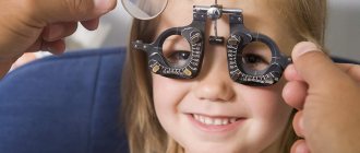
Correction in children
In the process of correcting pathology in children, it is necessary to monitor refractive error. As the child grows, it decreases and the need to replace lenses arises. In pediatrics, the following treatment methods are used:
- Contact lenses are most often used, especially for congenital aphakia. Hard and soft gas permeable lenses are used.
- Glasses are more effective for unilateral forms of the disease. Also prescribed in case of intolerance to contact lenses. The disadvantages include the difficulty of choosing comfortable glasses - for small children they are too heavy and slide off the nose.
- Intraocular lenses are often used to correct postoperative aphakia. The operation is performed after the growth of the eyeball has stopped.
The choice of method depends on the age of the child, the form of the disease, and the degree of decreased visual acuity. Over time, a specialist can replace it with a more effective one.
Treatment
There are several methods to restore eye health to patients suffering from aphakia; the choice of one approach or another is determined by the results obtained when making a diagnosis and depends on the degree of advanced disease.
Since after the loss of the lens, the refraction of the eyeball (the ability to refract light) changes significantly, therefore the main goal of the fight against aphakia is to correct the refractive ability of the eye that has lost its natural lens. Below we will look at the existing possibilities.
Treatment with medications
Unfortunately, humanity and world science have not invented drugs that allow us to grow a lost organ. In nature, there are no tablets or injections capable of growing a lens inside the eyeball of the desired shape and properties.
| Therefore, drug therapy for aphakia is devoid of all common sense and is not carried out. Medicines are used as an adjunct to other methods. |
Glasses
After surgery to remove the lens, the eye's ability to refract light directed into it changes significantly. The patient's farsightedness increases significantly. The refractive power of the amputated organ is compensated by auxiliary optical means - glasses.
To correct aphakia of the eye, a glass of at least +10.0 ÷ +12.0 D is required, which is significantly less than the refractive power of the lost lens, which, as a rule, is +19.0 D. This difference is due primarily to the fact that the lens of the glasses located differently, in a different place in the rather complex optical system of the eye.
Secondly, the lens installed in glasses is surrounded by air with completely different optical properties in terms of refraction, in contrast to the lens, which is physiologically immersed in the vitreous fluid, which has a similar refractive index of light.
For a person suffering from farsightedness, the optical power of the glass must be increased by the required diopter values; for a person suffering from myopia, it must be reduced, ultimately using glass of a smaller thickness.
For example, if before the removal surgery myopia was close to -19.0 D, then after surgery, due to the bringing of the optical system into a state of equilibrium, the patient may be able to do without glasses necessary for seeing objects located at a considerable distance.
An aphakic eye loses its ability to adapt, so for near work, 3.0 D glasses are prescribed more powerful than lenses for distance. Correction with glasses is not done if there is no lens in one of the organs of vision.
An eyepiece of +10.0 D or more is a fairly powerful magnifying glass. In the case when it is present only in front of one eye, the resulting images in both eyes are too strikingly different from each other in size and geometric dimensions, and they will never merge into a solid picture.
In other words, the patient will lack binocular vision, which is fraught with serious dangers for his life: the inability to estimate distances to objects, the inability to see the whole picture of the world around him here and now.
Contact lenses
Previously, a fairly advanced method of vision correction is used precisely in cases where the patient is diagnosed with monocular aphakia. As you know, in such a situation, the use of glasses is contraindicated, for the reasons discussed earlier. And the solution is to use individually made lenses.
Surgery
| A modern method of getting rid of aphakia is vision correction, carried out by implanting an intraocular lens (IOL) into the diseased eye. The procedure is prescribed for patients with monocular aphakia. |
The essence of the intervention is the replacement of a lost or amputated lens with an artificial lens of the required optical power, implanted into the eyeball through surgery. This method is recognized as the best in getting rid of the disease.
Folk remedies
As in the case of drug treatment, alternative medicine against aphakia of the eye is powerless. No decoctions, healing herbs, conspiracies or rituals can return the removed lens to the eye.
If you come across offers of help to overcome an illness in one or another “grandmother’s” way, by making an appointment with a “healer,” you should know that these are charlatans who are only interested in your money.
What is lens atrophy?
Atrophy of any organ is a decrease in its volume due to lack of nutrition. In other words, atrophy is an extreme form of the dystrophic process. Due to the lack of normal tissue nutrition, the number of working cells in the organ decreases,
In this article
- What is lens atrophy?
- Why does atrophy of the eye and lens occur?
- How is lens atrophy treated?
- How is the lens replaced?
- How does recovery occur after eye lens replacement?
- Complications after eye lens replacement
- What lens is used to replace the lens during atrophy?
ensuring its functioning. When tissues and cells begin to receive less oxygen and nutrients, their metabolism slows down. Over time they die. A dead cell does not survive in the body. It gradually breaks down into tiny molecules that are excreted from the body.
In place of a dead cell, a new one should appear, but due to atrophy, that is, lack of nutrition, this does not happen. The remaining cells begin to move closer to each other in order to fill the resulting voids. Thus, the gradual death of cells without their replacement by new tissues leads to a decrease in the size of the organ. Eye atrophy occurs according to the algorithm described above. In this case, atrophic processes can affect the entire eyeball or its individual structures. Unlike other organs, not all parts of the eye are composed of the same cells. However, atrophic changes occur in exactly the same way as with cellular structures. Often, atrophy affects the entire eye. Particular manifestations of dystrophy in the organs of vision are atrophy of the lens, vitreous body, retina, and cornea. Atrophy of the lens of the eye as its component is a rare example of dystrophic changes affecting non-cellular structures. The lens does not decrease in size, but its optical functions change greatly and are subsequently completely lost. All other parts of the eyeball atrophy according to the standard algorithm of cell death without their replacement. Lens dystrophy is not an independent disease, but a process that results from one or another pathology of the organ of vision. Atrophy of the lens of the eye as a symptom of an eye disease indicates the neglect of the disease, its course in an acute and severe form.
Other diseases from the group Diseases of the eye and its adnexa:
| Orbital abscess |
| Adenoviral conjunctivitis |
| Albinism |
| Amblyopia is binocular |
| Amblyopia hysterical |
| Amblyopia obscuration |
| Retinal angiomatosis |
| Optic nerve development abnormalities |
| Accommodative asthenopia |
| Muscular asthenopia |
| Optic atrophy |
| Blepharitis |
| Blepharochalasis |
| Myopia |
| Bourneville disease |
| Sjögren's disease |
| Internal stye |
| Inflammatory diseases of the choroid (uveitis) |
| Eversion of the lower eyelid, ectropion |
| Hemianopsia |
| Hemophthalmos |
| Herpes eyes |
| Herpetic eye lesions (herpetic keratitis) |
| Herpetic conjunctivitis |
| Heterophoria |
| Hypertension, eye manifestations |
| Hypofunction of the lacrimal glands |
| Glaucoma |
| Glaucomocyclic crises |
| Optic nerve glioma |
| Dacryoadenitis |
| Dacryocystitis |
| Retinal pigmentary degeneration |
| Senile retinal degeneration |
| Central retinal degeneration |
| Diabetic retinopathy |
| Iritis and iridocyclitis |
| Cataract |
| Keratitis |
| Keratoconus |
| Molluscum contagiosum |
| Strabismus |
| Xanthelasma of the century |
| Lagophthalmos |
| Macular degeneration |
| Maybolit |
| Iris melanoma |
| Choroidal melanoma |
| Mucocele of the accessory sinus |
| External stye (hordeolum externum) |
| Optic neuritis |
| Neurofibromatosis of the eye and its appendages (Recklinghausen disease) |
| Eye burns |
| Paratrachoma (conjunctivitis with inclusions) |
| Upper eyelid ptosis |
| Injuries to the eyeball |
| Syphilis of the eye and its appendages |
| Scleritis |
| Tenonite |
| Trachoma |
| Trichiasis |
| Thrombophlebitis of the orbit |
| Tuberculosis of the eye |
| Blunt eye injuries |
| Cellulitis of the orbit |
| Chalazion |
| Chalazion (hailstone) |
| Chalcosis eyes |
| Chlamydial conjunctivitis |
| Cellulitis (phlegmon) of the orbit |
| Exophthalmos |
| Endophthalmitis |
| Enophthalmos |
| Enteroviral hemorrhagic conjunctivitis |
| Entropion, entropion (entropium) |
| Epidemic hemorrhagic conjunctivitis |
| Episcleritis |
| Epiphora |
| Barley (Hordeolum, hordeolum) |
Causes
From birth, the lens has a regular spherical shape, high elasticity, soft consistency, and transparency. In adults, the front surface of the lens becomes flat. After 40 years (in most cases), the color changes from transparent to yellowish and with age the yellowness intensifies and elasticity decreases.
Such changes in parameters lead to presbyopia. This is a natural, not pathological condition, in which it is difficult to focus the gaze on nearby objects; near objects become slightly “blurry”.
People who encounter the anomaly for the first time mistake it for a vision pathology. In fact, the lens loses its accommodative abilities with age. Accommodation is what visual acuity depends on. Due to its high elasticity at a young age, the lens easily changes its shape when the distance from the eyes to the object changes.
The older a person is, the denser and harder the lens becomes, the less elasticity it has, the more difficult it is for it to change shape and focus an object. Poor vision with age is the result of sclerosis of the nucleus (phacosclerosis of the lens of the eye) - a functional change in the normal parameters of the lens.
The core begins to thicken at the age of 14. After age 40, core stiffness increases approximately 450 times, and in some people 60 years of age and older, up to 1,000 times. The reason for the changes is not exactly known. It was previously thought that this was due to dehydration of the eyeballs as we age. However, research has revealed that the eyeball does not lose fluid with age.
Sclerosing changes in the lens nucleus are possible with the following diseases:
- diabetes;
- glaucoma (increased intraocular pressure leading to impaired trophism);
- myopia;
- corneal ulcer (destruction of corneal tissue as a result of various injuries);
- iridocyclitis (inflammatory process in the iris and ciliary body).
Myopizing phacosclerosis can be a complication of a pathology such as initial cataract, caused by refraction (impaired refraction of light in the lens). The provoking factor in the development of the disease is family history.
Symptoms
Patients with aphakia complain of the following symptoms:
- A sharp decrease in visual acuity;
- Healthy and diseased eyes do not receive a uniform picture;
- Having difficulty focusing.
It is not difficult for an ophthalmologist to recognize the absence of a lens. As a result of the slit lamp examination, the doctor will notice the following signs of the disease:
- Deepening of the anterior chamber of the eye;
- The presence of iridodonesis, that is, trembling of the iris when moving the eyeball;
- The presence of a corneal scar or limbus (if aphakia was preceded by surgery);
- Cloudiness in the pupil due to protrusion (hernia) or leakage of vitreous contents into the anterior chamber
Due to the absence of a lens, the contents of the vitreous penetrate into the anterior chamber of the eye, resulting in the formation of a hernia.
Even interviewing the patient is enough to make this diagnosis. Instrumental diagnostics are necessary to exclude other pathologies, since symptoms of aphakia can be observed with dislocation or subluxation of the lens.
Types of pseudophakic lenses
Anterior chamber (ACL)
These lenses are installed in the anterior chamber of the eye, between the cornea and the iris (the color of the eyes is determined by its color). To implant it, only 1 small incision is made, and after the operation a suture is applied. Anterior chamber products are made of polymethyl methacrylate.
The fabric is fully compatible with eye tissue and does not change for 100 years. This type of lens provides lifelong good vision.
PCL is implanted if laser vision correction is not possible. They are indicated for myopia, hypermetropia, and astigmatism.
Implantation of an anterior chamber device facilitates the surgical technique. But some models cause significant complications. For example, damage to the posterior part of the cornea, bullous degeneration, iridocyclitis.
Posterior chamber
ZK IOLs are implanted more often. In 90% of cases, this type of pseudophakic lenses is installed, since they cause fewer complications.
Posterior chamber products are invisible to others and are not felt by the patient. They take the correct shape after implantation and do not cause discomfort.
ZK IOLs are installed in the posterior chamber, the lens bag. It is a narrow space limited by the peripheral part of the iris, the ligament of Zinn, the ciliary body and the lens itself. A posterior chamber lens can only be implanted after removal of the natural nucleus.
This type is considered the most optimal option when maximum quality of vision of objects is required.
Implantation of an artificial intraocular lens into the posterior chamber prevents the development of glaucoma (a chronic eye disease with increased intraocular pressure) and retinal detachment, which is more likely when PCL is installed.
Pupillary (pupillar)
At first, this method of IOL fixation caused severe complications. Nowadays, pupillary IOLs have been improved; they are distinguished by the relative availability of secondary implantation.
Pupil products are not related to the diameter of the anterior chamber and do not cause pressure on the cornea. However, they have many disadvantages.
Pupillary IOLs are known for their lack of stability and are therefore not as popular among ophthalmic surgeons. They are prone to dislocations.
Skolkovo talked about the possibility of complete restoration of vision
A new drug for the treatment of vision was presented at the innovation center. The medicine is not commercial and will not be advertised...
Read completely
There are few clinical examples of pupillary IOL implantation. The operation is performed through a small 2.2 mm incision using a cartridge or injector.
Posterior capsular (PCL)
Intraocular lenses are used in exceptional situations.
Posterior capsular IOLs are used when the natural lens is completely removed. The implant is secured into the capsule. The only posterior capsular lenses approved by the FDA (a standard confirming product quality and safety for health) are models from STAAR.
This is a Swiss company that has proven its products. Their models are used in ophthalmology clinics around the world.
Multifocal
These are products with several optical zones. After implantation of multifocal IOLs, visual perception is restored by 100%.
This type of implant is used most often. A relative indication for the use of multifocal SCLs are pathologies of the optic nerve, which in the future will affect the state of visual functions.
Accommodating
AIOLs are products with a pair of parallel lenses. They are able to move during tension of the eye muscle, while changing the distance from the optical center to the focusing point.
Accommodating products are implanted for myopia, cataracts, retinal diseases and in patients who have undergone Lasik surgery.
Toric
This type of lens is indicated for astigmatism and cataracts. Can be used in patients who have had a cornea transplant.
Toric IOLs are implanted using the phacoemulsification technique. They not only replace the optical power, but also correct the original corneal astigmatism.
Monofocal
Implanted if necessary to correct distance or near vision. Monofocal IOLs still require the use of spectacle correction.
Carrying out pseudophakia using these lenses is contraindicated up to 3 years, in case of benign or malignant tumors in the eyes, in case of decompensated diabetes mellitus.
Consequences and complications
Unilateral aphakia is often accompanied by aniseikonia. This pathology is characterized by obtaining images of different sizes in the healthy eye and the diseased eye, which greatly complicates the life of patients. If left untreated, the disease can cause significant deterioration in vision, loss of ability to work, and disability.
Anyone can experience aphakia. If you do not ignore the problem and consult a doctor in a timely manner, a person has every chance of restoring the affected eye and maintaining normal vision. Otherwise, you should expect a gradual deterioration of the condition and loss of performance.
Diagnosis of aphakia
If signs of aphakia are detected, you should immediately consult a doctor (delaying a visit is dangerous!). To diagnose aphakia, a comprehensive examination is used, the completeness of which depends on the cause of the disease. To detect the absence of a lens, the following examinations are carried out:
- refractometry;
- Ultrasound of the eye;
- biomicroscopy (using a slit lamp);
- examination of the eye chambers.
Additional tests may be ordered, depending on the specific case. This allows the ophthalmologist to determine the presence of a lens and, in its absence, the shape and degree of aphakia. If aphakia is caused by trauma, hematoma or scarring (an old wound) will be visible. If the ophthalmologist is not sure of the diagnosis, he may prescribe additional consultations with specialists in other fields.
Features in children
In most cases, young patients are diagnosed with a congenital form of aphakia. However, sometimes such a disease can occur due to injury or the development of cataracts.
Surgical treatment
Corrective glasses and lenses are used to treat patients under 2 years of age, since surgery is contraindicated at this age.
Intraocular correction is performed only for children over 2 years of age. In this case, the operation is most often performed after cataract surgery.
Due to the rapid growth of the child and his eyes, it is very difficult to correctly calculate the required lens power. In addition, aphakia in children often occurs against the background of microphthalmos. Many ophthalmologists believe that surgery to implant a lens in young children is fraught with disruption of the physiological growth and proper formation of the eyeball. Correct calculations of lens power can be made only after the patient reaches 2 years of age, when his visual organs have already formed and their growth has slowed down.
Use of glasses and lenses
Vision correction in children suffering from aphakia should be aimed at eliminating existing refractive errors and achieving maximum quality of visual perception. Since as a child grows there is a constant increase in the size of his eyeball, young patients should undergo regular examinations by an ophthalmologist. This is necessary so that if the slightest changes occur, the doctor can promptly replace glasses or contact lenses.
For children with aphakia, lenses are preferable. At the same time, it is recommended that they wear soft CLs, since they have good moisturizing properties and high gas permeability. Children under 1 year of age are recommended to use only soft, high-quality silicone contact lenses.
The use of glasses may be indicated for children with bilateral aphakia. Although glasses are much cheaper compared to high-quality contact lenses, it is very difficult for children under 5–6 years old to wear heavy glasses all the time.
Contact correction for children is quite expensive. This is due to their rapid growth and the need for constant selection and replacement of lenses. In addition, all young children lead a very active lifestyle, which is why they often lose their lenses.
It must be remembered that CLs require careful care, which consists of daily treatment of their surface with special disinfecting and moisturizing solutions. In addition, parents should ensure that the child does not touch the lenses with dirty hands. Otherwise, the baby may develop an inflammatory process, which is fraught with complications.
Causes
The reasons leading to aphakia boil down mainly to trauma to the visual apparatus. The lens can fall out and cause blindness due to trauma or penetrating injury.
It is extremely rare for ophthalmologists to encounter a congenital form of aphakia, when a child is born without a lens in the eye.
In most cases, aphakia is a consequence of surgery, most often after cataract removal. The lens is also removed due to its dislocation.
If there is no lens in only one eye, binocular vision is disrupted, and the perception of objects in both eyes changes.
Ways to fight
Aphakia is a rather complex ophthalmological disease that requires mandatory treatment. Only with a competent approach will you be able to restore natural visual acuity and prevent the occurrence of any serious complications. If there is a binocular deviation, then the patient is prescribed mandatory surgical intervention. If optical vision correction is not carried out in a timely manner, a person may lose the ability to see and work ability. The fact is that aphakia leads to damage to nerve fibers.
If you start treatment in a timely manner, the likelihood of complications will be minimal.
Wearing glasses
Wearing corrective glasses is the most common method of treating aphakia. They are prescribed to absolutely all patients with damage to both eyes. For greater convenience, many attending physicians prescribe two types of glasses at once - for near and for distance. If the patient suffers from myopia, then you can get by with one pair. Despite this, in the vast majority of cases, aphakia occurs together with astigmatism. Children are not always prescribed glasses - sometimes they are replaced with corrective lenses.
To return the patient to full visual acuity, it is necessary to select positive convex glasses. If this is not done, the person may experience numerous complications. Everything comes to the point that the patient cannot correctly assess the distance to objects. If the patient has unilateral aphakia, then corrective lenses are selected exclusively for one eye.
Contact lenses
Contact lenses are a common method of correcting aphakia. They are used to treat both monocular and binocular lesions. When selecting contact lenses, the doctor needs to evaluate a lot of features: the patient’s age, the presence of chronic diseases, the degree of damage to the eye. Young patients are prescribed to wear soft silicone lenses, adults - soft or hard. If surgery has been performed before, you can wear lenses no earlier than a month later. Remember that wearing contact lenses is a fairly expensive correction method. You should regularly purchase new lenses and lens care products.
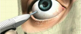
Surgery
Quite often, therapy for aphakia requires mandatory surgical intervention. Its essence is to install an artificial implant. After conducting a detailed diagnosis, the specialist will select the necessary corrective lenses that will allow you to return full visual acuity. Intraocular lenses are the most effective treatment for aphakia. Surgery is performed in a hospital setting under local anesthesia. Within a few hours the patient returns to his normal lifestyle. The first results of the intervention can be assessed within a few days.
Types of aphakia
Based on its localization in one or two eyes, the defect is immediately called monocular (single) or binocular (bilateral). Simple logic dictates that it is easier to live with a monocular one, but this is not so: the monocular form is more dangerous in terms of possible complications. This happens because a healthy eye and an eye with pathology perceive and distinguish the same visual image in different ways.
This is manifested in the fact that the right and left eyes (conventionally, the right eye is sick and the left healthy) will “draw” the same object on their retinas with different sizes and with different degrees of reliability. As a result, the information entering the visual center of the brain will not allow one to correctly estimate the size and distance to the object in question. This phenomenon is called aniseikonia. The discrepancy between the images triggers feedback and the eyes begin to adjust to one another. And due to the fact that they do not have the correct criterion for assessing the correctness of the projection, a general deterioration of vision occurs, and the healthy eye sharply loses the ability to see normally.
A problem in one eye automatically sooner or later affects the other, so methods of influencing the affected eye should facilitate the work of the healthy one.
In monocular aphakia, both eyes are unable to focus on the object in question equally. In cases of binocular pathology, objects can be seen clearly - but only at a certain, fixed distance from the eyes.
Stages of the disease
- Unilateral (monocular) aphakia. Vision may drop to 0.4 - 1.0 diopters. Surgical intervention can be avoided; vision is corrected with a contact lens or selected glasses.
- Monocular or binocular, but with the same (and significant) decrease in visual acuity. The way out is contact lenses, glasses or transplantation of artificial lenses in both eyes. If there are medical indications for it.
- Only monocular aphakia, but accompanied by other diseases affecting the healthy eye.
- Binocular form, which is accompanied by significant ophthalmological problems with a general deterioration in the condition of both eyes.
Depending on the severity of the disease, the patient may be assigned a disability from group 1 to limited VKK.
In the congenital form of the pathology, the decrease in visual acuity will be progressive. Lack of timely treatment almost always leads to complete blindness.
Symptoms of aphakia
Signs of the development of the disease are:
- significant deterioration in visual function;
- the patient’s lack of binocular vision, the patient’s inability to estimate the distance to objects located at different distances;
- a clear sign is iridodonesis - involuntary contraction of the iris when moving the eyeball;
- deep anterior chamber, lacking support from the lens;
- it is difficult for the patient to focus his gaze on an object.
It will not be very difficult for a doctor to diagnose the absence of a lens using a slit lamp; in addition, it can be used to see the scars of the limbus or cornea left after its removal.
Diagnostics
A specialist can easily recognize this disease by its symptoms. Signs of aphakia:
- the iris trembles during eye movement;
- binocular vision is impaired;
- there is no accommodation;
- visual acuity decreases;
- the image appears double.
The patient complains of headaches, periodic fog before the eyes, and general deterioration of his condition. Laboratory tests are not informative; they are usually performed to identify concomitant diseases.
To clarify the diagnosis, the following instrumental studies are carried out:
- gonioscopy;
- Ultrasound;
- refractometry;
- visometry;
- ophthalmoscopy;
- biomicroscopy.
With their help, it is possible to determine the presence of remnants of the lens in the eye, the formation of a hernia, and changes in the shape of the anterior chamber of the eyeball. Based on the data obtained, the doctor determines treatment tactics.
Read in a separate article: Subluxation of the lens of the eye: causes, symptoms and treatment
Prevention
There are no methods that would prevent or reduce the risk of developing congenital aphakia. However, in order to avoid the development of an acquired form of this pathology, you should periodically visit an ophthalmologist. Annual routine examinations allow timely detection of diseases that may require surgical removal of the lens. The most common such diseases include cataracts, which are characterized by clouding of the lens and require its replacement with an artificial implant.
People whose work involves a high risk of eye injury should take care of their eyes and follow safety rules. When carrying out welding work, using an angle grinder or other traumatic tools, you must wear safety glasses.
It is important to remember that contacting an ophthalmologist when the first signs of aphakia appear will help avoid the development of unpleasant complications and preserve vision
voice
Article rating
How does recovery occur after eye lens replacement?
The operation is performed on only one eye. The second one can be operated on no earlier than in six months. During this time, the eyeball will be completely restored. The most important month for the patient is the first month of rehabilitation. It is important to follow the following rules:
- put antibacterial drops into the eye;
- rinse the eye every day using a swab or gauze;
- do not drink alcohol;
- do not drive a car;
- do not sleep on the side of the operated organ of vision;
- do not lift weights or engage in strength sports;
- limit yourself to watching TV, reading and working on the computer;
- refuse to visit the pool.
You will need to wear a patch over your eye for the first two weeks after surgery to prevent germs from getting into your eye. Throughout his life, a person with an artificial lens will have to avoid intense physical activity and high temperatures, which can impair the functionality of the prosthesis. Compliance with all these rules and other instructions of the ophthalmologist will help to avoid complications. They don't happen often, but they are also worth mentioning.
Correction methods
The problem of aphakia can be solved using conservative methods or surgical intervention. The latter is more preferable, as it allows you to completely restore binocular vision.
Conservative therapy
You can increase the visual acuity of an aphakic eye with the help of:
- Points. They are indicated only for patients with bilateral aphakia. Usually you need two pairs: for near and far. Although, if visual impairments, for example, severe myopia, were diagnosed before the removal of the lens, then patients may not need distance glasses. To correct vision, the glass must be strongly positively convex (from +8 to +17 diopters).
- Contact lenses. This is a modern and most common way to solve vision problems. A huge advantage of the method is the possibility of application for both unilateral and bilateral aphakia. The type of material from which the lenses are made depends on the age of the patient. Thus, special soft silicone lenses are produced for newborns, and soft, breathable or hard ones for older children. But you can resort to their help no earlier than 4 weeks after surgery to remove the affected lens.
Strong plus glasses can cause annular scotoma, that is, the patient loses an entire area of vision, which leads to painful sensations from the regular sudden appearance of one or another object in front of the eyes. This significantly complicates the lives of patients, as it is difficult for them to cross busy intersections, etc.
Surgery
Surgery to install an intraocular lens (IOL) is used after lens removal.
The presence of an artificial lens in the eye is called pseudophakia, and the eye is called pseudophakic
This type of vision correction is a surgical procedure for installing an artificial plastic lens of a certain strength, the value of which is calculated using specially created computer programs and tables, taking into account:
- lens thickness;
- refractive power of the cornea;
- depth of the anterior chamber;
- length of the eyeball.
Its implementation is possible only after the final formation of the eye structures, which usually occurs by 2 years. Therefore, when treating children with congenital aphakia before reaching this age, it is necessary to use glasses or contact lenses.
There are 4 different types of IOLs, differing in how they are inserted:
- with fixation in the corner of the anterior chamber;
- iris clip lens (pupil);
- extracapillary;
- posterior chamber.
The optimal option is a posterior chamber IOL, which does not interfere with normal pupil dilation and provides better quality of vision, since it occupies the natural place of the lens.
Previously, other operations were used to restore the quality of vision in patients with aphakia, but due to their imperfections and the frequent lack of positive changes, they have not been carried out in recent years.
Causes of aphakia
The absence of a lens in the eye can be congenital or acquired; the latter type is more common. The congenital form is divided into 2 categories: primary and secondary. In the first variant, the lens did not develop due to genetic disorders, in the other, its rapid destruction occurred after initiation. If the lens is lost in one eye, aphakia is called monocular; if both organs are affected, it is called binocular.
The causes of the congenital form are determined by two factors:
- failures at the genetic level in the process of separation of the lens vesicle from the outer ectoderm at the stage of embryogenesis;
- idiobatic absorption of the lens due to random mutation.
The risk of getting the disease increases with age; one of the reasons for the acquired form is surgical removal of cataracts. It can also occur due to spontaneous dislocation of the lens, deep injuries to the eye.