Indications for pachymetry
Glaucoma is a common pathology affecting millions of people. It can lead to complete loss of visual perception if timely assistance is not provided. Several studies have confirmed that TR is an important parameter of disease progression. Patients with a TR of 550 µm or less are 3 times more likely to develop glaucoma. This indicates the initial stage of development of the disease.
Pachymetry is an important diagnostic procedure for the preoperative monitoring of a patient who will undergo refractive surgery with an excimer laser. The result of the diagnosis, combined with the degree of visual impairment to be corrected, determines the choice of refraction method to be followed during surgery. If the thickness is insufficient, LASIK and Z-LASIK (PRK is used) methods are usually chosen.
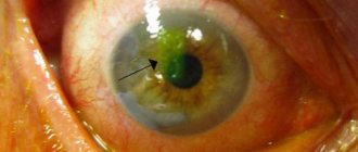
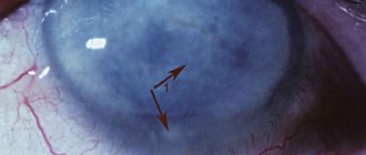
The diagnostic test is also indicated for swelling of the transparent outer membrane of the eye with deformation, ulcerative tissue lesions that are the result of an infectious or inflammatory disease. Pachymetry is performed for corneal dystrophy, which is inherited.
Indications and contraindications for pachymetry
This diagnostic method is used for pathologies that are characterized by changes in thickness and deformations of the cornea. During the appointment, if the patient has complaints, the ophthalmologist evaluates the results of simpler diagnostic tests - and, if they do not allow a diagnosis, he prescribes pachymetry.
| Indications | Contraindications |
|
|
How is corneal thickness measured?
Pachymetry does not require prior preparation from the patient. The ophthalmologist can perform it directly on the day of the first visit, if necessary. If the patient wears contact lenses, they will need to be removed. The price of pachymetry depends on how it is performed.
| Method of implementation | Its features |
| Optical pachymetry | The examination is carried out using a slit lamp, which directs a light beam into the patient's eye. In the process, different light filters are used, changing the length and width of the beam. In addition, two special lenses are used, which allows you to determine the thickness of the cornea. The steps of the procedure are as follows:
The technique is non-contact, so there is no risk of infection or damage to the cornea. |
| Pachymetry using ultrasound | The process uses ophthalmic ultrasound units. The technique is contact, as it involves contact between the ultrasound sensor and the cornea. The steps of the procedure are as follows:
After the procedure, the sensitivity of the cornea is restored quite quickly, and the patient does not experience discomfort. |
| CT pachymetry | The study is carried out using a tomograph, which allows one to obtain an image of the cornea and other tissues of the eyeball by shining them with infrared rays. In the process, their reflections are recorded, followed by processing and obtaining an image. The steps of the procedure are as follows:
The procedure lasts no more than 10 minutes, after which the patient receives the results in his hands. |
Contraindications
Diagnosis is not carried out for patients with corneal injuries if the person arrives for examination under the influence of alcohol or drugs.
Also, the procedure is contraindicated in patients with a serious mental disorder and diseases of the visual analyzer, which are accompanied by the formation of purulent contents. The latter include:
- conjunctivitis;
- herpes of the organs of vision;
- fungal keratitis;
- ulcer;
- dacryocystitis;
- blepharitis;
- stye or chalazion.
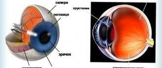
Normal corneal thickness
Each patient has a cornea of different thickness, it depends on the anatomical features of the organs of vision. Normally, this parameter should be in the range of 0.44–0.56 mm. At the edge, the TR increases; normally it is 0.7–0.9 mm.
Is a thick cornea good or bad?
The cornea is considered thick at parameters of 561–600 microns. This is bad. The thickness of the cornea affects intraocular pressure.
If the cornea is between 561 and 600 µm, then the IOP will actually be lower than that indicated by tonometry. The relationship between thickness and measured IOP does not change significantly over the lifespan in healthy populations.
If the TP parameters are greatly elevated, the IOP also appears elevated. This leads to incorrectly prescribed treatment that may not have been required.
In addition, the thickness-to-ophthalmotonus relationship should be considered when evaluating a patient with glaucoma.
Carrying out pachymetry
The normal thickness of the cornea in the center of the eye is 0.49 - 0.56 mm. The thickness in the limbus area is slightly greater and is 0.7-0.9 mm. The average thickness of the cornea in women (0.551 mm) is greater than in men (0.542 mm). The average daily change in corneal thickness is possible within 0.6 mm; if a specific indicator is higher, this indicates disturbances in its structure and requires careful examination.
Optical pachymetry
Non-contact method for measuring corneal thickness. When it is performed, a special attachment is put on a slit lamp (ophthalmological microscope), with the help of which the thickness of various parts of the cornea is measured. To do this, the patient's forehead and chin are placed in a special device in a sitting position, on the other side of which there is a doctor examining the eye. A special nozzle consists of two glass plates installed in parallel. In this case, the lower one is fixed motionless, and the upper one is capable of rotating along a vertical axis. The optical axis of the slit lamp is given a certain direction, perpendicular to which a special attachment is installed. The doctor, examining the patient's eye, moves the illumination to a given segment and, rotating the pachymeter handle, takes measurements of the thickness of the cornea, noting the indicators on a special scale. One degree of rotation of the nozzle plate corresponds to 1 mm of cornea.
Ultrasound pachymetry
This is a contact research method. Its results are more accurate compared to optical pachymetry (up to 10 microns). It is performed as follows: the patient is placed on a couch near the ultrasound machine, and a drip of anesthesia is given to the eye being examined. After this, they touch the surface of the eyeball with a hardware attachment, trying to have as little impact on the cornea as possible (this can slightly distort the results). The final results of the study are displayed on the monitor.
In our clinic, the cost of determining the thickness of the cornea of the eye is 1000 rubles. (you can find out the cost of diagnostic procedures in the Prices section), this study is mandatory when determining indications and contraindications in the case of laser vision correction.
All questions you are interested in can be asked to specialists by calling 8 800 777-38-81 and 8 (499) 322-36-36 or online, using the appropriate form on the website.
Dagaev Adam Huseinovich
Types of pachymetry
There are three ways to conduct the survey. They differ in execution technique and information content.
For diagnosis use:
- Optical method. A slit lamp is used for examination. It directs a beam of light into the patient’s visual analyzer, the length and width of which is adjusted by the ophthalmologist. This method allows you to accurately diagnose the thickness of the cornea.
- Ultrasonic method. It is carried out using ultrasonic devices. They not only allow you to study the thickness, but also the structure of the eyeball.
- Computer technique. This type of pachymetry is performed using a tomograph. The device illuminates the structures of the visual analyzer and produces an image.
Types of examination methods
Pachymetry of the eye
Currently, 2 methods have been developed for studying the cornea of the eye. These are optical and contact pachymetry. They differ in the methodology of the procedure.
Optical pachymetry
The examination is carried out using a slit lamp. This device works on the principle of a microscope. For pachymetry, additional equipment is installed on the lamp. These are 2 glass plates located parallel to each other.
The patient sits on one side of the lamp, the head and chin are fixed on special stands. The doctor takes a seat on the other side. He places the lamp at a certain angle and, observing the condition of the patient’s eye, begins to rotate the pachymeter handle. On the scale, 1 degree is equal to 1 mm of corneal layer.
The procedure is painless and non-invasive, but is performed only if the cornea is clear. Complications are unlikely. If after the procedure irritation of the mucous membrane of the eye occurs, it is recommended to use anti-inflammatory drops as prescribed by a doctor.
In addition to the optical examination method using a slit lamp, the coherent pachymetry procedure is used. For this you need a specialized coherence tomograph. The procedure, in essence, resembles the preparation of histological sections, but is carried out non-invasively and for life.
During an OCT study, the depth and size of a beam of light quanta are recorded, which are reflected from tissues with different optical properties. The reliability of the results may be affected by opacity of the cornea, other eye media, and the patient’s physical activity during the procedure. The duration of the study is 2–2.5 seconds.
The infrared radiation emitted by the tomograph is harmless to the patient. Therefore, there are no restrictions on the study. Recommendations for behavior after pachymetry are similar to non-contact examination using a slit lamp.
How is pachymetry performed?
Optical, ultrasound and computer pachymetry are performed in different ways. When using all techniques for one person, the results will be different on each device. Therefore, interpretation should be based on the normal values for each device.
Table. Diagnostic method depending on the technique
| Type of pachymetry | How it goes |
| Optical | The determination is carried out in a non-contact manner. The measurement is carried out using a special attachment that is placed on the slit lamp. Stages: place the chin on the stand; fix your head; open your eyes wide; the doctor directs the light to the desired area of the organ and rotates the pachymeter handle. Then the thickness is measured on a special scale. |
| Ultrasonic | Contact diagnostic method. Therefore, the ophthalmologist receives more accurate results, with an accuracy of up to 10 microns. The duration of the procedure is 1-3 seconds. The procedure is painless, but the patient will feel discomfort. After ultrasound diagnostics, wash the eyes with an antiseptic solution or water. |
| Computer | This procedure is considered the most accurate. The idea is to illuminate the eyes with infrared radiation and record reflections from different structures of the eyeball. Stages of implementation: the patient sits in front of the tomograph; the chin and forehead are fixed; the working part of the device approaches the eye and scans. The diagnosis lasts no more than 10 minutes. The use of anesthetic drops is not required. The patient’s task is to sit upright, not move or blink. |
Complications after pachymetry
Diagnostic measures are well tolerated by patients. Computer and optical pachymetry are considered the safest, since there is no contact with the cornea. When using ultrasound, the sensor comes into slight contact with it, so hyperemia of the mucous membrane, a feeling of a foreign body in the eye, and lacrimation may occur.
These complications are considered mild and resolve on the day of testing. Complex consequences include infection. This is only possible with ultrasound diagnostics, when the device’s sensor has not been disinfected after its use on another patient.
To prevent the development of complications, the ophthalmologist may recommend using anti-inflammatory drops for several days after the examination. You should also not touch your eyes until the unpleasant symptoms subside.
Pachymetry procedure
The process of computer, ultrasound and optical pachymetry differs. At the same time, examining even the same patient in different ways can give different results. As a result, normal values for each device should be taken into account when interpreting examination results. Corneal pachymetry is the norm when diagnosing vision pathologies in every patient.
Table. Pachymetry technique depending on the type:
| Type of pachymetry | Description of the procedure |
| Optical | It happens in a contactless way. The measurement is carried out using a special attachment placed on the slit lamp. Stages: · Place the patient's chin on a support; · Fix your head; · Widen your eyes as much as possible; · The light is directed to the desired area of the eye, and the doctor rotates the pachymeter manipulator; · Thickness is measured using a special scale. |
| Ultrasonic | Occurs by contact. Due to this, this type of diagnostics gives more accurate results, up to 10 microns. The duration of the procedure is 1-3 seconds. It is painless, although it is accompanied by some discomfort for the patient. After the examination, the eyes are washed with an antiseptic solution or running water. |
| Computer | This type of examination is considered the most accurate. In the process, the eye is illuminated with an infrared source and reflections from different elements of the eyeball are recorded by a computer. Stages: · The subject is placed opposite the tomograph; · The subject's face is recorded; · The scanner is brought closer to the eye and the doctor examines it. The duration of the scan is within 10 minutes. Does not require anesthesia. The patient should not move or blink. |
Pachymetry video
Zolotorevsky Kirill Andreevich tells how the examination is carried out and what is the norm:
Author's rating
Author of the article
Alexandrova O.M.
Articles written
2031
about the author
Was the article helpful?
Rate the material on a five-point scale!
( 2 ratings, average: 3.00 out of 5)
If you have any questions or want to share your opinion or experience, write a comment below.
Decoding the results
The thickness of the cornea is normal if its readings on the device are 490–560 micrometers in the central part and 700–900 micrometers along the edge (periphery). With keratoconus, thinning of the cornea is observed, that is, its thickness is less than 490 microns. If this procedure is prescribed before eye surgery, then the thickness of the cornea to be approved for laser vision correction using the LASIK method must be at least 490 microns.
If the indicators deviate from the norm in one direction or another, the doctors at our clinic will prescribe the necessary examinations and further treatment. Measures taken in time will help prevent diseases of the cornea and the accompanying loss of vision. To avoid this, we recommend that you visit an ophthalmologist at least once a year for preventive diagnostics.