Definition and code according to ICD-10
Heterophoria refers to a vision pathology in which the directions of movement of the eyeballs are mismatched with each other exclusively at rest.
There are no conditions for binocular vision in this case. The disease code according to ICD-10 is H50.5. Hidden strabismus can be visually diagnosed as follows: the patient must look at the object and close one eye. The presence of pathology is indicated by a rapid shift in the direction of gaze of the eyeball after it is opened.
Causes
Uncoordinated activity of the visual muscles is when the axis of one of the eyes deviates from a single point of fixation, the picture of perception is distorted. That is, in fact, the brain receives different images from each eye. When the eyes are healthy, they are both focused at a single point and the same image from each organ is transmitted to the brain.
The causes of this pathology are:
- pathological conditions of the nervous system;
- severe fear or constant stress;
- diseases of an infectious nature. Childhood diseases such as measles, scarlet fever and diphtheria greatly reduce the tone of the eye muscles;
- abnormalities of the ocular motor muscles;
- eye injuries;
- cerebellar damage;
- disturbances in visual acuity;
- paralysis and paresis;
- a sharp decrease in vision in the eye;
- growths in the brain, ears, sinuses, or eyes;
- hereditary predisposition;
- genetic abnormalities (for example, Down syndrome);
- negative effects on the fetus of medications, alcohol or drugs during pregnancy;
- birth trauma, low baby weight.
Etiology. Uneven force of action of the extraocular muscles.
Pathogenesis. Under normal conditions, thanks to the fusion ability of the visual analyzer, muscle imbalance does not manifest itself. When the eyes are separated (for example, by covering one eye or placing a prism on it with the base up or down), the relative weakness of any muscle begins to be revealed and the visual line of one of the eyes deviates inward (esophoria), outward (exophoria), upward (hyperphoria) ) or downwards (hypophoria), sometimes there is a deviation of the upper end of the vertical meridian of the cornea inwards (incyclophoria) or outwards (excyclophoria).
Causes and symptoms
Strabismus can be either congenital or acquired. Pathology develops due to the following conditions:
- microphthalmos;
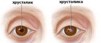
- cataract;
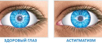
- astigmatism;
- coloboma of the iris;
- uveitis;
- improper development of eye muscles;
- fright;
- paralysis;
- some infectious diseases (scarlet fever, influenza, measles);
- traumatic brain injuries;
- somatic diseases such as asthenia and neurosis;
- stress.
Acquired strabismus is manifested by clinical symptoms such as decreased quality of vision and double vision.
Symptoms of acquired strabismus include:
- sharp deterioration in visual functions;
- double vision;
- drying out of organs;
- eye pain;
- hemorrhages.
To diagnose strabismus, sometimes not only ophthalmologists are involved, but also neurosurgeons, neuropathologists, and other specialists.
Essential infantile convergent strabismus
This is an idiopathic condition that develops in healthy infants during the first six months of life in the absence of restrictions on eye mobility and refractive pathologies.
As a rule, a large (more than 30D) constant angle is observed. Most patients in the primary position have alternating fixation of the right eye and cross fixation when looking to the left, and of the left eye when looking to the right. This may give the wrong impression of bilateral abduction deficiency, similar to bilateral sixth nerve palsy. However, abduction can usually be demonstrated by spinning the baby or using the doll's head maneuver. If difficulties arise with this, unilateral occlusion is possible, revealing the abduction ability of the other eye. With essential infantile convergent strabismus, horizontal nystagmus, latent or manifest-latent, can be observed. Depending on the child’s age, the refractive error is approximately +1.5 D.
In addition, there is asymmetry of optokinetic nystagmus and hyperfunction of the inferior oblique muscle, which is detected initially or develops later. By three years, most patients have dissociated vertical deviation. The possibility of developing binocular vision is extremely low.
During an ophthalmological examination at the Eye Clinic on Kurzenkova, a primary diagnosis is made to exclude congenital bilateral palsy of the sixth pair of cranial nerves using one of the above methods and sensory convergent strabismus resulting from organic eye pathology.
Also, nystagmus block syndrome, fixed strabismus, Duane and Mobius syndromes should be excluded.
Types: hyperphoria, exophoria, esophoria, hypophoria
The following types of heterotrophy are distinguished according to the direction of deviation:
- hypophoria (gaze directed downwards);
- hyperphoria (up);
- exophoria (outward);
- esophoria (inside).
In some cases, a deviation of the upper part of the vertical meridian of the cornea is observed, and incyclophoria (gaze directed inward) and excyclophoria (outward) are simultaneously diagnosed. Minor deviations in the angle of heterophoria do not affect the performance and quality of visual function. Find out more about divergent strabismus here.
Types of Heterophoria.
Conventional boundaries at which compensation for violations is required:
- exophoria – up to 6 diopters;
- esophoria – up to 3 diopters;
- hyper-/hypophoria – up to 1 diopter.
Types of strabismus in adults
Strabismus is divided into several types.
By time of occurrence:
- Congenital. In this case, the pathology begins when all the vital organs of the embryo are formed.
- Acquired. This type can develop at any stage of life for various reasons.
Read what penalization is in ophthalmology at this link.
What is purchased is:
- permanent;
- temporary, it is also called hidden strabismus. This pathology is periodic in nature and can go away on its own, or, on the contrary, it can become permanent;
- sensory. This means that the eyes squint to the side for a short period of time due to physical activity, alcohol consumption or some kind of disease. Children have seizures even due to exposure to very bright sun.
By eye involvement:
- one-sided;
- intermittent - the eyes squint alternately.
By severity:
- hidden;
- compensated, it can only be detected during examination;
- subcompensated – you can control it;
- decompensated – uncontrollable.
Towards:
- horizontal – the eye is deviated towards the bridge of the nose or temple;
- vertical with an offset up or down;
- mixed.
Due to the occurrence:
- friendly: movements of the eyeballs are preserved, there is no double vision, but binocular vision is impaired;
- paralytic - due to lesions and paralysis of the motor eye muscles.
How to determine strabismus in a child at home?
Source: zdorovyeglaza.ru
The most effective way to determine all visual impairments is an ophthalmological examination, but strabismus can be detected at home. To do this you will need a flashlight and a camera with flash.
Home diagnostic method
Of course, congenital strabismus is diagnosed already in the first days of our birth. But with acquired things the situation is different: small deviations are not always noticeable immediately, and medical examinations are not so frequent.
And I would like to determine the tendency to strabismus before visible symptoms appear: deviations of one or both eyes towards the nose or to the side, as well as “floating eyes” syndrome (when it is difficult to “catch” the patient’s gaze).
You can take a test for signs of hidden strabismus (or ask your child to do it) now, it only takes a few minutes.
Rules for performing the test
Lean back in a chair so that your head does not move and look out the window at some small immovable object (for example, a store sign or a satellite dish) and try to focus your gaze on this object for two seconds.
Then close your palm, first one, then the other eye, looking at the object for 1-2 minutes. If the object of fixation remains in place and does not jump from side to side when you open each eye, you can be calm.
Well, or almost calm... After all, only modern diagnostic equipment and professional examination can give a 100% result.
Causes, symptoms and types of strabismus
Why can strabismus occur in children under one year of age? There are quite a lot of risk factors.
These include:
- Genetic predisposition. The disease can be transmitted from parents and from more distant ancestors.
- Prematurity, congenital eye defects.
- Infectious diseases of a pregnant woman or baby that cause complications on the organs of vision.
- Birth injuries.
- Neoplasms in the organs of vision, hearing, and brain.
- Diseases of the nervous system (hydrocephalus, cerebral palsy).
- Mechanical damage to the organs of vision.
There are other causes of strabismus in children under one year of age. For example, improper placement of rattles above the crib or a serious fright or stressful situation in a child.
In older children, strabismus can be caused by infectious diseases, stress, trauma, as well as too much stress on the visual organs.
There are several classifications of this vision defect. Thus, one can distinguish congenital (such strabismus is found in children under one year old) and acquired disease, as well as permanent and non-permanent, that is, occurring periodically. Strabismus in children can be intermittent (the problem affects both eyes) or unilateral (the child has one eye squinting).
Types of disease according to the method of deviation:
- converging (direction of the pupil towards the nose);
- diverging (towards the temple);
- vertical (the eye looks up or down);
- mixed.
Divergent strabismus is characterized by concomitant myopia, while convergent strabismus is characterized by hyperopia.
Vertical strabismus occurs due to paresis of the vertical muscles of the organ of vision. It is difficult to treat and often requires surgical intervention. The secondary development of this type of disease can appear after surgery to treat horizontal strabismus.
Different types of disease are identified according to the degree of its severity.
Here are the possible:
- heterophoria (hidden disease);
- compensated type (only a doctor can detect it);
- subcompensated (occurs under the pressure of aggressive external factors);
- decompensated (uncontrollable).
A hidden disease or heterophoria is not immediately detected, because vision remains binocular if both eyes are open, and the displacement of the pupil is imperceptible.
This disease is also divided into subtypes due to its occurrence. Thus, the disease can be unfriendly (paralytic) and friendly. The first option usually appears after illness or injury, the second is most often caused by a hereditary factor.
Concomitant disease has its own subtypes:
| Variety | At what age does it begin to form? | Causes | How to get rid |
| Accommodative | About 3 years, earlier in weakened children. | Refractive errors (myopia, farsightedness, astigmatism). | Using correction glasses in combination with hardware treatment. |
| Partially accommodative and non-accommodative | During the first 24 months of life. | In addition to refractive errors, there are other visual deviations. | In most cases, the child needs surgery. |
A paralytic disease is characterized by insufficient or completely absent motor response of the eyeball towards the affected muscle. He is always accompanied by the child's complaints about dizziness and the fact that he began to see things in two. Paralytic strabismus occurs most often after infectious diseases or injuries, but can also be congenital.
Additional symptoms include:
- looking at an object with one eye, covering it with the other with your palm;
- excessive sensitivity to bright light;
- dizziness;
- decreased visual acuity in the affected eye;
- bifurcation of objects;
- developmental delay, behavioral deviations.
Another subtype of strabismus is false (imaginary). Its causes lie in the special structure of the eye. Sometimes people have an increased angle between the visual and optical lines. This does not interfere with vision, but visually creates the feeling of a “braid”. Also, false strabismus sometimes appears due to an asymmetrical arrangement of the eyes or general asymmetry of the face. There is no need to treat the imaginary variety; it often disappears as the child grows older.
Diagnostics
External deviation is obvious. Exotropia should not be confused with transient outward wandering of the eyes, which occurs in approximately 60–70% of normal newborns and resolves by 6 months of age.
Diagnosis of divergent strabismus consists of a series of tests:
- Measurement of visual acuity - may be normal or abnormal. The reduction may be caused by refractive error, pathology such as cataracts or retinal disorders, suppression, or a combination of these factors. Measuring visual acuity in young children is more challenging. For this, there are special circuits and devices that require a lot of patience.
- Cycloplegic refraction. This is an objective determination of the true refractive error by eliminating the effect of accommodation. Achieved by paralyzing the ciliary muscle with cyclopentolate eye drops or atropine eye ointment. Medicines also cause pupil dilation.
- Slit Lamp Test – This test checks for any disease or abnormality in the front of the eye.
- Examination of the fundus (retina) - fundoscopy - is used to examine the inside of the eye, involving the retina, optic nerve, blood vessels and allows you to measure the magnitude of the deviation, evaluate eye movements in different directions of gaze, check for the presence of diplopia, if present, determine the type, degree binocular vision.
Diagnosis of hidden strabismus
Hidden strabismus can be diagnosed using both hardware methods and more accessible “manual” ones. The most common is the Meddox method.
The Maddox scale includes two bars: a horizontal one 2 meters long and a vertical one 1.5 m high. At their intersection there is a switched-on electric lamp. The patient is positioned at a distance of 5 m from the Maddox scale. The scale has numbers corresponding to the tangents of the visual angle from a distance of 5 m. A special device - a “Maddox wand” - is a series of soldered cylinders of red glass. The patient is asked to look at the light source through the “Maddox wand”. Its properties are such that the luminous point extends into a red line.
This unnatural visual complex, unusual for the brain, causes a short-term disorder of binocular vision - one eye begins to see the light source, and the other - a vertical red stripe.
Therapy for hidden strabismus
A slight deviation of latent strabismus from the norm does not require specific therapy. In other cases, it is recommended to undergo examination and treatment.
To treat heterophoria, conservative treatment methods are used: glasses, the Synoptophore apparatus and visual gymnastics. Next, let's look at each of them in more detail.
For strabismus, it is very useful to perform gymnastics. The following exercises are suitable for this:
- straining your eyes, try to look at the tip of your nose for at least 7 seconds. Then comes relaxation with the eyes returning to their normal position for 3-4 minutes and everything repeats again;
- follow with both eyes an object that will be slowly moved by another person. In general, with strabismus it is very useful to observe moving objects;
- mentally drawing an infinity sign in front of your eyes or a figure eight - this greatly tightens your visual muscles;
- eye movements to the sides, in a circle;
- as you inhale, focus both eyes on the tip of your nose, and as you exhale, look into the distance;
- fast and strong repeated squinting gives a good load on the eye muscle;
- Try, if possible, to focus your gaze alternately on a close object, and then immediately on a distant one.
With hidden strabismus, the work of the extraocular muscles is impaired. The function of binocular vision forces them to remain in the correct position, but as soon as one eye is turned off from the visual process, the muscles of the other eye begin to work in a way that is more comfortable for them. In other words, hidden tension or weakness in certain extraocular muscles is compensated only when both eyes are working.
To detect hidden strabismus, it is enough to block the possibility of binocular vision, that is, close one eye. At the same time, it will deviate in the direction corresponding to the type of heterophoria. At the moment of restoration of binocularity, the pupil makes a characteristic adjustment movement and returns to the correct position. With orthophoria (absence of strabismus), the eyeballs remain in a consistent position under any conditions.
Due to constant overstrain of the oculomotor muscles in the process of synthesizing a binocular image, systematic headaches, a feeling of pressure and eye fatigue, nausea and dizziness may occur. To relieve such stress, it is recommended to constantly wear glasses with prismatic lenses. A prism with parameters of 2-3 degrees should be positioned with its base in the direction opposite to the direction of heterophoria.
If hypermetropia and myopia occur, sometimes corrective decentering glasses (changing the distance between the pupils upward or downward) are sufficient.
Synoptophore exercises give good results - training, relaxing and normalizing fusion reserves.
In some cases, hidden strabismus requires surgical intervention - the same as for obvious strabismus. However, the appropriateness of surgical assistance is determined by many factors (in particular, the degree of influence of the pathology on the patient’s quality of life).
What are the causes of strabismus in children?
The causes of strabismus in children can be different - genetic, a consequence of birth trauma or even mental disorders. We will look at the main ones. In addition to genetic factors, the most common cause of strabismus in a child is pathology of pregnancy and childbirth.
Due to fetal hypoxia, as well as due to birth trauma to the cervical spine or brain, innervation is disrupted and the extraocular muscles are deviated from the visual axis. At the same time, myopia, farsightedness and astigmatism can provoke the development of strabismus in a child.
Head injuries, eye surgeries, mental disorders and brain diseases can also cause strabismus in children. There are cases when this pathology occurs in a child after he has had the flu, measles, diphtheria or scarlet fever.
Apparent strabismus
Often, when parents go to the doctor, they complain about their child’s strabismus, but after examination the doctor does not detect it. This happens, as a rule, due to the congenital epicanthus, the structure of the skull or the wide bridge of the nose.
Apparent strabismus is more likely to disappear with age as soon as the skeleton begins to change. To determine hidden strabismus, you can try a cover test.
In this case, when the child has both eyes open, strabismus is not observed, but as soon as one of them is closed, the other begins to move to the side, and when opened, returns to its place. The main condition for this method is this: the child must look at the object that is being shown to him.
At 3 years of age, in addition to the above methods, visual acuity is tested using a table with or without glass correction. The state of binocular vision can be determined using a color test.
Color test technique
The study is carried out using a special disk with luminous circles of different colors located on it (1 red, 1 white and 2 green). The child wears glasses specially designed for this purpose with red glass on the right and green on the left.
Thus, the eyes see the color that is in front of them, that is, the right one is red and the left one is green. The white ball appears as one of two colors due to the filters placed in front of the eyes.
If the baby does not have any visual impairments, he will see 4 circles (either 2 red and 2 green, or red and 3 green). If one eye of a child turns off, he sees 3 green or 2 red circles (monocular vision). If the baby has alternating strabismus, he will see either 3 green or 2 red.
Treatment
In most cases, treatment for hidden strabismus in adults and children is not required. The congenital form often goes away on its own with age. When myopia develops, glasses or contact vision correction and microsurgical interventions are performed.
Glasses or contact lenses are used to correct vision. Diopters are determined individually, depending on the degree of decrease in visual function. Children are prescribed glasses with decentered lenses that correct the displacement of the eyeball.
Drug treatment of latent strabismus is ineffective. Eye drops are used for diagnostic purposes or to temporarily reduce the function of a healthy organ of vision.
Watch the video and find out what type of problem does not require treatment:
Penalization
This treatment method involves turning off the healthy eye so that the squinting eye begins to work more actively. Due to this, the eye muscles are strengthened. The healthy eyeball is covered with a special bandage - an occluder. At first it is put on for 10–15 minutes a day, gradually the time of wearing the bandage is increased to two hours.
It is recommended to wear a headband while walking. Using an occluder while reading or working at a computer causes fatigue, discomfort, and headaches.
Synoptophore
This is a special device on which treatment is carried out using gymnastics. The device consists of a pair of eyepieces. A person looks at them, trying to see all parts of the picture. In this case, the eyepieces are brought together and moved apart until ghosting appears.
With the help of a synoptophore, normal binocular vision is restored. The effect for latent strabismus is quite good, but is observed under long-term treatment.
Surgical treatment
The operation is performed if conservative treatment methods are ineffective or if complications develop. The intervention consists of normalizing the length of the extraocular muscles. If necessary, laser correction of myopia is prescribed.
Hidden strabismus is a non-hazardous condition for life and health. In most cases, it does not require special therapy. If a person is concerned about the aesthetic side or vision begins to deteriorate, consultation with an ophthalmologist is recommended.
Share the article on social networks, leave comments and describe your experience of dealing with an imaginary form of strabismus. All the best.
Symptoms
In the presence of hidden strabismus, the functionality of the eyeballs decreases in the patient. Visual acuity often decreases, which leads to the following symptoms:
- nausea, dizziness, poor health;
- pain in the eyes when concentrating;
- progression of decreased visual acuity;
- violation of the symmetry of the eyeballs, sometimes the affected organ of vision is slightly displaced from the orbit;
- lack of clear focus on a specific object;
- rapid eye fatigue, which occurs even in the middle of the working day.
These symptoms suggest strabismus. But you need to see a doctor to check the condition of your vision organs. Using various research methods, he will be able to make a diagnosis.
Therapy for hidden strabismus
Up to 6 months, slight strabismus is considered a physiological norm; this is due to the fact that the eye muscles are still very weak. Over time, this phenomenon will correct itself. However, if persistent strabismus occurs as the child gets older, this is a reason to consult a specialist. Older children can talk about their problems themselves, but kids cannot understand what is happening to them.
If such alarming symptoms are present, parents should definitely take their child to the doctor.
The same methods are used for treatment in children. Of course, this is much more difficult, because it is very difficult to convince a child, for example, to constantly wear glasses or occlusion. At home, parents can themselves monitor whether the instructions are being followed correctly, but in kindergarten the child is often left to his own devices.
It is very convenient to use soft contact lenses for a child. Firstly, they are not visible on the face and the baby will not be teased. Secondly, the child will not be able to remove lenses, such as glasses. Of course, the lenses must be selected by the attending physician.
Surgical treatment of congenital strabismus is used before the age of 3 years. For acquired strabismus, surgery is indicated if there is no effect of therapy.
How to determine strabismus in a newborn and one-year-old child?
By the end of the first week of a baby’s life, you can independently diagnose the pathology in question. To do this, you need to take a rattle and remove it from the child’s eyes at different distances, moving it from side to side.
Carefully monitor the reaction of the child's eyes when observing a moving object and draw a conclusion about how mobile the baby's pupils are. In newborns, the gaze may be discoordinated until 3-4 months; after this age, both eyes become aligned.
In some cases, in children with a wide bridge of the nose, strabismus may be apparent. You should consult a doctor and sound the alarm only if, after 4 months of life, the child’s eyes do not look at the same point most of the time.
Strabismus in one-year-old children can be recognized by the following signs:
- the child cannot direct his eyes simultaneously to one point in space;
- eyes do not move together;
- one eye squints or closes in bright sun;
- the child tilts or turns his head to look at the object;
- the baby bumps into objects (squint impairs the perception of depth in space).
Let us recall once again that true strabismus is characterized by the deviation of only one eye from the joint point of fixation. At the same time, for a newborn child, a slight defocus of the eyes is considered a completely normal phenomenon, which is observed in all babies.
Moreover, the absence of slight strabismus in a small child is rather an exception to the rule. Firstly, the eye muscles of children are very weak, so they need training. Secondly, the child has not yet learned to use these muscles, so sometimes it is not possible to look in different directions.
That is why small eyes, not listening to their owner, either converge to the bridge of the nose, or scatter in different directions. As soon as the baby learns to control the movements of his eyeballs, the squint will go away.
This pathology in infants is inextricably linked with weakness of the eye muscle. The most common causes of strabismus in newborns are:
- injuries and infectious diseases of the brain;
- changes in the eye muscles of an inflammatory, vascular and tumor nature;
- untimely treatment of myopia, astigmatism, farsightedness;
- congenital diseases and birth injuries;
- increased physical and mental stress;
- placing children's toys too close to the baby's face.
Heredity also quite often causes the development of strabismus in newborns. If one of the parents has this pathology, then there is a high probability that their child will inherit the disease.
Sometimes strabismus manifests itself as a symptom of other congenital diseases or as a result of illnesses suffered by the baby’s mother during pregnancy.
Accommodative convergent strabismus
Accommodation and convergence are two essential elements of the act of vision at close range. At the same time, accommodation is the process of focusing the eye on a nearby object with a change in the curvature of the lenses. To achieve bifoveal fixation of an object, the eyes converge simultaneously. Both the process of accommodation and the process of convergence are related quantitatively to the distance to the visible object with a very constant ratio relative to each other. Therefore, the cause of some types of strabismus is a change in the AK/A index.
Refractive accommodative convergent strabismus
With it, the AK/A index does not change, and strabismus occurs in response to severe hypermetropia, ranging from +4.0D. The accommodation tension required in this case for focusing even distant objects occurs with an increase in convergence, which is much higher than the negative reserves of fusion. At the same time, a loss of control occurs and convergent strabismus occurs, often in manifest form. It appears between the ages of 6 months and 7 years. When fixing the gaze on near and far objects, the difference in the squint angle is small and is less than 10 D.
In the process of optical correction of existing farsightedness, such strabismus is completely eliminated. In the case of partial convergent strabismus, optical correction of hypermetropia can reduce it, but will not completely eliminate it.
Non-refractive accommodative convergent strabismus
It is explained by a high AC/L index in the absence of serious hypermetropia, which is why increased accommodation occurs with a disproportionately large increase in convergence. There are 2 types of non-refractive accommodative convergent strabismus:
Kurtosis of convergence. In this case, a high AC/A index is noted due to an increase in AC (i.e., normal accommodation with increased convergence).
- Norm of the nearest point of accommodation.
- When fixating an object at a far distance, the eye position is normal; convergent strabismus occurs when fixing an object at a close distance.
With impaired accommodation. Low accommodation is explained by a high AK/A index, which occurs with a decrease in A, since the weakening of accommodation requires serious efforts, which arise with increased convergence.
Removing the nearest point of accommodation. Additional accommodative effort is required when fixating a close object, which leads to convergence excess.
Mixed accommodative convergent strabismus
It is characterized by a combination of hypermetropia with a high AC/A index, when when fixing an object at a far distance, the angle of deviation significantly increases (over 10 D) to fix an object at a close distance. When fixating a distant object, deviation is usually corrected with glasses. If there is no correction with bifocal glasses, convergent strabismus will persist even when fixing an object at a close distance.
Causes of heterophoria
The main reasons contributing to the development of hidden strabismus are:
- individual features of the anatomy of the eye (size of the orbital sockets, structure of the skull, etc.);
- difference in the strength of the eye muscles, which ensure the mobility of the eyeball;
- disturbances in the functioning of the endocrine system (for example, pathology of the thyroid gland);
- past infectious diseases, which often have a significant negative impact on the nervous system, including the activity of the visual apparatus;
- mental disorders;
- frequent stress;
- paralysis of the eye muscles;
- traumatic eye injuries;
- tumors affecting the organs of vision and surrounding tissues.
Often hidden strabismus is observed in recently born children. Heterophoria in this case occurs due to the fact that the baby’s eye muscles are not yet strong and cannot fully control the movements of children’s eyeballs. Typically, such hidden strabismus disappears within 3-4 months after birth, that is, as the muscles acquire normal strength and tone.


