Why is mechanical eye injury dangerous?
In accordance with the location, there are wounds and blunt injuries of the eyeball, as well as damage to the bone bed and adnexa: eyelids, conjunctiva, tear-producing organs, etc. Damage can occur for a variety of reasons, for example, when accidentally hitting the eye with a nail or finger, and also when exposed to any sharp object: wire, knife, glass, sharp pencil. In addition, when carrying out construction and repair work, for example, drilling, penetrating injuries often occur with the entry of fragments. Serious mechanical damage to the organs of vision leads to unfavorable disturbances in the functioning of the visual system.
In the most severe cases, they can lead to complications, including weakened vision, functional death of the eyeball and blindness. This leads to disability, so it is extremely important to provide first aid and promptly go to the emergency room.
Pre-medical measures
First aid for an eye injury is extremely necessary, especially if there is visual contusion. Apply a dry bandage from the very beginning, and after that you can apply cold to it (for no more than 5 minutes with a break of 10 minutes). Then urgently call an ambulance. If necessary, take a pain reliever, for example, Ketanov, Analgin or Panadol.
No rinsing or instillation should be done, since the exact clinical picture is not known.
Unskilled actions can only aggravate the situation and lead to loss of vision.
What types of mechanical eye injuries are there?
The method of treating eye injury depends on its nature and extent. In ophthalmology, several types of damage to the organs of vision are distinguished, depending on the mechanism of their occurrence, as well as the location and presence of developed complications. The most common are various eye injuries, which can be superficial, penetrating and through. In addition, they can be infected or uninfected, as well as with or without the penetration of a foreign body. Blunt injuries to the organs of vision, which include concussions and concussions, should be included in a separate category. Depending on the location, injuries to the eyeball, damage to the appendages, bone bed and internal structures of the organs of vision, as well as combined injuries are distinguished. According to the conditions and circumstances of occurrence, it is customary to distinguish between domestic, industrial (in the workplace), agricultural, military and children's types of injuries.
Other orbital injuries
In addition to damage to the eye by foreign bodies, there are several other types of injuries:
- chemical - the protein shell is affected by caustic toxic fumes from lyes, chemical reagents, or by direct contact of these substances with the retina;
- thermal – injury occurs when exposed to frost or contact with hot objects;
- radiation – damage of this type occurs from bright flashes and radiation waves;
- mechanical - the cornea is damaged by a blow to the eye or bruise.
Also in medical practice there are injuries of a mixed nature (thermochemical and thermoradial).
Superficial wounds of the eye
The most common occurrence. This category of injuries includes minor wounds of the eyelids, conjunctiva or cornea of various types. They often occur when a nail, finger, or tree branches accidentally enter the eye, as well as when using damaged contact lenses. Superficial wounds are characterized by a violation of the integral structure of the skin, therefore, in such cases, initial disinfectant treatment of the wound is required. In some cases, it is necessary to apply sutures and excise tissue along the edges of the injury site. In addition, in parallel, the patient is prescribed a course of drug treatment aimed at antibacterial and antiseptic effects.
In the most severe cases, in the presence of hemorrhages, procedures such as subconjunctival injections of the drug dionin, electrophoresis, and autohemotherapy are indicated.
These measures help accelerate blood circulation and have a resolving effect.
Symptoms of superficial wounds:
- Redness of the conjunctiva (mucous membrane) occurs;
- The patient feels the presence of a foreign body under the eyelid;
- There is a sharp cutting pain in the affected eye;
- There is profuse lacrimation and photophobia;
- Some herbs are characterized by swelling of the eyelids;
- In severe cases, visual acuity decreases.
Injuries involving foreign objects
If you suspect that foreign bodies have entered the internal structures of the eye, a thorough diagnosis of the pathology should be carried out. A distinctive feature of such wounds is the presence of a gaping hole in the outer membranes of the eyeball.
Foreign objects provoke the development of purulent processes, the appearance of infiltrates, and clouding of the cornea. The complexity of the situation lies in the fact that with significant damage to the eye it is quite difficult to visualize a foreign body.
If the object has large linear dimensions, then complications such as loss of internal structures of the eye may occur. Mandatory procedures when diagnosing an injury:
- biomicroscopy – examination of eye structures using a slit lamp;
- ophthalmoscopy – examination of the fundus of the eye using an ophthalmoscope;
- X-ray examinations if it is impossible to detect a foreign object using the first two methods;
- Ultrasound - to determine the location of a foreign object, identify other pathological processes in the internal structures of the eye that develop when a foreign body enters;
- CT – multiple high-precision images to determine further tactics for patient management.
Treatment is carried out surgically. The foreign body is removed using needles and spears with magnetic tips. Surgery is performed either through a wound or through an additional incision in the sclera at the location of the foreign object.
If the lens is damaged or a foreign body has penetrated into the biological lens, then removal of the lens and replacing it with an artificial one is indicated. After the intervention, massive antibiotic therapy is indicated to prevent the development of purulent processes.
Penetrating injuries
This type of mechanical injury occurs when the eyeball comes into direct contact with sharp objects such as metal, wood or glass fragments, as well as wire, cutlery or office utensils. In this situation, it is often diagnosed that a foreign body has gotten inside. Severe cases of injury are accompanied by loss of the contents and membranes of the eye. The most characteristic symptom of a penetrating wound is acute pain. In addition, lacrimation, photophobia, redness of the conjunctiva and eyelids, as well as severe hemorrhage in the damaged areas may be present.
Patients with this type of injury usually require immediate surgical attention.
After this, antibiotics, anti-inflammatory and antifungal drugs are prescribed to prevent the development of various pathologies. If foreign bodies are detected, for example, fragments in the conjunctiva or cornea, they are removed using jet rinsing.
Signs of penetrating eye injury:
- The presence of a through wound in the cornea or sclera;
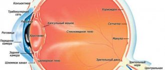
- Wound channel in the lens;
- The presence of an air bubble in the vitreous body;
- Displacement of the iris;
- Tear of the pupillary edge of the iris;
- Prolapse of the vitreous or ciliary bodies;
- Marked drop in intraocular pressure;
- Partial clouding of the lens.
Principles of treatment
Any penetrating eye injuries are an emergency and require urgent surgical treatment. Surgical intervention is aimed at restoring the integrity of the anatomy of the eye and eliminating the possibility of infection. In case of minor damage to the internal membranes and their loss, the structures are repositioned back. The injured, clouded lens is usually removed to avoid the development of an inflammatory reaction and increased intraocular pressure. The issue of implanting an artificial lens during surgical treatment of a penetrating wound with removal of a traumatic cataract is decided in each case individually. The main factors at this moment are the condition of the injured eye, the patient’s well-being, the extent of the eye injury and the severity of its inflammation. If there is a high risk of complications (which happens very often), lens implantation is postponed for several months. In the postoperative period, it is imperative to prevent infectious complications. It includes antibiotic therapy (intravenous and intramuscular injections), injections into the tissues adjacent to the eye, as well as long-term instillation of agents with anti-inflammatory and antibacterial properties. If necessary, tetanus vaccination is performed. After 1.5-3 months, the sutures from the cornea can be removed, which depends on the size of the injury to the eyeball, its location and the course of the recovery period. The sutures from the sclera are not removed, since they are covered by the conjunctiva.
Blunt mechanical damage
Due to a blow to the eyeball, bone bed or facial skeleton with a voluminous object, for example, a ball, fist, stone, stick, etc., blunt mechanical injury may occur. Often such injuries are accompanied by contusion of the organs of vision, hemorrhages in the tissue of the eye and eyelids, as well as fractures of the walls of the eye orbit. In some particularly severe cases, such injuries are accompanied by traumatic brain injury.
It is worth noting that mechanical injury of this type is extremely dangerous for the eye. The consequence of this injury may be partial retinal detachment.
In case of blunt mechanical impacts, a comprehensive examination of the visual system is mandatory and surgical assistance is provided as necessary. After this, a protective binocular bandage is applied to the affected eye. To prevent infectious and fungal complications, antibiotics are prescribed. To speed up the process of resorption of hemorrhages, electrophoresis using potassium iodide may be required.
Signs of blunt mechanical damage:
- Swelling of the organs of vision;
- Hemorrhages in the tissue of the eye and eyelids;
- Acute pain;
- Abnormal position of the damaged organ (displacement and problems with mobility);
- Headaches and dizziness.
Types of drops for eye injury
The effect of eye drops is based on the components contained in the medicine. Most often, patients are prescribed drops for eye injury with an antibacterial, anti-inflammatory or regenerating effect.
Anti-inflammatory
In addition to the direct effect of relieving the inflammatory process or preventing it, these drops can relieve acute pain, and therefore they are often included in therapy for injury. The active components of anti-inflammatory drugs are able to reduce the synthesis of prostaglandins - mediators of inflammation, as a result of which tissue swelling is also relieved. Nerves pinched by edematous tissue no longer cause pain, and the person’s condition improves significantly.
Antiseptics
Antiseptics are substances that are used to disinfect and disinfect tissues. This will significantly reduce the risk of developing an infectious process here. In addition, antiseptic drugs will help if an infection has already appeared in the eyes. In such a situation, it is necessary to remove particles (waste products) and pus only with the help of an antiseptic, and only then use medicinal products for the eyes.
Antiseptic compounds can also be used to treat hands before the procedure of instilling drops into the eyes.
Restorative
One way or another, the eye tissue will gradually recover. But it is also possible to speed up regenerative processes. Thus, the use of compositions with keratoprotective properties allows you to create a reliable protective shell, thanks to which tissue hydration will occur more intensively. In addition, restorative drops accelerate blood flow, which means that nutrition will flow to the cells faster and in full. Finally, some restorative eye products may contain vitamins and minerals that are so necessary for the tissues of the organs of vision to normalize visual processes.
Antibacterial
Why are drops with an antibacterial effect needed if a person has mechanical damage to the tissues of the organ of vision? Everything is very simple. Violation of tissue integrity is a provoking factor for the development of the infectious process. There are a huge number of pathogens in the air, and an open wound provides an opportunity for their direct penetration into the organ of vision. Therefore, antibacterial drops are used to prevent infectious diseases.
Don't be afraid of using antibiotics. Unlike tablets and intramuscular or intravenous injections, the active component here acts locally, which means it will not cause harm to other organs.
You should not try to reduce the duration of the therapeutic course. If the patient finishes dripping antibiotics into the eyes earlier than indicated in the recommendations of the attending physician, then he may encounter an infection that will be insensitive to this antibacterial drug.
First aid for mechanical eye damage
Many people experience mechanical damage to the eye. What to do in such a situation? It should be remembered that all injuries to the organs of vision are dangerous, so you should not hesitate to go to the doctor. It is impossible to treat an injured eye at home - this can cause adverse consequences, including loss of vision. This is especially true for serious penetrating wounds. However, before going to the emergency room, it is advisable to give yourself first aid and numb the eye.
In this case, it is strictly forbidden to put pressure on the injured organ of vision and try to independently remove the foreign object.
In addition, if a penetrating injury occurs, do not wash the eye to avoid the entry of microorganisms and the development of infection. When applying a bandage, it is not recommended to use cotton wool, as small fibers can penetrate inside and cause an inflammatory process.
Introduction
Eye injuries have firmly taken a leading place among the causes of visual disability in Russia, accounting for 22.8% of the number of persons recognized as disabled for the first time. The main task of healthcare is to prevent eye injuries and ensure optimal medical and social rehabilitation of victims. A retrospective analysis of the problem indicates the dependence of visual injuries on the social, domestic, military-political and criminal situation in the country and in the world. Analysis of the political and economic situation in the country allows us to predict the high relevance of scientific research in the field of ophthalmology in this direction (R.A. Gundorova, 1997). Injuries to the organ of vision can be divided into industrial, agricultural, household, and children's. Each type of injury has its own characteristics. Where to begin examining a patient with an eye injury?
First of all, it is very important to correctly and carefully collect an anamnesis of the injury: where, when and under what circumstances it occurred, what was the traumatic agent, and what is the mechanism of injury, the time elapsed from the moment of injury to the patient’s admission, vaccinations that were previously administered to the patient. Then they begin directly to study the ophthalmological status of the patient, which includes: 1. External examination of the eyelids and palpation of the bony edges of the orbit. 2. Inspection of the conjunctiva of the eyelids and transitional folds using the method of focal illumination of the eye and eversion of the eyelids. 3. Inspection and palpation of the lacrimal sac, determining the nature of its contents. 4. Detection of superficial and penetrating wounds of the cornea using fluorescein test and Seidel test. 5. Study of pupillary reactions. 6. Assessment of the color of the pupillary zone in transmitted color. 7. Checking visual acuity (at least approximate). 8. Approximate assessment of intraocular pressure. 9. Examination of the ocular media and fundus (if possible). The variety of injuries is difficult to fit into classification frameworks. There are injuries to the orbit, appendages of the eye and eyeball. It is rational to divide injuries into mechanical, thermal, chemical, radiant energy, vibration, toxic, etc. Mechanical injuries, in turn, are divided into blunt injuries (contusions) and wounds; the latter are penetrating and non-penetrating, and they separately distinguish through injuries. According to the severity of the injury, injuries are divided into mild, moderate and severe, but for the eyeball this classification is to a certain extent arbitrary, since it is difficult to predict the course of the wound process in the eye. With relatively mild injuries, especially penetrating ones, the course of the wound process in the eye can be severe.
First aid: foreign body in the eye
- If the foreign object is large, it is necessary to fix a protective frame over the eye. It can be made from improvised means, for example, a paper cup. This will prevent the foreign body from dislodging.
- It is recommended to cover the fellow eye with a napkin to prevent simultaneous movement of the eyeballs. This will prevent the foreign body from moving and causing additional damage.
- For disinfection, it is recommended to instill any antibacterial drops that are on hand: “Vitabakt” 0.05%, “Levomycetin” 0.25% or “Albucid” 20%.
- Next, you should consult a doctor as soon as possible.
What to do
- Calm the victim.
- Wash your hands thoroughly before any manipulations described below.
- Take the victim to a specialized medical facility as soon as possible.
Wounds of the eyelids
- Carefully clean the area damaged by contamination with water or an antiseptic solution.
- If you need to apply cold (without putting pressure on the eye), protect the wound with a clean bandage. In case of severe bleeding, you can make a bandage of gauze and cotton wool or put a hemostatic sponge under the bandage on the wound.
- Contact a specialized emergency room.
Sensation of a speck (foreign body) in the eye
Active blinking, accompanied by lacrimation, usually washes away small specks from the eye. If this does not happen, you need to do the following:
- Carefully examine the eye in bright light, pull down the lower eyelid - usually the specks remain there.
- If you find debris, try to wash it with water (do not try to remove it with a handkerchief, tweezers, or cotton wool!).
- Regardless of the result, drop an antibacterial agent into the eye (for example, Albucid 20%, Vitabact 0.05%, Levomycetin 0.25%).
- The scratching sensation will remain in the eye even after removing the trapped speck; they will go away on their own within a couple of days. If the speck cannot be removed, you need to contact a specialized medical center. institution.
A chemical got into the eye
- It is necessary to rinse the eyes and eyelids with sufficient running water.
- Sit the victim near the sink, tilt his head back or tilt his head towards the injured eye. Try to open your eyelids and rinse with running water for 20-30 minutes.
- If the solution gets into both eyes, both eyes must be washed at the same time. If the substance gets on the face or other parts of the body, in addition to washing the eyes, the victim should take a shower.
- Contact a specialized medical facility immediately after washing your eyes.
IMPORTANT! You should absolutely not wash your eyes if you come into contact with quicklime powder. Crystals must first be completely removed from the surface of the eyeball and eyelids (by interacting with water, lime produces heat and can worsen the burn). In this case, you should try to remove all the powder with a dry, clean cloth, and only then thoroughly rinse the damaged tissue under running water. Visiting a specialized emergency room is mandatory!
Flame burn
- Remove dirt from the skin of the eyelids and around the eyes by wiping everything with alcohol (be careful not to get alcohol into the eye!)
- Place dry ice on the eye (for example, ice in a bag, wrapped in a clean cloth on top).
- Apply an antibiotic ointment (for example, tetracycline ophthalmic) to the skin of the eyelids and place behind the eyelid.
- Contact a specialized hospital.
Eye burns from ultraviolet radiation (in a solarium, with a quartz lamp, during welding work)
- Darken the room, as the victim usually develops photophobia.
- Place an antibiotic ointment (for example, tetracycline ophthalmic) behind the eyelid.
- Place dry ice on the eye (for example, ice in a bag, wrapped in a clean cloth on top).
- Give an anesthetic (Pentalgin, Solpadeine, Nurofen tablet).
- If the pain does not go away within an hour or more, go to a specialized hospital.
Superglue hit
- Try to remove the glue present on the skin of the eyelids. The procedure will be made easier by applying an antibacterial ointment (for example, tetracycline eye ointment)
- Try to open your eye (often you need to cut off your eyelashes to do this)
- Apply antibacterial drops (Albucid 20%, Vitabact 0.05%, Levomycetin 0.25%).
- If possible, go to a specialized hospital as quickly as possible.
Bleeding from the eye
Bleeding may be due to penetrating injury or severe eye contusion. In this case it is necessary:
- Place antibiotic drops into the eye (Albucid 20%, Vitabact 0.05%, Levomycetin 0.25%)
- Cover the eye with a clean (ideally sterile) bandage. Don't put pressure on your eyes!
- Immediately contact a specialized hospital.
Foreign body protruding from the eye
- If the foreign body is large, to prevent its displacement above the eye, you can fix a protective frame (for example, using a disposable paper cup).
- Cover the fellow eye with a napkin, because simultaneous movements of the eyeballs can provoke the displacement of part of the foreign body inside the eye, which will cause additional damage.
- Instill antibiotic drops (Albucid 20%, Vitabact 0.05%, Levomycetin 0.25%).
- Immediately contact a specialized hospital.
In the medical department, everyone can undergo examination using the most modern diagnostic equipment, and based on the results, receive advice from a highly qualified specialist. The clinic is open seven days a week and operates daily from 9 a.m. to 9 p.m. Our specialists will help identify the cause of vision loss and provide competent treatment for identified pathologies.
You can make an appointment at the Moscow Eye Clinic by calling 8 8 (499) 322-36-36 (daily from 9:00 to 21:00) or using the online registration form.
Mironova Irina Sergeevna
