Keratoconjunctivitis sicca is a disease whose main cause is dry eyes. It occurs quite often and usually does not cause serious consequences, but due to the complete lack of treatment it can lead to weakened vision or even loss of vision. Let's consider the features of this disease.
In this article
- What is keratoconjunctivitis sicca?
- Features and functions of the tear film
- Causes
- Symptoms
- Diagnostics
- Keratoconjunctivitis sicca: treatment
- Treatment of keratoconjunctivitis with folk remedies
- Forecast
Keratoconjunctivitis sicca, also called dry eye syndrome and keratitis sicca, is widespread throughout the world. This is one of the most pressing eye diseases, which affects 5-6% of the world's population and 12% of Russians under the age of 40. Moreover, it is detected in 34% of older people and almost 10% of women during menopause. According to Russian scientists, today the syndrome is diagnosed 4.5 times more often than 30 years ago.
What is keratoconjunctivitis sicca?
Sicca keratitis is associated with dry eyes. It can be caused either by insufficient production of tear fluid or by its too rapid evaporation. Poor corneal moisture causes irritation. There may be microcracks on it, which are a good environment for microbes.
It cannot be said that keratoconjunctivitis sicca is a very dangerous pathology, but it can greatly reduce a person’s quality of life, cause other eye diseases, and, in the absence of treatment, partial or complete loss of vision. This happens rarely, so many consider this disease to be harmless and do not pay attention to it. This is what causes complications.
Classification of keratoconjunctivitis
According to the duration of the inflammatory process, acute and chronic keratoconjunctivitis is distinguished.
Depending on the cause of inflammation, the following are distinguished:
- infectious keratoconjunctivitis (herpetic, adenoviral, epidemic, chlamydial, acanthamoeba, bacterial, etc.);
- dry keratoconjunctivitis;
- allergic;
- tuberculosis-allergic;
- hydrogen sulfide;
- autoimmune, etc.
Of all these types, epidemic forms attract special attention, which are worth dwelling on in more detail, since these are quite common pathologies.
Epidemic keratoconjunctivitis
Epidemic keratoconjunctivitis is a highly contagious hospital-acquired infectious disease.
Attention. In approximately 65-70% of cases, infection with epidemic keratoconjunctivitis occurs in medical institutions. The source of infection is a patient with keratoconjunctivitis.
The causative agent of the disease is adenoviruses of the eighth, eleventh and nineteenth serotypes. The incubation period of infection is about four to seven days, in some cases the incubation period can be reduced to three days or extended to fourteen days.
After infection with epidemic keratoconjunctivitis viruses, the patient remains infectious for fourteen days. The greatest risk of infection through contact with a patient is observed in the first ten days of illness.
For reference. Infection with epidemic keratoconjunctivitis occurs mainly through contact. The virus can be introduced by infected hands, instruments, drops (when one bottle is used by several people), and contact lenses.
Much less frequently, transmission of the virus can occur through airborne droplets (during a conversation with a patient, when he coughs or sneezes).
Features and functions of the tear film
The tear film consists of three layers: mucin, aqueous and lipid. The first covers the corneal and conjunctival epithelium and is also responsible for the production of mucosal goblet cells. The mucin layer is very thin - only 0.5% of the total film thickness. But it performs an important function - it increases the hydrophilic properties of the epithelial surface of the cornea.
The aqueous layer, whose thickness is 7 microns, that is, 98% of the volume of this structure, helps remove waste and dying cells. In addition, it contains organic components and electrolytes that deliver oxygen to the cornea and connective membranes. The lipid (fat) layer is formed due to the work of the meibomian glands, which are located at the edges of the eyelids. Its main task is to prevent the evaporation of tear fluid from the eye.
The film is constantly updated. In just 1 minute it manages to change by about 15%. Another 8% evaporates during the same time, resulting in gaps appearing in it. It forces us to blink. The eyelids smooth out the renewed tear film, removing dead cells from the eyeball. Due to this, it is constantly protected from environmental factors. This natural defense process can be disrupted. If the film does not have time to renew itself, dryness occurs and keratoconjunctivitis sicca develops.
Chlamydial keratoconjunctivitis
In this case, the use of antibiotics is also indicated. Elimination of symptoms and treatment of this type of keratoconjunctivitis is carried out with the use of tetracyclines, macrolides and fluoroquinolones.
Local therapy involves the use of eye drops (ciprofloxacin solution and ofloxacin solution), anti-inflammatory drugs (dexamethasone solution and indomethacin solution) and ointment applications over the eyelids.
The treatment of this disease cannot be called simple. It is carried out comprehensively. That is, they carry out therapy aimed simultaneously against all pathogens identified during the tests.
Causes
So, the two main causes of keratoconjunctivitis sicca are decreased tear secretion or increased tear evaporation. Various factors can provoke this:
- physiological;
- external;
- eye diseases;
- non-ophthalmological diseases.
Physiological factors mean causes that are not associated with pathological processes. Violation of the integrity of the tear film can be caused by heavy physical activity, constant coughing, taking contraceptive medications, using low-quality cosmetics, pregnancy and old age.
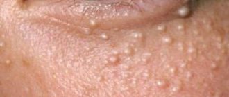
Environmental factors include poor ecology, smoking, working in conditions unfavorable for the eyes, that is, in the wind, in the sun, in air-conditioned rooms, etc. This group also includes one of the most common causes of dry keratitis today - long-term working on a computer. When a person looks at the monitor for a long time, he blinks less, as a result of which the tear film is not renewed, and the conjunctiva and cornea dry out.
A similar effect can be caused by contact lenses if used incorrectly. Modern models are made from soft materials with hydrophilic properties. They are worn even with dry eye syndrome. If contact vision correction products are selected incorrectly or are used for 14-16 hours a day without the use of moisturizing drops, keratoconjunctivitis sicca may develop.
In some people, it occurs after laser surgery to correct refractive errors. During the procedure, the doctor uses a laser to vaporize the tissue of the patient's eyeball, which can cause dry keratitis. This outcome is usually predicted.
Infectious eye diseases can provoke dry keratitis:
- conjunctivitis;
- dacryocystitis;
- blepharitis;
- iridocyclitis;
- keratitis;
- episcleritis;
- uveitis, etc.
In addition, dry keratitis can be a consequence of ophthalmopathologies of an aseptic nature. These include keratoconus, keratopathy, flabby eyelid syndrome, corneal ulcer, glaucoma, trichiasis, etc. If these diseases are present, treatment of keratoconjunctivitis sicca can be complicated, since you will first have to get rid of the primary pathology.
The fourth group of factors are non-ophthalmological diseases. Systemic diseases can cause dry eye syndrome:
- hypertension;
- allergy;
- diabetes;
- ARVI and influenza;
- lupus erythematosus.
In addition, autoimmune diseases and hormonal changes lead to dry keratitis.
Viral keratoconjunctivitis
We cannot ignore the disease of this form. Treatment of viral keratoconjunctivitis is aimed at eliminating the cause that caused it. So the doctor prescribes antibiotics and broad-spectrum drops. Only these drugs are capable of influencing a large number of bacteria known to science.
If a patient is diagnosed with a severe disease that is also progressing, then parenteral antibiotics are prescribed.
In parallel, it is necessary to use drugs that can protect the normal microflora of the intestines and other organs. Because with such treatment, against the background of changes occurring in it, the risk of developing fungal diseases and dysbacteriosis begins to increase.
As a rule, the elimination of symptoms and treatment of keratoconjunctivitis in adults is carried out with Tobrex and Sofradex drops. Acyclovir is also used. This drug prevents the infection from becoming chronic.
Symptoms
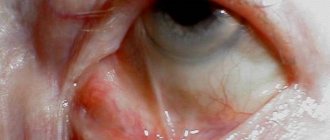
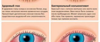
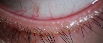
The main symptoms of keratoconjunctivitis sicca are redness and irritation. However, he can progress. Doctors distinguish three stages of the disease. In the first, superficial, hyperemia is observed on the periphery of the conjunctival sac. Further development of the disease leads to the second stage, ciliary, in which a bright red border forms around the junction of the cornea and sclera. It indicates that inflammation begins inside the eye. The mixed, third, stage is accompanied by even greater redness and damage to the inner membranes of the eyeball.
These are just some of the symptoms. Typically, patients complain of itching, stinging, and scratching sensations. It seems to them that there is a foreign body on the conjunctiva, and a feeling of “sand” arises. Some patients have eyes that hurt and get tired quickly, especially when working at a computer.
There may also be such a seemingly unusual symptom for dry keratitis as increased lacrimation. It is a consequence of a protective reaction of the body, which produces more tear fluid to moisten the surface of the eyeball. But it is very watery, and therefore does not lubricate the conjunctiva and does not alleviate the condition, but, on the contrary, causes even more discomfort. Moreover, a person begins to rub his eyes and introduces germs into them, as a result of which the risk of infection of the eyelids or eye tissue increases.
Symptoms of dry keratoconjunctivitis increase with visual strain, so it is better not to read, not sit at the computer, or use the phone. Also, symptoms intensify when a person is in the wind, in a dry room, uses a hairdryer, etc.
If two or more of the listed symptoms do not go away within a few days, you should definitely visit an ophthalmologist.
Rickettsial (infectious) keratoconjunctivitis
Rickettsial keratoconjunctivitis (Keratoconjunctivitis rickettsiosa), rickettsial conjunctivitis, general keratitis, ophthalmia, general eye disease, is an acute infectious disease of animals, mainly cattle, caused by rickettsia and accompanied by fever, catarrhal conjunctivitis and purulent-ulcerative keratitis.
Historical reference . Rickettsial keratoconjunctivitis was first described by Coles in South Africa in 1931. In 1937, A. Donatien and F. Lestocard discovered this disease in Algeria, Cordier and Mengiers in Tunisia. In the USSR, rickettsial keratoconjunctivitis was described by G.F. Panin (1953), K.A. Dorofeev (1954), V.P. Gromov, I.P. Vershinin et al. (1963), M.V. Plakhotin (1966), A.A. Kadyrov (1969). Rickettsial keratoconjunctivitis is recorded in Africa, America, Asia, and Europe.
The disease in individual farms can cause great economic damage due to the loss of vision by animals.
Etiology . The causative agent of the disease is Ricolesia bovis (Rickettsia conjunctivae) - a small polymorphic (cocco-, disc- and rod-shaped) microorganism 0.5-1.0 microns in size, cultivated in the yolk sac of 6-7-day-old chicken embryos. It is stained blue according to Romanovsky. The causative agent of rickettsial keratoconjunctivitis is poorly resistant to environmental factors and chemicals. At room temperature it dies within 24 hours. In a 0.85% sodium chloride solution at a temperature of 20-22°C, the microbe retains its pathogenic properties for 24 hours. On sterile sheep wool, the pathogen dies after 96 hours; a 5% solution of collargol inactivates rickettsia in 15 minutes.
Epizootology . Large and small cattle, camels, pigs and poultry are susceptible to rickettsial keratoconjunctivitis. In this case, calves aged 5 months and older are most sensitive. up to 1.5 years and lambs over 15 days of age. The source of the infectious agent is sick animals and rickettsia carriers, which secrete it with conjunctival secretions and mucus from the nose. The main route of transmission of the disease is airborne droplets. Mechanical transmission of the pathogen by flies (Musca domestica, Stomoxys calcitrans) is possible. The disease is characterized by exceptionally rapid spread; the disease spreads especially quickly when owners keep their animals in large groups. Rickettsial keratoconjunctivitis is recorded at all times of the year, but most often in spring and summer. The disease is characterized by its stationarity. The degree of damage to animals is negatively affected by poor zoohygienic conditions of their keeping and a lack of vitamin A in the diet.
Pathogenesis . A.A. Kadyrov found that after injecting a suspension from the diseased conjunctiva of calves into the anterior chamber of the eye of rabbits, 90% of the animals became ill within 2-4 days. When post-mortem examination of infected rabbits occurs on the 20-30th day, A.A. Kadyrov found catarrhal pneumonia in rabbits, from which he came to the conclusion that keratoconjunctivitis is a general disease of the body.
Immunity . After contracting rickettsial keratoconjunctivitis, the animal becomes immune to this disease for a period of one year.
Clinical picture . The incubation period (latent period) is from 2 to 12 days. The course of the disease can be acute and subacute. The main symptom of the disease is conjunctivitis. Most often, one eye is affected. During a clinical examination of a sick animal, a veterinarian notes discharge from the diseased eye, the eyelids become swollen, and the animal has a strong reaction to light (photophobia). When examining the surface of the edematous conjunctiva, we note fine granularity. In the 3rd century, abundant granulations develop. The inflammatory process that has begun can spread to the cornea, causing keratitis, ulcerations, and iridocyclitis. When examined, the cornea is cloudy, has a yellowish tint, and an abscess may form in it. The body temperature of a sick animal is increased, the condition is depressed, and appetite decreases. After some time, the abscess opens and an ulcer forms. Mucous-purulent discharge comes from the affected eye. After 8-10 days the animal usually recovers, but in some animals the disease can last 20-35 days. After recovery, the sick animal is left with a thorn in the eye.
Diagnosis . The diagnosis is made by veterinary specialists based on the analysis of epizootic, clinical data and the results of laboratory tests of smears - scrapings from the surface of the damaged conjunctiva. At the beginning and middle of the disease, erythrocytes, many polymorphonuclear neutrophils and epithelial cells containing the pathogen are found in smears. As the inflammatory process weakens, the number of neutrophils decreases and they are replaced by lymphocytes; At the same time, the amount of pathogen in epithelial cells decreases. Rickettsiae are most often found on the 2-5th day of illness, but they cannot be detected in recovered animals.
Differential diagnosis . Veterinary specialists must differentiate rickettsial keratoconjunctivitis from keratitis due to chlamydia, thelaziosis, pasteurellosis, smallpox, as well as keratitis caused by traumatic eye injuries.
Treatment . Owners should isolate sick animals in a dark room and carry out treatment. When treating sick animals, a 5% solution of collargol, 3% chloramphenicol, aureomycin and penicillin ointments, and a 0.1% solution of zinc sulfate are used. The use of syntomycin emulsion with dibiomycin ointment promotes healing of affected eyes on days 9-24. To resolve the resulting opacities of the cornea, practical veterinary specialists successfully use 10% ointment or calomel powder (half and half with sugar) and 1% ointment or hydrocortisone solution. For keratoconjunctivitis in cows and sheep, furazolidone powder in the form of an aerosol is effective. In severe cases of the disease, novocaine blockade is used.
For the purpose of prophylaxis in disadvantaged herds, conditionally healthy animals are injected into the conjunctival sac of both eyes (once a week) with dibiomycin ointment, syntomycin emulsion or powder according to the prescription: biomycin, sulfantrol, syntomycin (equal parts). The powder is blown into the eyes of animals 4-5 times for 1.5 months. Simultaneously with the preventive treatment of animals, livestock premises are disinfected once a week with a 0.1% chlorophos emulsion. Disinsection is carried out periodically. In farms unaffected by this disease, veterinary specialists examine animals for this disease monthly in winter and daily in summer.
Diagnostics
To identify dry keratoconjunctivitis in a person, a doctor needs only an external examination or a Schirmer test. It is carried out as follows: the ophthalmologist places a thin strip of filter paper on the edge of the eyelid of the subject. By how wet it is, a specialist can determine how many tears are produced by the glands and what their composition is.
Next, you need to determine the etiology and stage of the disease. This is done using ophthalmoscopy or slit-lamp biomicroscopy. Be sure to check intraocular pressure, visual acuity and oculomotor activity. If infection is suspected, a bacteriological analysis is performed. In rare cases, gonioscopy, ultrasound and MRI are required. If dry keratoconjunctivitis develops against the background of systemic pathology, the patient is sent for consultation to an appropriate specialist. After collecting all the information, treatment is prescribed.
Diagnosis of the disease
The disease is identified by the attending physician (preferably an ophthalmologist) during an examination and announces it as a preliminary diagnosis. This is facilitated by the presence of the following symptoms:
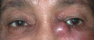
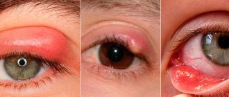
- Localized excess fluid accumulation in the eyelids.
- The presence of discharge from the eyes of a varied nature.
- Detection of the growth of lymph nodes, for the detection of which the neck area is palpated.
- Diagnosis of acute or chronic inflammatory process with the development of infection that affects the mucous membrane.
- The presence of a set of manifestations of conjunctivitis with blood overflowing the vessels of the circulatory system of the mucous membrane.
In the laboratory, to confirm the correctness of the diagnosis, the following studies are prescribed:
- A method in which specific Ags are detected using Abs conjugated to a fluorochrome. This is one of the ways to determine the presence of viral antigens at the initial stages. To do this, smears are taken from the mucous membrane of the conjunctiva.
- RSK (Complement Fixation Test) and ELISA (Enzyme Immunoassay) are common serological reactions that detect the titer of antibodies in the blood to adenoviruses. For the majority of infections, this indicator becomes informative when the increase occurs no less than four times. However, this technique cannot be classified as urgent or quickly feasible.
- PCR (Polymerase Chain Reaction). This method allows you to identify the adenovirus DNA molecule, if any, in the scraping. This technique is very expensive and is carried out only in specialized laboratories.
- A bacteriological study in which the growth of adenovirus is obtained in a special medium for identification. The method cannot be called fast.
We must not forget about the similarity of the course of adenoviral conjunctivitis with bacterial inflammation of the eyes. This means that it is impossible to exclude the possibility of prescribing the wrong course of treatment, which can provoke quite serious complications.
Very often, there is a risk of the process moving to the anterior most convex transparent part of the eyeball. The consequence of this may be its defeat, and this, in turn, can lead to serious visual impairment.
Keratoconjunctivitis sicca: treatment
Keratoconjunctivitis sicca can be treated with medications, physical therapy, and surgery. The method is chosen by the doctor based on the symptoms and the presence of concomitant pathologies. The choice of drugs is determined by etiology. For allergies, antihistamines are prescribed, for a bacterial infection - antibacterial drops or antibiotics, in case of severe inflammation - non-steroidal or steroidal medications.
Regardless of the root cause of dry keratitis, moisturizing solutions are always used to treat it. They help to quickly relieve its symptoms: redness, irritation, lacrimation, etc. Drops such as “Artificial tear”, “Systane”, “Systane Ultra” or “Oxial” are used as moisturizers. These drugs do not contain preservatives. They are based on components that imitate natural human tears, so they do not cause allergies or other side effects. Drops can be instilled several times a day. Some people try to relieve the symptoms of dryness with folk remedies. We'll talk about their effectiveness later.
Usually, with dry keratitis, you have to abandon contact correction products. But the doctor may prescribe therapeutic soft lenses if conservative therapy does not bring results and you need to protect the cornea from erosion and ulcers. Physiotherapy can also be used.
Physiotherapeutic treatment of keratoconjunctivitis consists of laser stimulation of tears and irradiation of the conjunctival fornix with a helium-neon laser. After a course of 5-7 procedures, stabilization of the tear film is observed. In parallel, the patient continues to be given drops and other treatments.
In rare cases, keratoconjunctivitis must be treated surgically. The following types of operations are used:
- Obstruction of lacrimal openings (canaliculi) - blocking the outflow of tears. It lingers on the surface of the eye and moistens it.
- Tarsorrhaphy is the formation of artificial ptosis aimed at narrowing the palpebral fissure. This helps reduce the rate of evaporation of tear fluid.
- Conjunctival plastic surgery according to Kunt - creating a flap of the conjunctiva to cover ulcers on the cornea.
- Gland transplantation. The sublingual salivary glands are transplanted into the conjunctival cavity. The patient's relatives can become donors.
These are the main ways to combat dry eye syndrome. Surgical methods are prescribed very rarely, only in cases of complete lack of effect from therapy and the risk of complications. Let's find out whether dry keratoconjunctivitis is treated with folk remedies.
Epidemic keratoconjunctivitis
In the case of this form of disease, therapy turns out to be very problematic. Talking about the symptoms and treatment of this type of keratoconjunctivitis, it should be noted that there are no drugs yet that have a selective effect on adenoviruses. Therefore, therapy is fraught with difficulties.
As a rule, broad-spectrum medications are used. These are interferons (ophthalmoferon and lokferon) and their inducers, installations 6-8 times a day. If the stage is acute, then you additionally need to take antihistamines and take antiallergic drops, for example, “Spersallerg” or “Allergoftal”.
In the subacute form, Lecrolin and Alomide drops are used. If films have formed, you will need to take corticosteroids - Maxidex, Dexapos and Oftan-Dexamethasone. When the cornea is damaged, “Coperegel”, “Vitasik”, “Korpozin”, “Taufon” help.
Treatment of keratoconjunctivitis with folk remedies
Doctors recommend not using folk remedies without examination. Moreover, they should not become an alternative to drug treatment. It is better not to resort to their help at all. Usually, supporters of traditional medicine make eye lotions from tea leaves, aloe, chamomile, etc. At best, they will not bring any benefit, at worst, they will lead to a worsening of the condition.
The fact is that dry keratitis can be accompanied by a bacterial or fungal infection. It is impossible to cure them with compresses. Moreover, warm lotions from various decoctions and infusions promote the growth of bacteria. If necessary, the doctor will prescribe folk remedies as an addition to the medication.
Treatment
A good preliminary study makes it possible to accurately diagnose and prescribe treatment. Almost always, when there are disturbances in the functioning of the glandular system, the doctor prescribes the use of certain drops. The list of them is extensive.
At the first stage, when the disease is not advanced, the doctor prescribes a regular tear substitute:
- Artificial tear;
- Natural tear;
- Oksial;
- Hyphenation.
Doctors also often prescribe moisturizing gels:
- Systane;
- Otagel.
The selection of drops should be taken especially seriously in cases where the manifestation of the disease is associated with infection, injury or disruption of the glands. Treatment at home is possible, but only with the medications prescribed to you by a qualified ophthalmologist.
Forecast
Keratoconjunctivitis can be treated quite quickly if you immediately consult an ophthalmologist. Within a few days the symptoms will disappear and the condition will noticeably improve. But the disease will recur if it is not prevented. You need to carefully monitor hygiene, do eye exercises, work at the computer with breaks every 40-50 minutes, use moisturizing drops, watch less TV and play computer games, etc.
Dry keratitis rarely causes complications. This often occurs due to the lack of therapy, the use of only folk remedies, or weak immunity. Hypothetically, dry eye syndrome can cause weakened vision or even blindness if a complication such as panophthalmitis occurs, but this almost never occurs in practice.
Vernal keratoconjunctivitis
As a rule, this disease occurs among boys 4-10 years old. Treatment of vernal keratoconjunctivitis primarily involves minimizing exposure to ultraviolet radiation to the eyes. Therefore, it is strongly recommended to wear sunglasses and not be outside during daylight hours.
The use of antihistamines, as well as mast cell stabilizers, is indicated. Sodium cromoglicate in the form of drops and Olopatadine are excellent. But this needs to be done systematically. Long-term use of these medications will help avoid exacerbation.
To reduce itching, you will have to use a 3% sodium bicarbonate solution. You can also make lotions from a solution of boric acid.
Complications
It is worth noting! Lack of appropriate treatment for the syndrome can lead to serious consequences:
- erosions and ulcers on the surface , with further formation of scars. This has a negative impact on the quality of vision;
- disruption of the process of self-cleaning of the eye from external contaminants . As a result, inflammatory processes develop: keratoconjunctivitis, keratitis.
In rare cases, the disease can lead to significant deterioration or partial loss of vision.