Development of glaucoma
The development of glaucoma can be different, depending on the type of disease; it can be primary, secondary, congenital. The pathogenesis of the development of this disease is very complex and not fully understood. It can be latent for a long time, that is, it can be hidden and debut with an acute attack, or it can gradually develop with an increase in symptoms.
It is often a slowly progressive eye condition that damages the optic nerve, resulting in loss of permanent vision. She has been called the "silent thief of sight" because she can slowly steal sight. By the time people become aware of vision problems, it is usually too late.
This is why annual eye examinations with an ophthalmologist are of great benefit, especially for at-risk groups.
Everyone is at risk from the disease, from infants to the elderly. Older adults are at higher risk, but children can be born with glaucoma (statistics show it affects about 1 in every 10,000 children born in the United States). Young people may also be susceptible to this disease. African Americans, in particular, are susceptible at a younger age.
The development of the disease in old age is associated with impaired formation and outflow of intraocular fluid, which can subsequently lead to its stagnation and increased IOP, which leads to optic nerve dystrophy. Senile glaucoma develops for the following reasons:
- for diabetes mellitus;
- for hypertension;
- with swelling cataracts;
- for problems of various types with blood circulation.
Most often, older people develop open glaucoma, less often - closed glaucoma, and even less often a special type of glaucoma develops - pigmentary glaucoma, more often in men.
Acquired glaucoma
It is characterized by the fact that its formation is provoked by both external factors and some internal pathologies formed during the formation and growth of the human body. Acquired glaucoma also includes two types:
- Primary;
- Secondary.
Primary glaucoma
How does fluid stagnation occur in glaucoma?
It is characterized by the fact that with it, processes leading to degeneration of a number of ocular tissues occur before the onset of pathology and, with further progression, are only the cause of increased ophthalmotonus.
Deformations of the visual apparatus, in this case, are a consequence of a number of diseases preceding the disease. However, they may not be the triggering mechanism.
Pathological eye damage develops as a result of ischemia of the outer part of the eye, which develops dystrophic processes in the trabecular apparatus and is characterized by an abnormal IOP state (possibly high without changes or spasmodic manifestation). With this type of disease, distortions in the visual field are possible, and the optic nerve tissue atrophies. These symptoms are accompanied by deterioration of the central part of vision (the person's vision becomes clouded), the final result may be blindness.
Ophthalmologists have adopted a general classification of primary glaucoma. The basis of the classification is the shape of the anterior chamber angle.
There are two forms of glaucoma:
- Open angle;
- Closed angle.
Open angle glaucoma
Primary open-angle glaucoma is divided into several forms according to nosological characteristics:
- Primary open angle. People over forty years of age are most susceptible to it. Pathogenesis – pressure inside the eyes is higher than established standards. Its manifestation leads to deformation of the optic disc, retina, and disruption of the functioning of the visual apparatus.
- Pseudoexfoliative open angle. Elderly people can get this type of infection. Signs include deposits of exfoliative material in the external compartment of the organs of vision, trabeculopathy, canalicular blocks, and high intraocular pressure.
- Pigmented. It usually occurs in young people and middle-aged people. With this form, a deep external chamber, a large angle of the anterior canal, retraction of the root processes of the iris, disruption of pigmentary processes, and deposition of pigmentary components are observed.
- Low (normal) pressure. The incidence of this pathology is especially high among people over 30 years of age. With it, intraocular pressure is normal, only the individual level is lower than the established reference value. The pathology can develop together with hypertension and vascular dystonia.
Angle-closure glaucoma
Among the closed-angle forms there are:
- Angle-closure glaucoma with pupillary block. It can occur in middle-aged and elderly people. It is expressed by manifestations characteristic of acute, subacute attacks, and subsequently develops into a chronic form. Hyperopia (farsightedness) is considered to be a characteristic precursor of the pathology. Pupillary block occurs due to a slight increase in pupil size.
- Angle-closure glaucoma with flat iris. Initially, it is expressed by rare manifestations, turning into a state of chronic disease. Provoking factors are: increased size of the roots of the iris; the ciliary crown and the base of the iris are placed in the anterior positions. Pathology appears due to the blocking of the angle of the outer chamber by a thick fold of the iris of the dilated pupil.
- "Creeping" angle-closure glaucoma. Its course is chronic, with the absence of any short-term manifestations. The periphery of the iris and the outer wall of the anterior chamber angle grow together at a low speed.
- Closed angle with glass lens block. It can be caused by increased size of the roots of the iris. Severe anatomical deformations are observed.
Secondary glaucoma
A type of disease that can be triggered by a large number of mechanisms that are disrupted during the development of the body (pathogenesis). Based on these pathogenesis, it can be divided into:
- uveal post-inflammatory;
- phacogenic;
- vascular;
- dystrophic;
- traumatic;
- postoperative;
- neoplastic.
Uveal post-inflammatory glaucoma
Possible with the appearance of keratitis. The course of the disease can be open-angle or closed-angle. It may affect the episcleral vessels and ocular drainage system.
Phacogenic glaucoma
One type of secondary glaucoma is divided into several forms:
- Phacotopic (caused by degenerative processes) is formed when the lens dislocates into the vitreous or outer chamber of the eye, retinal detachment, uveopathy, phakomatosis.
- Phacomorphic - the result of an increase in lens tissue. Due to the increased size of the lens, pupillary block occurs. Accompanied by acute and subacute attacks.
- Phacolytic often occurs with cataracts. Complex organic structures are removed through the deformed outer capsule, which leads to blockage of the trabecular filter. The pathology manifests itself in attacks of the form
Vascular glaucoma
It is divided into:
- Neovascular – a consequence of side effects manifested after retinal disease, characterized by insufficient oxygen supply. Due to the formation of new fibrovascular fibers, incomplete obliteration of the angle of the external chamber is formed. Possible swelling of the cornea, hemorrhages inside the eye, rubeosis of the iris.
- Phlebohypertensive – expressed as the consequences of increased pressure in the venous network of the ocular apparatus. Identified by a highly dilated and tortuous venous network.
Dystrophic glaucoma
Pathologies of a dystrophic nature play a decisive role in the development of this form. These include:
- iridocorneal endothelial syndrome;
- severe hemorrhages inside the eyes;
- retinal detachment and, as a result, ocular hypertension;
- primary systemic amyloidosis.
Traumatic glaucoma
Caused by mechanical, chemical and radiation injuries to the eyes. The causative mechanisms of the disease are:
- intraocular hemorrhages;
- violation of the integrity of the anterior chamber angle;
- blockage of the drainage network of the eyeballs.
The disease can manifest itself an indefinite time after traumatic exposure.
Postoperative glaucoma
After surgery, a complication may occur in the form of a temporary or permanent increase in IOP. The most common causes of this type of glaucoma include operations:
- corneal transplantation (keratoplasty);
- for cataract extraction;
- with retinal detachment;
This pathology can be of both open-angle and closed-angle forms. Sometimes secondary malignant glaucoma with vitreolens block can develop.
Neoplastic glaucoma
It is the result of complications of orbital and intraocular formations. The main reason is tumor decomposition products blocking the angle of the anterior chamber.
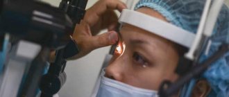
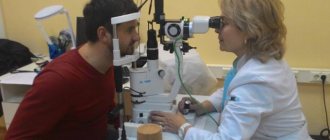
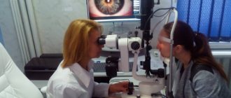
Diagnosis of glaucoma
If you are at high risk of developing glaucoma, you should be examined at least once every two years. To make a diagnosis, an ophthalmologist or optometrist will perform a comprehensive eye examination, which may include the following diagnostic steps:
- Visiometry – testing visual acuity.
- Tonometry according to Maklakov and Goldman consists of measuring the pressure inside the eye using a special tonometer instrument, which measures pressure indirectly, that is, the strength of the eye’s response to the force that was applied to it is measured.
- Examination of the pupil, which includes: reaction to light, condition of the pigment border, presence of exfoliation. This examination is carried out using biomicroscopy.
- Gonioscopy: examination of the anterior chamber angle, determination of the width of the angle, areas accessible to inspection, and the presence of pigmentation.
- Ophthalmoscopy: examination of the optic nerve and retinal nerve fibers (the fundus of the eye is examined with an ophthalmoscope)
- Static perimetry: assessment of changes in visual fields, makes it possible to assess visual fields.
- Assessment of eye hydrodynamics (tonography): state of formation and outflow of intraocular fluid, Becker coefficient.
Also, desirable, but not mandatory, research methods include:
- dynamic perimetry;
- keratopachymetry (the thickness of the central zone of the cornea is determined);
- ultrasound biometry (determining the topography of structures and topometry);
- optical coherence tomography, MRI.
Differential diagnosis is also important when making a diagnosis.
After conducting all the research and testing data, a diagnosis of glaucoma can be formulated. Early diagnosis is the key to successful treatment.
Treatment methods for terminal glaucoma
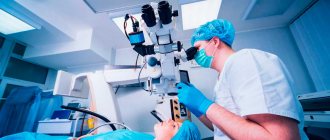
At this stage of the disease, laser therapy and surgery are used for treatment.
If the patient has the first symptoms of the disease, you should not try to cure him on your own, but urgently need to contact a specialist. The doctor will collect complaints, conduct an eye examination and prescribe special treatment methods. To cure terminal glaucoma, patients are prescribed drug therapy, surgical treatment methods and folk remedies. Each patient is prescribed a specially selected dietary diet.
Drug treatment
Patients with absolute glaucoma are prescribed the drugs listed in the table:
| Group | Name |
| Cholinomimetics | "Pilocarpine", "Fotil" |
| Sympathomimetics | "Tyramine", "Ephedrine", "Clonidine" |
| Prostaglandins | "Xalatamax", "Glauprost", "Xalatan" |
| Adrenergic blockers | "Arutimol", "Ocumed" |
| Carbonic anhydrase inhibitors | "Azopt", "Trusopt" |
| Antihypertensive drops | "Mexidol" |
| Combination drugs | "Cosopt", "Xalacom", "Azarga" |
Surgical methods of treating absolute glaucoma
Laser treatment is widely used in the treatment of glaucoma.
Laser methods are widely used. These include traction laser operations, transscleral laser cyclocoagulation, laser iridotomy, peripheral iridoplasty and papilloplasty. All these operations are based on the use of a laser beam, with which it is possible to perform surgery without cutting the walls of the eye. This operation is not painful and is performed quickly.
Features of operations:
- Traction laser operations are performed in the area of the trabecular network of the corners of the anterior chambers of the eyes. They are based on the effect of a laser coagulant in the trabecular area, which leads to improved outflow of intraocular fluid.
- Transscleral laser cyclocoagulation is based on thermal destruction of part of the ciliary body. As a result, the production of aqueous humor and the pressure inside the eyes are reduced.
- Laser iridotomy is used as an additional surgical intervention after intraocular surgery. It is not used if there is swelling or clouding of the cornea, or if the patient has a shallow anterior chamber of the eye.
- Peripheral iridoplasty and papilloplasty are based on the fact that light laser coagulants are applied to the periphery of the iris. As a result, the angle of the anterior chamber expands.
Other surgical methods
The following methods are also used:
- Diathermocoagulation and cryopexy of the ciliary body. With the help of these operations, the production of aqueous humor is reduced, eye pain is reduced and intraocular pressure is reduced.
- Optociliary neurectomy. It allows you to relieve pain in a short time, reduce intraocular pressure and preserve the eyeball.
- Removal of the eye if organ-preserving operations are not possible.
Causes of glaucoma
Most often, the formation of glaucoma is the result of exposure to high fluid pressure in the eye. This occurs when the fluid in the anterior chamber does not circulate properly.
Typically, fluid, called aqueous humor, drains from the eye through the trabecular meshwork. If this channel is blocked, the latter accumulates. The cause of this process is unknown, but it is known that it is transmitted from parents to children and is genetically determined, and if the family history is burdened with glaucoma, then you need to be wary and regularly examined for its presence.
Less common causes include blunt trauma or chemical damage to the eye, severe eye infection, and blocked blood vessels inside the eye. Quite rare, but sometimes the cause may be eye surgery. Also, many scientists believe that psychosomatics influences the development of this disease, since everything in the human body is interconnected, and the burden of stress negatively affects eye health.
We recommend the article: Causes of glaucoma
What kind of disease is this?
Glaucoma is a whole group of chronic eye diseases (about 60), which are characterized by:
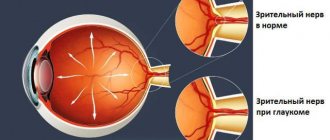
- disruption of the normal circulation of aqueous humor in the chambers of the eye and ophthalmic hypertension (increased IOP - intraocular pressure);
- gradual loss of vision from the periphery to the center;
- tunnel vision syndrome;
- blindness due to optic nerve atrophy.
Translated from ancient Greek, “glaucoma” means “green water.” This pathology name is given because during an attack the pupil dilates and acquires a greenish tint.
The eye has two chambers - the anterior one, in which the outflow of intraocular fluid (IOH) occurs, and the posterior one, in which aqueous humor is produced by ciliary cells from the blood. Between them are the iris and lens.
Normally, the liquid constantly circulates through 2 chambers, renewed and maintained in a certain volume by the hydration system. But if an obstacle to outflow appears on its way and it becomes difficult, then the pressure rises. As the process progresses, it compresses the fibers of the optic nerve (glaucoma neuropathy), ending in its atrophy. Signals from the eyes no longer reach the brain, and this means blindness.
Important! The insidiousness of the pathology is its asymptomaticity at all stages except the terminal stage. The vast majority of cases are primary open-angle glaucoma (POAG).
Causes
Today there are no common ideas about the causes of pathology. Only risk factors have been identified - general and local.
Local ones include:
- refractive errors (with OAG this is myopia);
- frequent excessive visual stress;
- thinning of the cornea.
General risk factors:
- heredity;
- endocrinopathies – thyroid disease and diabetes;
- age after 40;
- head and eye injuries;
- overripe cataract;
- long-term use of corticosteroids (their suppression of immunity causes metabolic disorders);
- Physical inactivity and obesity are of some importance.
In the body, after 40, aging processes are activated and this can also cause changes in IOP. Ignoring an examination by an ophthalmologist at this age increases the risk of glaucoma significantly.
Glaucoma can be considered a vascular disease, because any circulatory disorders - local and general - can also be considered risk factors for it. Patients with atherosclerosis, hyper-, dys-, and hypotension are at risk.
The more factors, the higher the risk of glaucoma. There may be many reasons, but the outcome is always the same - loss of vision.
Useful video
Open Angle Glaucoma - 5 Facts about Open Angle Glaucoma:
Forms
The classification of pathology is complex and extensive. Glaucoma happens:
- Congenital (very rare, occurs in the embryo as a result of dysgenesis of the anterior chamber angle of the eye).
- Secondary or symptomatic (occurs against the background of various other eye diseases, injuries, tumors and lack of insulin).
- Primary open angle (POAG) is the most common type. Women over 40 are most susceptible to it. It accounts for more than 90% of all cases of glaucoma. The essence of its occurrence is the appearance of an obstacle to the outflow of fluid, its accumulation and a gradual increase in intraocular pressure.
- Angle-closure glaucoma is less common. More typical after 30 years with hypermetropia. Its course is often paroxysmal. With this form, the deeper layers of the eye are damaged.
Symptoms and signs of glaucoma
There are different forms of glaucoma, the most common being primary open-angle and closed-angle (malignant). Open-angle glaucoma is often called the “silent thief of vision” because it may not cause any symptoms until significant and irreversible vision loss has already occurred. The symptoms of these forms are different.
Symptoms of open angle glaucoma. Usually there are no early signs and symptoms that would warn of the development of the disease; it is compensated for a long time. It develops slowly, and sometimes without noticeable loss for many years. Most people with open-angle glaucoma do not see changes in their vision at first glance because peripheral vision loss occurs first and visual acuity is maintained at normal levels until the end. By the time the patient notices visual disturbances, the disease is usually quite advanced and already decompensated.
Symptoms that force a patient to see an ophthalmologist are:
- the appearance of rainbow circles that appear when the gaze is fixed on direct rays of light;
- decreased accommodative ability;
- the appearance of fog and flickering before the eyes;
- When IOP is exceeded, patients complain of a very severe headache that radiates to the eyes and brow ridges.
Vision loss is irreversible with any treatment, even with the most modern surgical interventions.
Because there are several signs that warn of the presence of open-angle glaucoma before permanent vision loss occurs, it is important to see your doctor for regular eye exams. If the disease is detected during a preventive examination, vision loss can be prevented.
Symptoms of an acute attack of glaucoma:
- blurred vision;
- the formation of rainbow circles that surround the lights of light;
- acute pain in the eyes and severe headaches;
- severe nausea and vomiting that accompany severe pain in the head;
- sudden loss of vision.
The symptoms of angle-closure glaucoma are usually very noticeable, and vision damage occurs very quickly. This form is very malignant. If any of these symptoms occur, you should immediately seek help from an ophthalmologist.
Related publication: Symptoms and signs of glaucoma
Symptoms of open angle glaucoma
The main problem with the pathology is damage to the optic nerve. The pathophysiology is not fully understood, but there is a progressive loss of retinal ganglion cells and axons. In the early stages it affects only the peripheral field of vision, but as it progresses it affects central vision, leading to loss of acuity, which can lead to severe impairment and complete loss of visual perception.
For most OAG, optic neuropathy is associated with elevated IOP. This has given rise to the hypothesis of retinal ganglion apoptosis, the rate of which is influenced by hydrostatic pressure on the optic nerve head. Optic neuropathy results in characteristic changes in the optic disc and loss of visual field.
In the open-angle form, the outflow through the trabecular meshwork worsens (the role of which is to absorb aqueous humor). This is a chronic degenerative obstruction that is painless.
IOP is not always elevated.
Early primary symptoms of open-angle glaucoma are rare. Typically, the patient becomes aware of visual field loss when optic nerve atrophy occurs. Signs of OAG:
- The head of the optic nerve (disc) is a slightly vertically elongated circle with a central depression called the cup. The sensorineural rim is the tissue between the rim of the cup and the rim of the disc and consists of the axons of retinal ganglion cells. Characteristic changes in the optic nerve include an enlarged cup, thinning or tearing of the neurosensory rim, and hemorrhage.
- Changes in the visual field caused by damage to the optic nerve are manifested by arcuate and paracentral scotomas. Problems with twilight vision and rainbow glare appear.
- Discomfort, pain in the eyes due to increased IOP and redness periodically occur in the eyes.
Forms of glaucoma
There are two main types: open-angle (the most common form, affecting approximately 70-90% of people with glaucoma) and closed-angle. There are also several other common forms of this eye disease, including normal-tension, chronic, congenital, secondary, pigmentary, and exfoliative glaucoma.
Open-angle glaucoma is the most common form, accounting for 70-90 percent of all cases. Often in the early stages there are no noticeable symptoms, and high IOP is the most significant risk factor or indicates that the disease is already developing.
This form of glaucoma is chronic and progresses slowly. Many factors, including age, structural defects, damage to the trabecular meshwork and/or other drainage systems in the eye, overproduction of aqueous humor or blockage of aqueous humor, can cause increased IOP.
Open-angle glaucoma most often occurs with age, in people with a genetic predisposition, and its development is also influenced by the environment and lifestyle. As we age, the cells of the trabecular meshwork may not function as well as before or may become smaller, so this type is more likely to develop in older people and is more likely to develop after age 40. Affects African Americans and Hispanics at higher rates than other ethnic groups.
Open-angle glaucoma is also divided into several more subtypes.
Chronic
Chronic open-angle glaucoma is a condition in which the optic nerve at the back of the eye is damaged. This is usually caused by increased pressure in the eye. If left untreated, the disease can lead to visual loss and even complete loss of vision, as this form of glaucoma is steadily progressive. Treatment can slow progression and help prevent these effects. All adults over the age of 35-40 should have their eyes checked regularly, including measuring their eye pressure. Older people, after 40 years, are most prone to this type of glaucoma.
Normal pressure glaucoma (normotensive)
In normal-tension glaucoma, the optic nerve is damaged, although the pressure in the eye is not very high. Doctors don't know why some people have optic nerve damage even if they have near-normal blood pressure levels. Normal pressure glaucoma is different from other forms. Most types of glaucoma are associated with increased intraocular pressure. In general, normal blood pressure ranges from 12 to 22 mmHg. Art. The causes of normal pressure glaucoma are still unknown. For some reason, the optic nerve is sensitive to damage even at normal pressure. Researchers continue to study why some optic nerves are damaged at these relatively low IOP levels. There is an increased risk of developing this form in people who have a family history of normal-tension glaucoma, people of Japanese ethnicity, and people with heart conditions such as irregular heart rhythms.
Congenital glaucoma
By definition, congenital glaucoma is present at birth in newborns, but its manifestations may not be recognized until infancy (may occur in infants) or early childhood. In adolescents, this disease is usually already diagnosed, because if there is no adequate treatment, children become blind by the age of 5-7 years. It is characterized by abnormal development of the ocular outflow system, which leads to an increase in IOP and subsequent damage to the ocular structures, which leads to loss of vision. Although the disease is relatively rare, the effects on the development of the visual system can be extremely damaging. Early recognition and appropriate treatment of glaucoma can significantly improve a child's vision. Occurs from birth to 3 years of age. If the disease appears after 3 or 4 years, it is called primary juvenile open-angle glaucoma.
Secondary glaucoma (acquired)
Secondary glaucoma is any form of glaucoma in which there is a known cause of increased pressure, resulting in significant damage to the optic nerve, which in turn leads to vision loss. Like primary glaucoma, secondary glaucoma can be either open-angle or closed-angle, and it can occur in one or both eyes. Secondary glaucoma can be caused by eye injury, inflammation (in which case glaucoma is inflammatory secondary), certain medications such as steroids, and common cases of cataracts or diabetes (diabetic glaucoma). The choice of treatment depends on the underlying cause, but usually includes medications, laser, or conventional surgery. The neovascular form of the disease is caused by the abnormal formation of new blood vessels in the iris and along the drainage channels of the eyes. Neovascular glaucoma is usually always associated with other diseases, most often diabetes. It never happens on its own. The new blood vessels block the flow of fluid flowing through the trabecular meshwork, causing an increase in eye pressure. Traumatic glaucoma: Trauma to the eye can cause post-traumatic glaucoma. This form of open-angle glaucoma can occur immediately after injury or develop years later. This can be caused by blunt trauma that bruises the eye (called blunt trauma) or by trauma that penetrates the eye. Glaucoma associated with uveitis (uveal) is a tumor, or inflammation of the choroid, the middle layer of the eye. It provides most of the blood supply to the retina. Increased visual pressure with uveitis may be the result of the inflammatory process itself or the medications (steroids) used to treat it. Secondary glaucoma can occur at any age, depending on the cause that causes the disease.
Pigmentary glaucoma
Pigmentary glaucoma is a hereditary open-angle glaucoma that is inherited more often in men than in women. Most often it manifests itself at the age of 20-30, which makes it especially dangerous for a normal lifestyle, since it causes disability in young people. Patients who have myopia are more likely to suffer. The anatomy of myopic eyes appears to play a key role in the development of this type of glaucoma. Nearsighted eyes have a concave iris that creates an unusually wide angle. This causes the eye's pigment layer to rub against the lens. This friction causes the pigment of the diaphragm to spill into the aqueous humor and surrounding structures such as the trabecular meshwork. The pigment can plug the pores of the trabecular meshwork, causing it to become clogged, resulting in an increase in IOP. It occurs most often at a young age, which makes it very dangerous; myopia is often the cause of the development of this disease.
Exfoliative glaucoma
Exfoliative glaucoma is caused by an abnormal accumulation of protein in the drainage system of the eye. This disease occurs in approximately 50% of eyes with pseudoexfoliation syndrome, which is usually seen in older people and in certain racial groups such as Russians, people from northern countries, Greeks, and Mediterranean populations. This form of the disease develops slowly, rarely ever causes any symptoms, and often goes undetected without regular screenings. Unlike other forms, exfoliative exhibits higher eye intraocular pressure, higher rates of progression, poor response to drug therapy, and increased need for surgical intervention. Exfoliative glaucoma occurs in old age and is the most common type of this disease.
Closed-angle glaucoma is a much rarer disease and is very different from open-angle glaucoma because the IOP usually rises very quickly. An acute attack of angle-closure glaucoma is an emergency, begins acutely, and will require immediate treatment.
An acute attack of angle-closure glaucoma is defined as at least 2 of the following symptoms: eye pain, nausea, or vomiting, and intermittent blurred vision with halos; and at least 3 of the following: IOP more than 21 mm Hg. Art., conjunctival injection, corneal epithelial edema, mid-season non-reactive pupil and smaller chamber in the presence of occlusion.
Fluid is constantly produced inside and leaks out of the normal eye. This fluid, called aqueous humor, is not associated with tears, which are found only on the outside of the eye. High blood pressure is caused by an imbalance in the production and drainage of fluid in the eye. If the channels inside the eye that normally drain fluid from inside the eye do not function properly or are blocked, the pressure increases.
In this case, more and more fluid is continuously produced, but cannot be removed due to improperly functioning or blocked drainage channels. This leads to an increase in the amount of fluid inside the eye, in the anterior chamber, which is a confined space, which increases intraocular pressure.
The canthus is an anatomical part of the eye that contains structures that allow fluid to drain from inside the eye. The angle is located between the peripheral cornea and the peripheral diaphragm. The angle contains a trabecular meshwork that acts as a filtration system to drain fluid from the eye.
In angle-closure glaucoma, the iris (the colored part of the eye) is pushed or pressed against the trabecular meshwork (or drainage channels) at the angle of the anterior chamber of the eye. When the diaphragm is pushed or pulled against the trabecular meshwork, the fluid (called aqueous humor) that normally drains from the eye is blocked and cannot drain, thereby increasing the IOP.
This type of glaucoma occurs most often in people aged 60-70 years, women and people with farsightedness are more prone. An acute attack will require emergency medical attention.
There is also juvenile glaucoma, which manifests itself in adolescence in young people; it has a poor prognosis and is often associated with vision loss at a very early age.
Congenital glaucoma
This form of the disease includes types that appear either after a certain period of time after birth, or after a long hidden interval. Ophthalmologists distinguish the following types of congenital glaucoma:
- Primary (hydrophthalmos);
- Infantile (IIH);
- Juvenile (South);
- Combined (SVG);
- Secondary (VVG).
Primary congenital glaucoma (hydrophthalmos)
An acute form of pathology of the visual apparatus, ultimately leading to complete blindness. It is caused by an excessive increase in the level of intraocular pressure (IOP) due to a number of genetically hereditary or intrauterine distortions during the formation of the eye drainage system. As a result, there is a change in the outflow of fluid contained inside the eyeball.
This form of the disease is extremely rare. It occurs in one child out of ten thousand. Boys are more often susceptible to it. In up to 15% of cases, the development of pathology is of a family-hereditary nature, due to disrupted developmental processes during the prenatal period, for example, due to infectious complications of the mother.
The main symptomatic signs for diagnosing the disease are:
- increased corneal diameter (more than 8 mm);
- manifestation of photophobia;
- blepharospasm;
- dilated pupils;
- slow responsiveness to bright light.
The risk group includes children under three years of age.
Infantile congenital glaucoma (ICG)
An ailment that occurs in a person during the period of formation of the body from three to ten years. Hereditary factors and causes of its appearance are identical to primary hydrophthalmos.
With this disease, an increase in IOP is manifested, which is not accompanied by an increased size of the cornea and the organs of vision themselves.
Juvenile glaucoma (JG)
Most often it manifests itself from eleven to fifteen years of age. The hereditary factor of the pathology is caused by disrupted relationships in chromosome pair 1 and TIGR. The main causes of the disease are trabeculopathy or goniodysgenes. A combination of both pathological changes is possible.
Children who have been diagnosed with it have increased intraocular pressure relative to the normal level, and deformation of the optic nerve head occurs.
Combined congenital glaucoma (CCG)
A disease combined with other anomalies expressed at birth. Among them the main ones are:
- dysgenesis of various types (peripheral, mesodermal);
- homocystonuria;
- chromosomal mutation processes.
Secondary congenital glaucoma (SCG)
Diagnostic signs:
- dilated and tortuous anterior ciliary vessels are observed;
- the body of the eye is stretched;
- corneal edema;
- processes atrophying the optic disc predominate over glaucomatous excavation.
It differs from other glaucoma in that it manifests itself in various forms, as it is a consequence of a number of visual pathologies. These include:
Axenfeld syndrome
In this disorder of the formation of the visual apparatus, increased intraocular pressure is explained by genetic structural disorders of the angle of the external chamber. After a short period of time, the following signs are detected in a person or immediately in a newborn:
- formation of posterior embryotoxon;
- mesenchymal tissue in the anterior chamber angle is represented by remnants.
Pictures of the eye with Axenfeld syndrome
Rieger syndrome
Symptoms appear at the time of birth of the newborn or after a short period of time.
It is classified as peripheral dysgenesis of the mesodermal type. It is diagnosed by the highly attached iris to the trabecula. Mutations occur during the formation of the scleral sinus and trabecular apparatus. This can lead to high blood pressure.
Symptoms of the disease:
- abnormal tooth growth;
- hernia in the navel area;
- heart defects are severe;
- strabismus.
Peters anomaly
The progression of the disease occurs immediately in the newborn.
The main reason for its appearance: ring anomaly of chromosome 210, alcoholic eye syndrome.
High intraocular pressure in humans is observed due to impaired development of the anterior canal angle.
Symptoms of the disease:
- clouding of the central part of the cornea;
- formation of fusions of the cornea, iris and lens;
- cataract.
In addition, the following physiological pathologies are detected:
- growth retardation;
- weak perception of sounds by the hearing aid;
- slow psychomotor development;
- cloudiness of the cornea.
Frank-Kamenetsky syndrome
The risk group is people from 10 to 20 years old.
Intraocular pressure with excessively high levels is a consequence of the incorrect genesis of the anterior chamber angle. It appears exclusively in men. Signs of the disease may appear after birth or after a certain period of time.
Among the symptomatic signs are:
- a certain type of iris (lightening and darkening along the periphery of the eyeball are superimposed on the pupillary area).
- increased pressure in the organs of vision;
- increased size of the eyeball.
Microscopic examinations help in diagnosing pathology.
Aniridia
Most often it appears between 5 and 15 years. It may appear either after a short period of time after birth or at the same moment.
An increase in pressure occurs for the same reason as with Frank-Kamenetsky syndrome, as well as with a disrupted process of formation of the eye drainage network.
Symptoms:
- root remains of the iris;
- the process of vascularization of the peripheral part of the cornea;
- displacement of the lens position.
Sturge-Webber-Krabbe syndrome
Pathological impairment of the visual apparatus is accompanied by angioamatosis of the skin and brain against the background of ocular abnormalities. Angiomatosis of the skin can appear at birth or in childhood with the formation of facial angioma.
This syndrome manifests itself from the first years of life as primary hydrophthalmos. The result of increased IOP is dysgenesis of the angle of the external chamber, a deformed formation of the drainage network.
Marfan syndrome
It is inherited in an autosomal dominant manner.
Increased IOP is caused by dysgenesis of the external canal angle and defective development of the ocular drainage network.
Symptomatic signs:
- deformations of bone components and joint capsules;
- the formation of elongated and dystrophic arms or legs;
- weak ligament-articular network.
Signs noted in the eyes:
- lens displacement;
- presence of cataracts;
- high degree of myopia;
- lack of accommodation.
Marfan syndrome: lens dislocation
Marchesani syndrome
It is caused by dysgenesis of the anterior canal angle and blocking of the pupil by the spherical lens.
The main symptoms of the disease are based on deformations in the structure of the skeletal system (small and wide fingers of the limbs, growth retardation).
Degrees of glaucoma
According to the clinical classification, there are four degrees of glaucoma:
- Initial degree (1st degree): IOP is within normal limits, the dynamics of visual functions are stabilized. At this stage, peripheral vision is normal, but central vision is impaired. The excavation zone of the optic disc (deep disc) is significantly expanded, but it does not reach its edges.
- Advanced degree (grade 2): intraocular pressure is moderately increased. On the nasal side, the field of view is narrowed by more than 10; in some sectors, the excavation reaches the edge of the optic nerve head.
- Advanced degree (grade 3): intraocular pressure is high. On the nasal side, the field of view is narrowed by more than 15, the peripheral field of vision is concentrically narrowed, and marginal excavation of the optic disc is observed during ophthalmoscopy.
- Terminal degree (4th degree): high intraocular pressure, unstabilized dynamics of visual functions. Leads to complete loss of vision, or to light perception with incorrect projection of light; the formation of a residual island of vision in the temporal part is possible. Absolute vision loss is incurable.
Prevention of glaucoma
Regular eye examinations are now the best form of prevention for significant vision damage from glaucoma. Early detection and careful, lifelong treatment will ensure long-term preservation of visual function in patients with glaucoma.
In general, it is necessary to check for the presence of this disease:
- up to 40 years, every 2-4 years;
- from 40 years to 54 years, every one to three years;
- from 55 to 64 years, every 1-2 years;
- after 65 years, every 6-12 months.
Anyone with high risk factors should be screened every year or two after age 35. People at increased risk include people of African descent, people with diabetes and people with a history of glaucoma. People who have close relatives with glaucoma are especially at risk. Timely diagnosis and appropriate treatment are the key to preventing this dangerous disease.
Although there is no known way to prevent glaucoma, blindness or significant vision loss can be prevented if the disease is recognized in the early stages. In the most common form, primary open-angle glaucoma, vision loss is silent, slow and progressive. It usually affects side vision (peripheral vision) and as it progresses, central vision is lost.
Glaucoma medications slow the progression of the disease by reducing elevated intraocular pressure (IOP), which means they prevent damage to the optic nerve. Surgical procedures are also available.
It is also recommended to exercise. A regular program of moderate exercise will benefit overall health, and scientific research as well as feedback from glaucoma patients has shown that it has an IOP-lowering effect.
Wearing safety glasses is important when playing certain sports or doing daily chores because eye damage can lead to post-traumatic, or secondary, glaucoma, so protecting your eyes from injury is another way to prevent the disease.
Proper diet plays a role in preventing glaucoma too, you need to increase vitamins A, , , foods containing carotenoids, lutein and zeaxanthin, as well as omega-3 fatty acids in your diet.
It is also very important to monitor the hygiene of the lighting in the room.
And we must remember that regular comprehensive eye examinations are the best form of prevention against this and other eye diseases.
Causes of open angle glaucoma
The open-angle form of the disease has many causes, with 60 to 70% of cases having no identifiable cause and called primary open-angle glaucoma (POAG).
⅔ of patients with this pathology have increased IOP - >21 mm Hg. Water drainage is insufficient, while production by the ciliary body is normal. Secondary OAG occurs due to developmental abnormalities, scarring, trauma or infection, blockage of channels with iris pigment (pigment dispersion syndrome) or abnormal protein deposits (pseudoexfoliation syndrome).
We recommend reading: Closed-angle and open-angle glaucoma
POH develops due to compression of the optic nerve by tumors or hematomas, dysfunction of the outflow of intraocular fluid and degenerative processes in the eye.
Glaucoma treatment
Most treatment for glaucoma is aimed at reducing ocular pressure (IOP). This helps preserve vision by slowing damage to the optic nerve. In adults, treatment cannot restore vision that has already been lost as a result of the disease process. But in some children, some of the damage caused by congenital glaucoma can be repaired.
Treatment options include medical and microsurgical treatments, including laser surgery. Research shows that medications and surgery are effective treatments, but risks and benefits may vary depending on the type of glaucoma, age, race and other factors. Only an experienced ophthalmologist can provide an introduction to the essence of treatment, explain the difference between these methods, and choose the right method. It is important to understand that treatment will most likely continue for the rest of your life. Regular examinations by an ophthalmologist will be required, and the doctor will decide whether to operate, or whether medication can be used.
Drug treatments include:
- eye drops that improve the outflow of intraocular fluid;
- eye drops to reduce fluid production;
- combination eye drops;
- pills.
The introduction of drops into the eyes must be carried out correctly for the desired effect.
Laser treatment methods:
- trabeculoplasty;
- iridectomy;
- goniotomy.
Quite often, after laser treatment for glaucoma, a repeat procedure may be required. Combination (mixed) therapy gives good results.
Surgical methods are used when other methods are no longer effective, when the disease is already quite advanced, or it is refractory to other treatment methods; also, the surgical method is the best choice in the treatment of congenital glaucoma, which is associated with impaired development of the drainage system in newborns. Surgical methods include non-penetrating deep sclerectomy (removal of part of the sclera), as well as implantation of artificial valves to filter fluid. The advantage of this method is the speed of onset of the effect.
There is treatment for glaucoma with various folk remedies, which include acupuncture, hirudotherapy, herbal treatment, they can be used at home as an addition to drug therapy.
If any changes in vision have occurred as a result of glaucoma, then vision correction is possible with the help of lenses and glasses, which can be selected by an ophthalmologist.
Detailed information in the article: Treatment of glaucoma
Contraindications for glaucoma
The following medications are contraindicated for patients with glaucoma:
- Corticosteroid eye drops can worsen open glaucoma by damaging the already abnormal trabecular meshwork in the eye. This can happen in most people who have open-angle glaucoma and use steroid eye drops. Corticosteroid drops should be avoided in people with narrow-angle glaucoma. Steroid eye drops cause an increase in IOP in people without glaucoma, which does not disappear even after stopping the use of the drops.
- Oral steroids, steroid creams, and steroid inhalers.
- Antihistamines can cause angle closure in people with narrow-angle glaucoma. All of these medications cause dilation of the pupil, which closes the angle and leads to increased pressure and severe pain in the eyes.
- Other medications that cause pupil dilation. Many medications cause the eyes to dilate. Any of these types of medications are of interest to people with undiagnosed or untreated narrow-angle glaucoma. These include tricyclic antidepressants, medications that treat Parkinson's disease, anticholinergics such as atropine, antispasmodics, and antipsychotics.
All patients should avoid physical exercises that involve bending the head below the level of the heart, such as a headstand or dog pose in yoga. Also, for a person who lifts weights during exercise, it is advisable to do this with lighter weights, up to 2-3 kg.
In everyday housework, you also need to avoid certain things, especially if they involve bending your head, such as washing floors, manual work in the garden, in the garden, and so on.
A similar breathing problem applies to those who play large wind instruments such as the French horn. One study suggested that symphony brass band members were more likely to develop glaucoma.
At late times of the day, when there is no lighting, patients should not drive a car, but only during the day, and with the use of sunglasses.
An important reminder to the patient: bright light is contraindicated. For patients with glaucoma, it is mandatory to wear special sunglasses, which can be purchased at a pharmacy or optician.
Just like bright light, complete darkness is contraindicated for patients with glaucoma, so it is recommended to sleep with a night light on.
Sudden changes in temperature are contraindicated; too warm or cold air can also trigger an attack.
At this time, there is no evidence that pregnancy is contraindicated, or can make the course of glaucoma worse; data from some studies show that normalization of IOP is possible during pregnancy.
Consequences of glaucoma
The consequences can be different, and they depend on when this disease was diagnosed and at what stage. Some people with glaucoma have very poor vision and are unable to carry out everyday activities, even with glasses and contact lenses. Consequences of glaucoma can include loss of contrast sensitivity (the ability to see shades of the same color), glare problems, light sensitivity, and reduced visual acuity (the ability to see fine details). The most serious consequence is complete loss of vision, or loss of vision before light perception.
If glaucoma is undiagnosed or not adequately treated, it is believed that the consequences may be unfavorable, since all damage to the optic nerve cannot be restored.
This means that all people should be alert and know their family history in order to prevent the occurrence of irreversible changes, since advanced glaucoma entails very serious consequences and threatens vision loss. People with this disease live quite a long time, but their quality of life can be severely limited.
FAQ
Do they take you into the army with glaucoma?
Best materials of the month
- Why you can't go on a diet on your own
- 21 tips on how to avoid buying stale food
- How to keep vegetables and fruits fresh: simple tricks
- How to curb your sweet cravings: 7 unexpected products
- Scientists say youth can be extended
Glaucoma is a disease that is not accepted into the army. When examining conscripts, the stage of the disease is taken into account, since this corresponds to the loss of vision of the conscript. Glaucoma in stage II in both eyes, or stage III and IV, is a contraindication to military service; the conscript is unfit for this and is issued a military ID under Article 32, paragraph “a”. A conscript is exempt from military service and enlisted in the reserves with glaucoma stages II, III and IV of the left or right eye. The conscript is obliged, if there is a suspicion of this disease, to undergo examination and treatment, after the full course he is diagnosed and then assigned a fitness category.
Is there any disability for glaucoma?
In the later stages, with developed irreversible processes, disability for glaucoma is assigned. Group III disability is assigned to people who have a decrease in visual acuity to 0.09-0.04, as well as a narrowing of their visual fields. Group II disability is assigned to those people who have developed atrophic changes in the optic nerve, or a pronounced narrowing of the visual fields by ⅕, and vision - 0.04 to 0.08. Group I is assigned if the patient’s vision decreases to zero.
Is it possible to work on a computer if you have glaucoma?
It is possible to work on a computer with glaucoma, but you need to follow some rules. For example, you need to limit your work time, take breaks while working, and try to do exercises that help relax your eyes. Glare from the monitor has an adverse effect and can be avoided by using new displays with a matte monitor. You also need to ensure proper lighting in the room; when working, it is better to use, in addition to the main overhead lighting, also side lighting.
And, even if the work involves using a computer, then the best solution would be to spend time at home without it, as well as without the TV, to give your eyes time to rest at least a little. This advice may seem difficult to follow, but it is the key to long-term vision preservation.
More fresh and relevant information about health on our Telegram channel. Subscribe: https://t.me/foodandhealthru
We will be grateful if you use the buttons: