Constriction of the pupil is not always a manifestation of destructive processes occurring in the body. In some cases, this is a completely normal physiological reaction, for example, when the lighting level changes or physical fatigue. However, in some situations, eye miosis can become an alarming symptom that a malfunction has occurred in the functioning of the internal organs. If in the first case the pupil returns to normal after some time without the intervention of doctors, then in the second the abnormal condition requires treatment.
What is miosis?
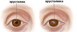
In the absence of deviations, the pupil size is two and a half millimeters.
The narrowing of an element is called miosis in medicine. This condition is not always a manifestation of pathological processes occurring in the body. Most often this is a natural reaction to bright lighting. In this case, the pupil changes its size to minimize the negative impact of intense light rays on the retina. Miosis is also considered a normal manifestation in the following situations:
- During sleep in people suffering from hypermetropia;
- In elderly patients and newborn infants;
- As a result of severe mental or physical stress;
- When the visual axes of the left and right eyes are brought together as a result of focusing the gaze on specific objects.
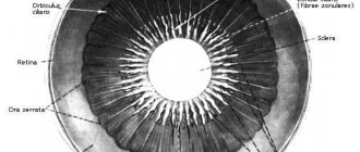
The internal muscles of the organ of vision are responsible for changing the size of the pupil. Due to the activity of the ocular sphincter and dilator muscle, the element becomes smaller or larger. If the pupil narrows under the influence of environmental factors and returns to normal after their influence ceases, there is no reason to worry.
| However, if the element remains in a “reduced state” and does not respond to light fluxes, then destructive processes are occurring in the body that require medical intervention. |
Forms of the disease
Based on the root cause of the deviation, the following types of pathological phenomena can be distinguished:
- Functional. It is a normal natural reaction in infants and elderly citizens, during physical and mental activity, with temporary exposure to bright light;
- Medication. Occurs due to the use of drugs that irritate the sphincter. This type of deviation is called reactive. Such drugs include: adrenergic blockers, anticholinergic stimulants with a nerve-paralytic effect, or agents containing bromine, morphine, and aniline dyes. Alcoholic drinks, coffee, nicotine and mushrooms also have a similar effect.
- Paralytic. It is a consequence of paralysis of the sympathetic trunk of the cervical spine.
- Spastic. The disease is caused by a disorder of the nervous system, followed by spasm of the sphincter of the pupil. A similar phenomenon is characteristic of diseases such as meningitis, multiple sclerosis, encephalitis, and brain tumors.
- Syphilitic. Characterized by symptoms of neurosyphilis.
Classification of the disease
Miosis is usually divided into unilateral (affects one eye) and bilateral (develops simultaneously in both eyes), depending on the degree of spread. In the first case, we can talk about disturbances in the body’s functioning and the development of pathologies such as syphilis and Horner’s syndrome. Also, unilateral miosis appears when a foreign body enters the eye and while taking a certain group of medications.
Bilateral constriction of the pupil occurs for the following reasons:
- Increased intracranial pressure;
- Pathological processes in the brain;
- Increased blood sugar levels;
- Syphilis.
Taking antiglaucoma medications can also provoke a change in pupil size. Return to contents
Classification of miosis
The contractile function of the pupil is carried out by two muscles, namely the sphincter and the dilator, they work from different groups of nerve fibers, these are the sympathetic and parasympathetic nervous systems. In medicine, it is customary to distinguish between two forms of miosis, namely unilateral and bilateral.
When the body is affected by a disease such as syphilis, unilateral damage to the visual apparatus is observed as a result of injury, after prolonged use of miotic drugs in one eye, which reflexively narrow the pupil, and also if the 3rd pair of cranial nerves is damaged, a unilateral form of miosis can be observed.
Physiological forms of miosis do not require treatment, since when the influencing factors disappear, the symptom disappears. Pathological forms of miosis disappear immediately after the underlying disease or the reason for which they arose is eliminated.
There is such a disease as mydriasis. Its main symptom is pathological dilation of the pupil. But the opposite can also happen, and it will be called ocular miosis. That is, under the influence of a certain number of reasons and factors, the apple of the eye stops responding to light and is constantly in a narrowed state (less than two millimeters in diameter). This phenomenon is considered the norm for some people:
- For older people;
- For recently born babies;
- For a person after serious mental or physical stress;
- For false myopia (that is, accommodation);
- With convergence of the eyes;
- For farsightedness during sleep.
Depending on the cause of miosis, several groups of the disease are distinguished:
- The functional form of miosis is a natural reaction in newborns, during physical and mental labor, the norm in old age, as a temporary phenomenon in bright lighting.
- The medicinal form of miosis occurs when taking certain types of drugs that irritate the sphincter or suppress the work of the dilator. It is also called reactive miosis. The drug that causes miosis may be an adrenergic blocker and anticholinergic stimulant, have a nerve-paralytic effect, contain bromine, organophosphorus inclusions, aniline dyes and morphine. The same effect can be achieved from alcohol, caffeine, nicotine and mushrooms (in case of poisoning with them).
- The paralytic form of miosis is caused by paralysis of the sympathetic trunk (in the cervical region), the spinal cord (in the region of the ciliospinal center), or directly in the dilator of the pupil.
- The spastic form of miosis is caused by lesions in the nervous system that lead to spasm of the sphincter of the pupil. This is typical for meningitis, multiple sclerosis, encephalitis and tumor processes in the brain.
- The syphilitic form of miosis is the defining feature of neurosyphilis.
Types of pupil miosis
Depending on the cause of miosis, several groups of the disease are distinguished:
- The functional form of miosis is a natural reaction in newborns, during physical and mental labor, the norm in old age, as a temporary phenomenon in bright lighting.
- The medicinal form of miosis occurs when taking certain types of drugs that irritate the sphincter or suppress the work of the dilator. It is also called reactive miosis. The drug that causes miosis may be an adrenergic blocker and anticholinergic stimulant, have a nerve-paralytic effect, contain bromine, organophosphorus inclusions, aniline dyes and morphine. The same effect can be achieved from alcohol, caffeine, nicotine and mushrooms (in case of poisoning with them).
- The paralytic form of miosis is caused by paralysis of the sympathetic trunk (in the cervical region), the spinal cord (in the region of the ciliospinal center), or directly in the dilator of the pupil.
- The spastic form of miosis is caused by lesions in the nervous system that lead to spasm of the sphincter of the pupil. This is typical for meningitis, multiple sclerosis, encephalitis and tumor processes in the brain.
- The syphilitic form of miosis is the defining feature of neurosyphilis.
Types of pathology
The disease is divided into several forms depending on the cause of the anomaly.
Drug-induced miosis
Develops as a result of long-term use of a certain group of drugs:
- Adrenergic blockers. These are drugs aimed at reducing the activity of the adrenergic system;
- Cholinergic stimulants;
- "Reserpine", cardiac glycosides;
- "Muscarine", "Pilocarpine" (M-cholinomimetics);
- "Aminostigmine", organophosphorus compounds (drugs that have an M-choline potentiating effect).
Also, constriction of the pupils occurs when the body is intoxicated with the following drugs and substances:
- Alcohol;
- Cigarettes;
- Caffeine;
- Mushrooms;
- "Morphine";
- Aniline dyes;
- "Muscarine", etc.
Paralytic miosis
The pathology develops as a result of paralysis of the dilator, which is responsible for the narrowing of the pupil. The cause of the anomaly is most often hidden in diseases of the cervical sympathetic trunk or damage to the plexuses of the carotid artery.
Spastic miosis
It develops as a result of spasm of the pupillary sphincter, as well as during pathological processes in the nervous system. The following diseases can cause miosis:
- Encephalitis;
- Meningitis;
- Neoplasms in the brain;
- Multiple sclerosis;
- Uremia.
Syphilitic miosis
One of the main signs of the tertiary form of syphilis. Pathology also develops as a result of the appearance of diseases such as:
- Getting a foreign object into the eye;
- Corneal ulcer;
- Anterior and posterior uveitis;
- Acute iritis;
- Horner's syndrome.
Return to contents
Types of miosis
Miosis is usually divided into the following types
:
- Medication. Occurs when using certain medications, as well as when taking alcohol, nicotine, morphine, caffeine, mushrooms and other substances with a nerve-paralytic effect.
- Paralytic. Occurs due to pathology of the dilator (the muscle that is responsible for dilating the pupil). Also, this feature may appear with the development of diseases of the ciliospinal center, carotid artery plexuses and in the cervical spine.
- Spastic. It occurs when there is a spasm of the sphincter (the muscle that is responsible for the constriction of the pupil) due to disorders of the nervous system and a number of diseases (meningitis, brain tumors, multiple sclerosis, etc.).
- Traumatic. Occurs when a foreign body enters the cornea and with ulcerative lesions, for example, with acute iritis, uveitis, etc.
- Syphilitic miosis is one of the common symptoms of the tertiary (late stage of the disease) form of syphilis or neurosyphilis (an infection of the central nervous system).
Diagnosis and treatment of miosis
With the disease, there is only one symptom that all patients complain about: constriction of the pupils, which is noticeable not only to the doctor, but to the patient himself. Therefore, a specialist does not have any problems making a diagnosis, since miosis has one symptom.
Physiological forms of miosis do not require treatment, since when the influencing factors disappear, the symptom disappears. Pathological forms of miosis disappear immediately after the underlying disease or the reason for which they arose is eliminated.
Causes of the disease
Since the muscles located in the iris function independently of human consciousness, they spontaneously contract when the brightness of the light increases and the eye accommodates. A normal variant of miosis is for:
- elderly people;
- children of the first year of life;
- persons suffering from farsightedness;
- a person in a state of sleep.
Constriction of the pupil may occur when taking certain medications.
Miosis is unilateral or bilateral. The latter occurs:
- against the background of age-related changes in the organs of vision and farsightedness;
- with increasing lighting brightness;
- during sleep;
- in certain forms of coma;
- in case of poisoning of the body;
- with local use of drugs;
- for some diseases.
Bilateral miosis often occurs when the cerebellum or pons of the brain is damaged. In this case, the pupils narrow to the size of a pinhead. This symptom manifests itself most clearly with damage to the lower parts of the midbrain and increased intracranial pressure, which contributes to compression of the brainstem.
The development of unilateral miosis is caused by the administration of drugs of the miotic group, damage to the autonomic nervous system, which causes Horner's syndrome. In addition to the narrowing of the pupil, there is a drooping of the upper eyelid, a deterioration in the reaction of the organs of vision to light, retraction of the apple, and dyshidrosis of the facial muscles.
Unilateral myositis occurs with local lesions, for example, penetration of a foreign object into the cornea. Constriction of the pupil occurs when the motor nerve or its nucleus is inflamed. Observed in meningoencephalitis, spinal cord compression, multiple sclerosis. Myositis can be combined with decreased pupil mobility.
Damage to the vagus nerve and sympathetic fibers of the cervical spine can also contribute to the occurrence of this condition. This is caused by tuberculous lesions of the posterior segments. Ocular myositis develops with acute iritis, uveitis, keratitis or trauma.
Types of miosis
Depending on the cause of this pathological condition, the following forms are distinguished. Functional is caused by natural factors and is considered a variant of the norm. Reactive miosis develops when taking or administering drugs that, in addition to direct action, have a stimulating effect on the eye muscles.
What is pupillary miosis and how to treat it
Constriction of the pupil is not always a pathological condition. This phenomenon is observed in absolutely healthy people with severe fatigue and during sleep.
But we must not forget that miosis, which is what narrowing of the pupillary space is called, often occurs in various diseases.
Miosis is observed when taking certain medications, when consuming alcoholic beverages and narcotic substances.
Reasons for development
The causes of miosis can be very different. This could be due to too much bright light, medications, or fatigue. This condition is often observed in serious diseases. The pupillary space can be greatly reduced in the following cases:
- For serious brain damage. This may be a consequence of injury or cancer.
- When taking Pilocarpine or morphine-based medications.
- For diabetes mellitus.
- As a protective reaction when a foreign body comes into contact with the mucous membrane.
- With damage to the fibers of the nervous system.
- With Horner's disease. But in this case, not only the pupil, but also the entire palpebral fissure decreases.
- For infectious diseases of the brain - encephalitis and meningitis.
- For Parkinson's disease.
- For epilepsy.
- With increased intracranial pressure.
- In a state of coma.
If the pupils are constricted in both eyes at the same time, then the reason for this lies in various neurological diseases. Brain tumors and strokes lead to this phenomenon. In most cases, against this background, the person’s consciousness is severely confused and coordination of movements is impaired.
It is noteworthy that with serious neurological problems, a person’s pupils become no larger than the head of a pin.
Types of miosis
Doctors identify several signs of miosis, depending on the root cause that provoked it:
- Functional. This is a normal reaction of the eye to bright light, under the influence of which the pupillary space is greatly reduced.
- Paralytic. It is observed with paresis of the dilator muscle.
- Drug. This form of the disease is also called reactive. It occurs as a reaction to taking certain medications. These may be barbiturates, opiates or cardiac glycosides. After stopping the medications, the symptoms of reactive miosis disappear.
- Toxic. This form is observed under the influence of various toxic substances that enter the body. This could be alcohol, drugs, various dyes and gases.
- Sympathetic. It happens with a spasm of the sphincter of the pupil. This condition is typical for meningitis, epilepsy, multiple sclerosis and various brain tumors. Any brain disease can lead to this condition.
- Syphilitic. In some cases, severe narrowing of the pupillary space is one of the symptoms of syphilis.
Treatment is necessary in all cases except physiological miosis. But even here it is necessary to eliminate the root cause of the violation as soon as possible.
If the constriction of the pupils is not physiological, then the patient is prescribed a detailed examination to identify the cause of the disorder.
Prognosis and prevention
With timely treatment, the prognosis is good. In most cases, it is possible to stabilize the patient’s condition and return the pupils to normal size.
To avoid miosis, you should promptly treat all systemic diseases and undergo an annual preventive examination by doctors. If you have any vision problems, you should consult an ophthalmologist.
Do not ignore the strong constriction of the pupils. In many cases, this condition indicates serious illness. Only a qualified doctor can make an accurate diagnosis.
Forms
Ophthalmologists divide miosis into bilateral and unilateral. The first manifests itself as a result of the development of hypermetropia. Often, bilateral constriction of the pupil is observed in elderly people. Also, a similar symptom is a normal reaction of the body to bright lighting. When the brain is damaged in the cerebellar area, the pupil becomes like the head of a pin. Bilateral miosis develops in patients in a coma and in people with increased intracranial pressure.
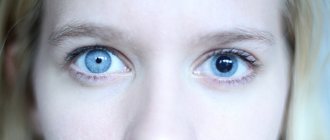
Anisocoria or unilateral constriction of the pupil is in 99% of cases a signal about the development of pathological processes in the body. It is a symptom of the following abnormalities:
- Horner's syndrome;
- Taking miotic medications;
- Unilateral damage to the anterior chamber of the eye;
- Irritation of the third pair of cranial nerves.
In exceptional cases, anisocoria signals the development of syphilis.
Miosis - causes and treatment
Miosis
- this is a slight narrowing of the pupil (less than 2.5 mm in diameter), which is not always a pathology. Constriction of the pupil, in general, is a natural physiological reaction of the eye, which reflexively occurs in bright light. As a normal variant, miosis appears, for example, in older people and newborns, as well as when looking at different distances and during strong physical or mental stress. This condition can also occur in people with farsightedness.
Symptoms
Miosis is a medical term describing a condition in which the pupil constricts to a significant degree. Such a condition may be a variant of the norm (observed in certain situations) or signal a pathology.
Why does the pupil size change? The fact is that the size of the pupil constantly varies depending on many factors. This process is regulated by the tone of the internal muscles of the eye. The sphincter, innervated by parasympathetic fibers, ensures contraction of the pupil (miosis), while the dilator, innervated by fibers, is responsible for enlargement (mydriasis).
The sphincter, located in the iris, is not controlled by human consciousness and contracts reflexively when:
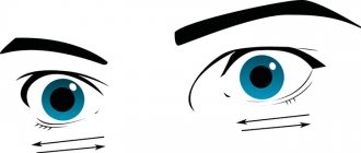
- Accommodation of the eye;
- Sudden increase in light intensity.
Miosis, as a physiological norm, is characterized by:
- For elderly people (over time, the accommodation of the eye changes, as the lens loses its elasticity);
- For newborn children;
- For persons suffering from hypermetropia;
- For all people in a state of sleep;
- In case of overstrain (physical and mental);
- As a reflexive reaction to bright lighting;
Miosis is not difficult to determine. To do this, just look a person in the eyes. The pupillary space is small, and this condition can persist for quite a long time.
Against the background of constriction of the pupils, headaches, increased intracranial pressure and impaired visual fields may occur. Depending on what caused the miosis, other symptoms may be observed. This includes dizziness, confusion and signs of intoxication.
In medicine, myositis is understood as inflammation of skeletal muscles of various origins. The disease has a specific etiology, course and symptoms. The danger of the pathology lies in possible complications on the intestines, joints, heart, lungs and skin.
Systemic involvement of all skeletal muscles is rare; cervical and lumbar myositis are more common. The disease disguises itself as a common cold, but after a couple of weeks the person cannot get out of bed.
Only timely diagnosis and treatment can help eliminate inflammation.
Miosis may be accompanied by additional symptoms that will indicate the root cause of the pathological abnormality.
For example, a diabetic patient experiences a feeling of thirst and frequent urination. If constricted pupils are a consequence of syphilis, the skin and mucous membranes will have peculiar lesions.
Organophosphate poisoning
Organophosphate poisoning
is an acute intoxication that occurs when certain insecticides (karbofos, chlorophos, thiophos) or chemical warfare agents (sarin, soman, VI gases) are ingested by inhalation, transdermal or orally.
Main signs: miosis, pain in the eye area, vomiting, increased sweating, abdominalgia and chest pain, bradycardia, ataxia, respiratory paralysis, convulsions. Pathology is diagnosed based on history, clinical picture and information obtained during laboratory examination.
Treatment includes anticholinergics, cholinesterase reactivators, and symptomatic therapy.
FOV - fluorides of phosphoric or methylphosphonic acid. Poisoning with them accounts for 1-2% of the total number of acute exotoxicoses. The poison is able to penetrate the skin without damaging it. At high concentrations of the xenobiotic, pathological symptoms appear within a few minutes.
Organophosphorus compounds dispersed in the air form a persistent cloud located near the surface of the earth, which persists for 4-6 hours. Military chemical agents are capable of maintaining toxic concentrations for several days.
Lethal doses vary greatly depending on the properties of the particular compound.
Organophosphate poisoning
The vast majority of cases are associated with violations of safety rules when treating fields or premises against insects.
Poisoning with organophosphorus substances occurs when you refuse to use a gas mask, insufficient tightness of protective suits, or point the spray at people.
Such intoxications are usually mild or moderate, since the dose of the drug received is relatively small. Other possible reasons:
- Substance abuse.
The peak of occurrence was in the 80-90s of the 20th century. Today there is a steady downward trend in the number of such cases. Derivatives of phosphoric acid can cause hallucinations and an altered state of consciousness, which to a certain point makes them popular among drug addicts. - Hostilities.
During military campaigns and terrorist acts, chemical warfare agents can be used: soman, sarin. Poisoning is severe because persistent forms of OPA are used, capable of long-term maintenance of high concentrations. The semi-lethal dose of sarin is 1.7 g, soman is 60 mg. - Suicide.
Attempts to commit suicide using organophosphorus compounds are relatively rare, their frequency does not exceed 3-4% of the total number of suicides. As a rule, common insecticides with moderate or low toxicity are used: karbofos, chlorophos, bromophos and demufos, so the effectiveness of the method is not too high.
The main pathogenetic mechanism of action of FOV is the suppression of cholinesterase activity by 30-80%, disruption of the processes of synthesis and hydrolysis of acetylcholine.
Poisoning with extremely severe organophosphorus substances leads to 100% inhibition of the enzyme.
Accumulation of ACH occurs in synapses, which is accompanied by their overexcitation, disruption of the brain, heart, smooth and striated muscles. Complete recovery of the central nervous system occurs after 147 days, the blood system - after 48 days.
The toxicant also has a direct stimulating effect on cholinergic structures, so the severity of intoxication does not always correspond to the amount of blocked cholinesterase.
FOV also sensitizes the body’s cholinergic receptors to its own ChE, which leads to a relapse of symptoms after complete cleansing of the toxic substance.
The latter mechanism involves the accelerated release of accumulated acetylcholine, which provokes its excessive accumulation on presynaptic membranes.
Poisoning with organophosphorus substances can be subdivided by the name of the toxicant (sarin, karbofos, soman); routes of poison intake (inhalation, transdermal, oral); mechanism of occurrence (unintentional injury, suicide, military action); period of intoxication (latent, acute, complications and consequences). In clinical practice, a common classification according to severity is:
- Easy.
No more than 20-30% of cholinesterase is blocked. The leading syndromes are: neurotic, myotonic, cardiac, gastrointestinal, respiratory disorders. Occurs during a short stay in an infected area, more often with careless use of household insecticides. - Moderate weight.
The amount of blocked enzyme varies from 30 to 80%. Main syndromes: psychoneurotic, bronchospastic. Signs consistent with mild intoxication may appear. Occurs during suicide attempts using low-toxic substances or during prolonged stay in an area treated with insecticides. - Severe and lethal.
The basis of the clinical picture is convulsive-paralytic syndrome. There is a pronounced disturbance of breathing and hemodynamics. Develops when exposed to combat gases or through oral or inhalation intake of highly toxic OPAs. Accompanied by early and late complications.
Poisoning with organophosphorus substances is manifested by symptoms of damage to several organs and systems.
On the part of the respiratory system, bronchospasm, expiratory shortness of breath, cough with large amounts of sputum, dyspnea, and pulmonary edema are noted.
Disruption of the gastrointestinal tract leads to vomiting, nausea, cramping abdominal pain, and involuntary defecation. The sweat and salivary glands react by increasing secretion - drooling and increased sweating are observed.
Damage to the cardiovascular system is accompanied by bradycardia, weakened hemodynamics, and a transient increase in blood pressure with its subsequent fall.
The reaction of the organs of vision is the appearance of miosis, anisocoria, spasm of accommodation (the victim loses the ability to see at a great distance). In severe intoxication, severe weakness, involuntary twitching of isolated muscle groups, paralysis and paresis are determined.
Convulsions, depression of consciousness up to coma, signs of suppression of the activity of the respiratory and vasomotor center are possible.
All complications are divided into early and late. Early ones occur 1-2 days after intoxication. Late ones can be diagnosed from 3 to 150-200 days.
The first include central respiratory paralysis caused by damage to the corresponding center of the brain, recurrent bronchospasm, acute pneumonia, myocardial dystrophy, intoxication psychosis, nephropathy and hepatopathy with the development of multiple organ failure. The incidence of early complications is about 5-7%.
In the later stages, peripheral paralysis of the respiratory muscles, toxic encephalopathy, polyneuritis, and asthenovegetative syndrome may develop.
In some patients, persistent paresis and hemiparesis of the limbs, nerve damage accompanied by dysfunction of internal organs, fecal and urinary incontinence are determined. A relapse of the clinical picture of intoxication is also possible.
Poisoning with organophosphorus substances is complicated by late pathological phenomena in 3-4% of cases.
The basis of diagnosis is the clinical picture and anamnesis of the patient. The diagnosis is made by an emergency physician or a toxicologist at a specialized center (if the victim applies independently).
Consultations with a gastroenterologist, neurologist and other specialists are required.
The pathology is differentiated from drug poisoning, neurotoxic poisons, substances that can affect the activity of cholinesterase (ovulation inhibitors, muscle relaxants, arsenic, glucocorticosteroids). Examination methods:
- Physical.
There is confusion, lethargy, and coma. On examination - cyanosis or pale skin, spontaneous twitching of individual muscle groups or clonic-tonic convulsions. Blood pressure is higher or lower than normal, breathing is difficult, pulse is slow. On auscultation of the lungs, moist rales may be heard and a productive cough may be present. - Laboratory.
The color change of the indicator during a laboratory test is 22-40 minutes. There is a shift in blood pH to the acidic side and an imbalance in electrolyte balance. The partial pressure of oxygen is reduced, the content of carbon dioxide increases. When kidneys are damaged, urea and creatinine accumulate. Against the background of liver pathology, an increase in AST and ALT is noted. - Hardware.
They are used primarily for diagnosing complications. Pulmonary edema manifests itself as flying shadows on an x-ray; with bronchospasm, the volume of forced expiration is sharply reduced. The ECG shows atrial fibrillation, tachycardia caused by direct myocardial damage or hypotension.
Before the arrival of doctors, the victim should be evacuated from the area of chemical contamination, laid down, and a supply of fresh air provided. If the toxicant gets on unprotected skin, this area must be wiped with a 10% ammonia solution.
Cases of eye damage require rinsing with 2% sodium bicarbonate followed by instillation of atropine. The consequences of oral consumption of poison can be reduced if, immediately after contact with it, the stomach is rinsed using a tubeless method and given activated charcoal.
The dose of sorbent is calculated according to the formula 1 tablet per 10 kg of weight.
First aid
First aid is provided by emergency medical services teams. The main measure is the administration of atropine, which neutralizes the effect of organophosphorus compounds. The drug is administered intravenously, in case of severe tachycardia - intramuscularly, 1 ml of 0.1% solution every 15 minutes.
For oral intoxication, gastric tube lavage, administration of enterosorbents, and saline infusion are performed. Symptomatic therapy includes anticonvulsants, pressor amines, bronchodilators, and diuretics.
If consciousness and breathing are impaired, tracheal intubation is performed followed by transfer to mechanical ventilation.
Hospital treatment
The basis of the therapeutic regimen is cholinesterase reactivators and anticholinergics. The latter includes atropine. Patients receive it at 15-minute intervals.
The assessment criterion for a sufficient volume of antidote therapy is the occurrence of atropine delirium; this may require 20-30 ml of the drug. ChE reagents (isonitrosine, dipyroxime) are used at all stages of poisoning.
The regimen is selected individually, depending on the severity of the pathology and the category of the patient (children, adults, old people).
To combat emerging disorders, the patient receives symptomatic treatment. Succinic acid preparations, respiratory stimulants, antidepressants, antipsychotics, antioxidants, anticonvulsants, and calcium channel blockers are prescribed.
To accelerate the elimination of the xenobiotic, the method of forced diuresis and intestinal stimulation (salt laxatives) are used.
If necessary, resuscitation assistance is provided (intubation, mechanical ventilation, hemodialysis, titrated administration of dopamine, monitoring).
Rehabilitation
After discharge, the patient is prescribed outpatient follow-up treatment. He should carefully monitor his condition and regularly visit a general practitioner or therapist.
Drug support includes taking nootropics, antipsychotics and cardiac medications. It is recommended to visit sanatorium-resort institutions located in coniferous forests.
In the presence of paresis, observation by a neurologist, taking medications that improve neuromuscular conduction, and physical therapy are required.
Full recovery takes about two hundred days. During this period, delayed complications may occur. The hospitalization time for patients with mild and moderate damage usually does not exceed 10-15 days.
The duration of inpatient treatment for severely injured patients depends on the course of the somatic stage of exotoxicosis and the regenerative capabilities of the body. The mortality rate is 12.9%.
The main cause of death is paralysis of the respiratory center and untimely medical care.
To prevent organophosphorus poisoning, it is necessary to strictly adhere to safety requirements.
When working with pesticides and herbicides, insulating gas masks and clothing are used that do not allow the toxicant to get on the skin.
Soldiers taking part in combat operations must be trained in chemical hazard recognition and provided with protective equipment. FOV houses should be stored in closed areas where children and people with unstable mental health do not have access.
Source: https://www.KrasotaiMedicina.ru/diseases/urgent/organophosphate-poisoning
Causes of the disease
The following factors can cause pupil constriction:
- Increased blood sugar levels;
- Penetration of a foreign object into the cornea or violation of its integrity;
- Vasoconstriction as a result of taking Pilocarpine;
- Damage to central nervous system fibers;
- Horner's syndrome. Accompanied by a decrease in the palpebral fissure and pupil, drooping of the eyelid.
Bilateral changes in pupil size are caused by neurological diseases, usually malignant or benign neoplasms. In this case, miosis manifests itself against the background of impaired consciousness. The following can also cause the development of pathology:
- Increased blood sugar levels;
- Increased intracranial pressure;
- Epilepsy;
- Parkinson's disease;
- Inflammatory processes affecting the cerebral cortex.
Anisocoria is characteristic of Horner's syndrome. In this case, simultaneously with the narrowing of the pupil, the eyelid droops. Since the main reason for the development of the disease lies in damage to the muscle responsible for dilating the pupil, it is most pronounced in the dark.
Also, the development of Horner's syndrome can be triggered by multiple sclerosis, neoplasms in the brain, abnormal conditions in the muscles, stroke and cervical osteochondrosis.
You will learn more about Horner's syndrome from the video.
Return to contents
Miosis: types, symptoms and treatment and what it is -
Constriction of the pupil is not always a manifestation of destructive processes occurring in the body. In some cases, this is a completely normal physiological reaction, for example, when the lighting level changes or physical fatigue.
However, in some situations, eye miosis can become an alarming symptom that a malfunction has occurred in the functioning of the internal organs.
If in the first case the pupil returns to normal after some time without the intervention of doctors, then in the second the abnormal condition requires treatment.
Causes of the disease
The following factors can cause pupil constriction:
- Increased blood sugar levels;
- Penetration of a foreign object into the cornea or violation of its integrity;
- Vasoconstriction as a result of taking Pilocarpine;
- Damage to central nervous system fibers;
- Horner's syndrome. Accompanied by a decrease in the palpebral fissure and pupil, drooping of the eyelid.
Bilateral changes in pupil size are caused by neurological diseases, usually malignant or benign neoplasms. In this case, miosis manifests itself against the background of impaired consciousness. The following can also cause the development of pathology:
- Increased blood sugar levels;
- Increased intracranial pressure;
- Epilepsy;
- Parkinson's disease;
- Inflammatory processes affecting the cerebral cortex.
Anisocoria is characteristic of Horner's syndrome. In this case, simultaneously with the narrowing of the pupil, the eyelid droops. Since the main reason for the development of the disease lies in damage to the muscle responsible for dilating the pupil, it is most pronounced in the dark.
Also, the development of Horner's syndrome can be triggered by multiple sclerosis, neoplasms in the brain, abnormal conditions in the muscles, stroke and cervical osteochondrosis.
You will learn more about Horner's syndrome from the video.
Diagnostics
If physiological factors for the appearance of miosis are excluded, then the doctor prescribes a series of examinations that will help determine the exact cause of the development of the deviation:
- Analysis of the condition of the eyeball. The examination is performed using a slit lamp;
- Magnetic resonance imaging of the organ of vision;
- Ultrasound examination of the eye;
- Checking the reaction of the pupil to a light source;
- Perimetry. Conducted to examine visual fields;
- Fundus analysis.
If the ophthalmologist was unable to detect any abnormalities in the eyes, he sends the patient for additional examination to a neurologist.
Prevention
To minimize the risk of developing miosis, you must follow simple rules:
- Any medications are used only as prescribed by a doctor;
- Do not start treatment for neurological diseases. These are very dangerous anomalies that can cause serious harm to health. Visit a neurologist regularly for preventive examinations;
- Elderly patients are advised to have their blood glucose levels checked more frequently;
- Wear safety glasses when working at the computer for long periods of time.
| Following simple rules will help maintain eye health for many years. |
Conclusion
Miosis is a constriction of the pupil. In some cases, such a manifestation is a natural reaction of the body, in others it signals the development of pathological processes that require urgent treatment. Under no circumstances should you choose medications on your own! Visit a doctor who, after a detailed diagnosis, will identify the cause of the anomaly and select effective treatment.
Causes of miosis
The size of the pupil is determined by the work of two muscles - the dilator and the sphincter. The dilator is responsible for the dilation of the pupil, and the sphincter is responsible for the constriction.
Miosis can be unilateral or bilateral in form.
Unilateral miosis occurs with local injection of miotic drugs into one eye, with Goner's syndrome (damage to the sympathetic nervous system) and with local damage to the eye (a foreign body entering the cornea, for example).
The causes of bilateral miosis can be
:
- Elderly age
- Farsightedness
- Intense lighting
- Using certain eye drops or injections
- Brain damage (in this case, the constriction of the pupils is especially pronounced)
- Diabetes
- Somatic and venereal diseases
- For some types of coma: diabetic, narcotic, pancreatic, etc.
- During sleep.
Diagnostics
If physiological factors for the appearance of miosis are excluded, then the doctor prescribes a series of examinations that will help determine the exact cause of the development of the deviation:
- Analysis of the condition of the eyeball. The examination is performed using a slit lamp;
- Magnetic resonance imaging of the organ of vision;
- Ultrasound examination of the eye;
- Checking the reaction of the pupil to a light source;
- Perimetry. Conducted to examine visual fields;
- Fundus analysis.
If the ophthalmologist was unable to detect any abnormalities in the eyes, he sends the patient for additional examination to a neurologist.
Treatment
Physiological miosis does not require therapy, nor does dilation of the pupils caused by taking appropriate medications for diagnosis require treatment.
In other cases, pathology must be fought. As part of the therapeutic course, the doctor prescribes the following procedures:
- Normalizing carbohydrate metabolism;
- Removing a foreign object from the organ of vision;
- Selection of medications for the treatment of syphilis;
- Therapy of pathological processes occurring in the vascular system of the brain. In this case, the doctor prescribes a vitamin-mineral complex, nootropics and vasoactive medications.
How to treat?
The physiological type of miosis does not require treatment. Reactive miosis is eliminated after stopping taking medications that cause disruption of the functioning of the muscles of the visual organs. Getting rid of a foreign object that has entered the eye, as well as correcting metabolic processes involving carbohydrates, have a positive effect on the dynamics of treatment of the pathology. More severe manifestations of miosis in some groups require the participation of specialized specialists for the treatment of cerebrovascular diseases, syphilis and other primary ailments that cause complications in the eyes.
Prevention
To minimize the risk of developing miosis, you must follow simple rules:
- Any medications are used only as prescribed by a doctor;
- Do not start treatment for neurological diseases. These are very dangerous anomalies that can cause serious harm to health. Visit a neurologist regularly for preventive examinations;
- Elderly patients are advised to have their blood glucose levels checked more frequently;
- Wear safety glasses when working at the computer for long periods of time.
| Following simple rules will help maintain eye health for many years. |