Pneumotonometry
Pneumotonometry is a diagnostic test in ophthalmology designed to measure IOP (fluid pressure inside the eyeball). Diagnostics is performed using a specialized device - a non-contact tonometer. The procedure is completely safe and painless and does not require special preparation or anesthesia.
Monitoring the IOP level allows early detection of the development of glaucoma, which, as it progresses, causes atrophy of the optic nerve up to complete blindness.
Formation of intraocular fluid
Intraocular pressure is formed under the influence of fluid inside the eyeball.
The fluid is initially formed in the posterior chamber of the eye, then through the pupil it enters the anterior chamber, from where it drains. The ratio of the volume of fluid formed to that removed creates pressure inside the eyeball.
In a healthy state, this process is fully proportionate, which indicates normal IOP values.
Any disturbances in the circulation of aqueous humor (the ratio of inflow and outflow) indicate the presence of pathological processes.
The essence of the technique is to expose the eyeball to air flow under pressure. The pressure of the air stream affects the cornea, resulting in its applanation (flattening). The obtained data is recorded by the optical system of the tonometer. The doctor who conducted the study deciphers the results.
Description of the IOP measurement technique
The procedure is performed without physical contact of the eyes with the equipment. The result is recorded in millimeters of mercury (mmHg).
The reliability of the results does not always correspond to reality, so if the thickness of the cornea exceeds the average, the results may be overestimated. This is a prerequisite for additional diagnostic methods.
Indications for pneumotonometry
In order to prevent glaucoma and other pathologies of the visual apparatus, annual pneumotonometry is recommended for people over 40 years of age.
Measuring intraocular pressure is performed for various diseases and problems:
- dysfunction of endocrine organs;
- retinal detachment;
- abnormalities in the development of tissue structures of the eye;
- pathologies of the cardiovascular system;
- in the period after surgery or in the presence of complications;
- glaucoma.
What diseases can be detected during the diagnostic process?
If the obtained indicators differ significantly from the norm, serious deviations in the functioning of internal organs and systems can be suspected. With increased pressure inside the eyeball, one can assume the development of glaucoma, as well as the presence of pathologies such as:
- CVD diseases;
- pathological processes in the kidneys;
- inflammatory and infectious eye diseases;
- history of TBI;
- dysfunction of the hypothalamic-pituitary system.
If during pneumotonometry indicators were recorded below the generally accepted norm, the presence of the following pathological conditions is assumed:
- previous injuries to the eyeball;
- the presence of inflammatory processes;
- persistent decrease in blood pressure;
Sometimes the cause of this condition can be regular use of alcoholic beverages and drugs.
Contraindications for the study
Non-contact tonometry is not performed in the following cases:
- bacterial and viral eye infections;
- violation of the integrity of the cornea as a result of injury;
- pathological changes in the eyeball
- in the postoperative period when using laser surgery and the presence of complications;
The study is not carried out in patients who are intoxicated or under the influence of drugs.
Stages of the procedure
An optical ophthalmic device measures the fluid pressure inside the eyeball in 10 seconds.
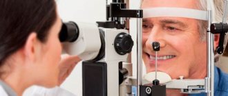
To perform the procedure, the patient must remove glasses and contact lenses. The subject is seated in front of the device, fixing his head. The gaze is directed in front of you to the illuminated point of the device.
Next, a stream of air is supplied at a certain speed, which leads to a change in the shape of the cornea. At this stage, the tonometer processes the results obtained, recording them on a form. Based on the indicators, the diagnostician interprets the results.
Interpretation of pneumotonometry: norm and deviations
After receiving the printout issued by the optical device, the ophthalmologist begins decoding using a special table. In a healthy state, intraocular fluid pressure varies between 15-24 mm Hg. Art.
- The results figures depend on many factors, so if the eyes are very tired or the procedure is carried out in the evening, the indicators may slightly differ from the accepted norm. In this connection, the value is below and above the norm by 10-20 mmHg. Art. is not considered a pathology. In such cases, repeated pneumotonometry is prescribed after some time.
- Deviations from the norm are indicators significantly exceeding the values of 15-24 mm Hg. Art. Also, the pathological condition is determined by the presence of additional symptoms with low or high pressure.
- In most cases, high IOP indicates glaucoma, which most often occurs in people of middle and older age.
- Low rates are extremely rare and are more often the result of insufficient competence of the diagnostician or incorrectly conducted research.
Given the high variability of results, a final diagnosis cannot be established based on pneumotonometry data alone; in the presence of significant deviations, a comprehensive examination is prescribed, as well as contact measurement of IOP.
Fill out an application on the website, we will contact you as soon as possible and answer all your questions.
Make an appointment
Pneumotachometry - what is it and what is it for?
Pneumotachometry is a test that measures the patency of the airways.
During the procedure, a special device is used to determine the speed of air passage during inhalation and exhalation.
Indications and contraindications for
Pneumotachometry is performed for diseases of the bronchi and upper respiratory tract. It is more often used to diagnose patients with chronic diseases.
Such pathologies include:
- Bronchial asthma;
- Atopic bronchitis;
- Pneumosclerosis;
- Chronic obstructive pathology.
Regular pneumotachometry allows you to determine the progress of the disease and evaluate the effectiveness of treatment.
The examination is also carried out:
- In patients with an increased risk of lung disease;
- In case of complicated heredity - in persons whose relatives suffer from bronchopulmonary and allergic diseases;
- In military personnel and athletes to assess the functional activity of the lungs.
In some situations, pneumotachometry is contraindicated or caution is required when performing it.
Such cases include:
- High blood pressure;
- Severe circulatory failure;
- Ischemic or hemorrhagic stroke suffered no more than three months ago;
- Previously suffered a heart attack;
- Aneurysm of the aorta or cerebral arteries;
- Severe airway obstruction;
- Marked decrease in lung function, which led to respiratory failure;
- General serious condition of the patient, which does not allow pneumotachometry;
- Acute respiratory infection;
- Epilepsy;
- Pregnancy.
Preparation for pneumotachometry
In order for the examination results to be as reliable as possible, it is worth going through simple preparation for pneumotachometry.
To do this you need:
- Stop smoking and drinking alcohol for 24 hours before the test;
- Stop taking medications that affect breathing by increasing airway patency (such as bronchodilators);
- Avoid physical and emotional stress on the day of the examination;
- Before undergoing pneumotachometry, wear loose clothing that does not restrict the chest.
It is recommended to take the examination no earlier than 2 hours after eating. If the procedure is carried out in the morning, you need to refrain from breakfast.
Methodology
Pneumotachometry is carried out in the treatment room, using a special device - a pneumotachometer.
The patient can be placed in a sitting or standing position; a clamp is placed on his nose, thanks to which only mouth breathing is possible. A sterile mouthpiece is placed on the pneumotachometer tube.
The patient takes several breaths in and out calmly, then draws in the maximum amount of air into the lungs and exhales it forcefully, then repeats this operation.
Pneumotachography is easily tolerated by patients, but in some cases complications may develop:
- Darkening in the eyes;
- Dizziness;
- Fainting.
At its core, this diagnostic technique is close to the forced expiratory spirogram.
To obtain additional information, bronchodilation tests may be performed when performing pneumotachometry. In this case, the patient takes drugs that dilate the bronchi, and the results obtained after this are compared with the original ones.
The main advantages of pneumotachometry:
- This examination method does not involve intervention in the patient’s body, that is, it is not invasive;
- Completing pneumotachometry does not take much time;
- The method is well suited for conducting mass surveys;
- Pneumotachometry is a well-studied and proven diagnostic method;
- No complicated preparation is required for the examination.
The disadvantages include the fact that the accuracy of diagnosis strongly depends on compliance with the procedure and on the qualifications of the doctor who conducts the examination and interprets the results.
Interpretation of results
The study evaluates:
- Vital capacity of the lungs;
- Maximum air flow rate;
- Peak expiratory flow;
- Average expiratory flow;
- Tiffno index.
The vital capacity of the lungs is the maximum volume of air that can enter them during inhalation and exit during exhalation. The Tiffno index allows you to assess airway obstruction.
The data obtained during the examination is compared with the norm. A low maximum expiratory flow rate indicates a deterioration in the function of the muscular system or a decrease in airway patency.
With a more detailed analysis of the air flow curves, it is possible to determine whether there is a violation of its passage in the small, medium and large bronchi and in the trachea. Pneumotachometry makes it possible to identify disturbances in the passage of air through the bronchi at an early stage, when the pathology does not yet manifest itself.
The results obtained during the examination are analyzed taking into account age, height, weight, level of physical fitness and other individual characteristics of the body. To do this, before the procedure, the doctor measures the patient’s anthropometric parameters.
Anyone who is at risk or already suffers from a respiratory tract disease needs to know what pneumotachometry is and how to prepare for it. This is an easy-to-conduct study that does not take much time and requires almost no preparation.
This diagnostic technique can be used to identify bronchial obstructions, including at an early stage. It allows you to assess the dynamics of the course of bronchopulmonary diseases and the effectiveness of the prescribed treatment.
Preparation for pneumotachometry
In order for the examination results to be as reliable as possible, it is worth going through simple preparation for pneumotachometry.
To do this you need:
- Stop smoking and drinking alcohol for 24 hours before the test;
- Stop taking medications that affect breathing by increasing airway patency (such as bronchodilators);
- Avoid physical and emotional stress on the day of the examination;
- Before undergoing pneumotachometry, wear loose clothing that does not restrict the chest.
It is recommended to take the examination no earlier than 2 hours after eating. If the procedure is carried out in the morning, you need to refrain from breakfast.
Pneumotachometry is a simple and effective method for studying the lungs
For some diseases associated with the bronchi and respiratory tract, pneumotachometry is necessary. Using this procedure, the patency of organs is measured, the speed and volumes of air during exhalation and inhalation are determined. In what cases is research recommended and how is it carried out?
When is pneumotachometry needed?
Pneumotachometry is recommended for people suffering from diseases of the bronchi and upper respiratory tract, especially if they are chronic. It is thanks to this method that a number of pathologies can be diagnosed at the earliest stages.
Such diagnostics are carried out in preparation for surgical intervention on the respiratory organs.
A pneumotachometer (spirometer) is used for the study. Through it, the patient takes several breaths and exhales, and the device sets the level of bronchial patency and the maximum speed of air movement.
The doctor’s final conclusion depends on:
- maximum air speed;
- average expiratory flow;
- peak expiratory flow;
- Tiffno index;
- vital capacity of the lungs.
When analyzing indicators, the individual characteristics of the patient’s body, height, age and level of physical fitness are taken into account.
Indications for use
Using regular pneumotachometry, conditions are monitored and the progress of the disease is assessed. This is necessary during active treatment and allows you to evaluate its effectiveness.
Clear indications include:
- bronchial asthma;
- pneumosclerosis;
- atopic bronchitis;
- chronic obstructive pathology.
In these pathologies, pneumotachometry is performed without fail and, as a rule, repeatedly. Only in this way can a specialist assess the condition of the respiratory system and select the appropriate treatment.
Additionally, to confirm the diagnosis, spirography may be prescribed - a method of measuring lung volume, taking into account indicators of forced and natural breathing. During the procedure, the patient must inhale atmospheric air or pure oxygen.
How the research is carried out
During the procedure, the patient can sit or stand; the body must be sufficiently relaxed. A special sterile mouthpiece is placed on the device, and a clamp is placed on the patient’s nose: during the examination, you can only breathe through your mouth.
To prepare for receiving data, take several calm exhalations and inhalations. Without holding your breath, deep forced inhalations and exhalations are performed and repeated several times. Using the pneumotachotra scale, the specialist analyzes the rate of forced expiration.
For a certain age and gender, there are standard pneumotachymetry indicators
In adult men, the expiratory flow rate should be from 5 to 8 l/s, in women -4-6 l/s. The volumetric velocity is calculated using the Badalyan formula. Actual lung capacity is multiplied by 1.2%. The result obtained must be more than 85%, otherwise a patency disorder is diagnosed. In the presence of a chronic violation, speed indicators are noticeably reduced.
Mild discomfort may be experienced during the examination. If coughing begins during the examination, the measurement must be interrupted and repeated after a few minutes. It is necessary to stop the procedure if you cough up blood or experience chest pain.
Pneumotachometry is absolutely painless and does not cause any discomfort to the subject. In some cases, it is recommended to postpone the procedure indefinitely or cancel it altogether. These include:
- arterial aneurysm;
- pregnancy;
- hypertensive crisis;
- epilepsy;
- severe respiratory tract disorders.
You will also have to refuse the study if the patient experienced a stroke, myocardial infarction or respiratory tract infection within three months before the date of pneumotachometry.
How to prepare
During the day before the study, the patient must completely stop smoking and drinking alcoholic beverages: all this can negatively affect the result, distorting it. If necessary, you need to take your inhaler with you. If the patient is taking short-acting bronchodilators, you need to tell the doctor about it in advance.
Four hours before tachometry, bronchodilators should be discontinued.
The patient's clothing should be loose, not restricting the movement of the chest, and not putting pressure on the throat. It is necessary to be in as calm a state as possible. Fast walking, physical exercise, or even excessive anxiety can affect the final result of the study.
The last meal is allowed two hours before the test, preferably on an empty stomach. Before the procedure, the specialist must be informed of your height, age and weight. If the study is repeated, the patient should be in the same position in which the original data were obtained.
Indications for use
The main medical indication for pneumotachometry is to assess the functional state, namely the patency of the airways. This study makes it possible to diagnose a pathology accompanied by a deterioration in the passage of air through bronchi of various sizes, which includes:
- Bronchial asthma is a specific inflammatory lesion of the bronchi, which is of allergic origin. It is accompanied by periodic development of attacks of shortness of breath, coughing with the discharge of viscous sputum. The mechanism of development of the pathological process is associated with a narrowing of the lumen of the bronchi (bronchospasm), caused by an increase in the tone of the smooth muscles of their walls against the background of the development of an allergic reaction, which is provoked by the body’s contact with an allergen (foreign compounds, often of a protein nature).
- Atopic bronchitis is also an allergic lesion of the lungs, accompanied by a milder course with the development of bronchospasm and inflammation in the bronchi of various sizes.
- Chronic obstructive pulmonary pathology is a long-term inflammation of the bronchial tree, which can be caused by various provoking factors (infections, systematic inhalation of dust or vapors of various chemical compounds, smoking) and is accompanied by a deterioration in bronchial patency.
- Pneumosclerosis is a severe lesion of the lungs, characterized by the replacement of lung tissue with connective tissue; it compresses the bronchi, causing their lumen to narrow. This pathological condition is a consequence of the long-term course of various pathologies of the respiratory system, as well as the influx of dust.
Carrying out pneumotachometry for these diseases allows the doctor to assess the functional state of the structures of the respiratory system and select the most optimal therapy.
Contraindications for carrying out
There are several pathological and physiological conditions of the patient’s body in which pneumotachometry is excluded, these include:
- A recent hemorrhagic or ischemic stroke of the brain (at least 3 months ago).
- Arterial hypertension (increased levels of systemic blood pressure), which often accompanies hypertension.
- Previous infarction (death of a section of the heart muscle) of the myocardium.
- Aneurysm of the arteries of the brain, as well as the thoracic aorta.
- Acute infectious process in the respiratory system.
- Respiratory failure, accompanied by a pronounced decrease in functional activity.
- Epilepsy (pathological development of seizures).
- Pregnancy at any stage.
Before prescribing pneumotachometry, the doctor must ensure that the patient has no contraindications.
Where and how is pneumotachometry performed?
Pneumotachometry is carried out in the treatment room of a medical institution. The patient lies on the couch.
The doctor gives him a pneumotachograph tube with a sterile mouthpiece attached to it, and a special clamp is put on his nose.
After this, he takes several calm breaths into and out of the tube, then inhales and exhales the air as much as possible several times (usually 2 times), and the doctor at this time records the readings of the pneumotachograph.
Preparing for the study
To obtain the most reliable results of pneumotachometry, the patient must follow several simple preparatory recommendations before performing it, which include:
- Stop smoking and drinking alcohol the day before the test.
- Cancel the use of certain medications, in particular the pharmacological group bronchodilators (drugs that improve airway patency) 4 hours before.
- Clothing should not restrict breathing movements.
- It is not recommended to be exposed to increased emotional or physical stress on the day of pneumotachometry.
- The study should be carried out on an empty stomach or no earlier than 2 hours after eating. Usually it is carried out in the morning, the patient does not have breakfast.
The doctor gives more detailed recommendations during the appointment. It is also necessary to measure anthropometric indicators (height, body weight, chest volume), which will allow the specialist to correctly determine the state of the structures of the respiratory system.
Basic information about pneumotachometry
The study using the pneumotachometry method makes it possible to identify impaired patency in the bronchi, both small and large, at an early stage, as well as diagnose bronchial asthma. The procedure itself resembles an exhalation spirogram.
A special device called a pneumotachometer or spirometer is used for the study. The patient will have to make two inhalations and exhalations through it with maximum effort. The result allows you to set the maximum speed of air movement, as well as the patency of the bronchi.
When making a conclusion, the doctor is guided by the following indicators:
- The Tiffno index is a test that allows you to identify obstructions in patency. The optimal figure should be at least 70%.
- Vital capacity is the volume of air that enters the lungs during maximum inhalation and leaves them during maximum exhalation. It is obtained by summing the inspiratory and expiratory reserve volume and the tidal volume. Normal vital capacity is about 3700 ml.
- Peak expiratory flow rate, normal - 0.5-1.5 l/s.
- The average expiratory flow rate is in the range of 25-70% of the vital capacity of the lungs.
- Maximum air speed (MOC - 25, 50, 70% of vital lung capacity).
All indicators are analyzed taking into account the individual characteristics of the patient: his age, height and physical fitness. The results obtained are of great importance for the diagnosis of airway obstruction.
Data acquisition and interpretation
The procedure for obtaining data is as follows:
- The patient sits comfortably in a chair, placing his hands on the armrests.
- The device is prepared for examination; a mouthpiece is put on it. The patient's nose is closed with a nose clip.
- After this, the patient should take several calm breaths and exhalations and prepare for the examination. Then he makes a forced inhalation and the same exhalation, without holding his breath.
- Then forced inhalations and exhalations are repeated several times.
- The doctor analyzes the forced expiratory rate on a scale that corresponds to the diameter of the pneumotachometer.
How the research is carried out
During the procedure, the patient can sit or stand; the body must be sufficiently relaxed. A special sterile mouthpiece is placed on the device, and a clamp is placed on the patient’s nose: during the examination, you can only breathe through your mouth.
To prepare for receiving data, take several calm exhalations and inhalations. Without holding your breath, deep forced inhalations and exhalations are performed and repeated several times. Using the pneumotachotra scale, the specialist analyzes the rate of forced expiration.
For a certain age and gender, there are standard pneumotachymetry indicators
In adult men, the expiratory flow rate should be from 5 to 8 l/s, in women -4-6 l/s. The volumetric velocity is calculated using the Badalyan formula. Actual lung capacity is multiplied by 1.2%. The result obtained must be more than 85%, otherwise a patency disorder is diagnosed. In the presence of a chronic violation, speed indicators are noticeably reduced.
Mild discomfort may be experienced during the examination. If coughing begins during the examination, the measurement must be interrupted and repeated after a few minutes. It is necessary to stop the procedure if you cough up blood or experience chest pain.
What is pneumotachometry, how is it carried out and what indicators are studied?
If certain diseases are suspected, patients are asked to undergo a certain medical procedure. Pneumotachometry is a study of the patency of the airways and bronchi, which involves determining the speed of air flow during inhalation and exhalation.
IT IS IMPORTANT TO KNOW! Fortune teller Baba Nina: “There will always be plenty of money if you put it under your pillow...” Read more >>
Indications and contraindications
Indications for the use of pneumotachometry are any diseases of the respiratory system. The procedure is necessary to identify pathologies that are accompanied by the appearance of certain symptoms: shortness of breath or prolonged cough.
Tachometry is needed to monitor patients with chronic obstructive pulmonary disease (COPD) or bronchial asthma, especially during treatment. Pneumotachometry is a necessary study when determining a person’s ability to work and in preparing for surgery.
The following are considered contraindications to the procedure:
- pregnancy;
- epilepsy;
- suffered a stroke (hemorrhagic, ischemic) less than three months before the study;
- respiratory tract infections suffered less than two weeks before tachometry;
- severe respiratory tract dysfunction;
- if shortly before the date of the procedure the patient suffered a myocardial infarction;
- hypertensive crisis;
- arterial aneurysm.
Discomfort may occur during pneumotachometry. If the procedure causes coughing, it is interrupted for a while and repeated after a few minutes. Chest pain and coughing up blood serve as signals to immediately stop tachometry.
Preparing for the study
At least one day before the procedure, the patient will have to give up smoking and alcoholic beverages, as they can negatively affect the final result.
If the patient is prescribed short-acting bronchodilators, this should be reported to the doctor. By agreement with him, these medications can be discontinued 4 hours before pneumotachometry.
If the patient is forced to periodically use an inhaler, you can take it with you.
A few hours before the procedure, it is recommended to calm down and spend some time in a calm atmosphere. Intense physical exercise, stress, even walking at a brisk pace can “knock down” your breathing, which will lead to inaccurate results.
It is prohibited to come to the procedure in tight or uncomfortable clothing, as this will restrict the movement of the chest.
It is recommended to stop eating at least 2 hours before pneumotachometry; it is better to undergo the study on an empty stomach. The patient must have a cloth or paper handkerchief with him.
Pneumotachometry does not have any restrictions; it can be performed in both children and adults. But to get accurate results, it is important to report basic indicators: weight, height and age. According to these data, the doctor will select normal indicators and identify violations based on deviations from them.
Loading…
Basic information about pneumotachometry
The study using the pneumotachometry method makes it possible to identify impaired patency in the bronchi, both small and large, at an early stage, as well as diagnose bronchial asthma. The procedure itself resembles an exhalation spirogram.
A special device called a pneumotachometer or spirometer is used for the study. The patient will have to make two inhalations and exhalations through it with maximum effort. The result allows you to set the maximum speed of air movement, as well as the patency of the bronchi.
When making a conclusion, the doctor is guided by the following indicators:
- The Tiffno index is a test that allows you to identify obstructions in patency. The optimal figure should be at least 70%.
- Vital capacity is the volume of air that enters the lungs during maximum inhalation and leaves them during maximum exhalation. It is obtained by summing the inspiratory and expiratory reserve volume and the tidal volume. Normal vital capacity is about 3700 ml.
- Peak expiratory flow rate, normal – 0.5-1.5 l/s.
- The average expiratory flow rate is in the range of 25-70% of the vital capacity of the lungs.
- Maximum air speed (MOC - 25, 50, 70% of vital lung capacity).
All indicators are analyzed taking into account the individual characteristics of the patient: his age, height and physical fitness. The results obtained are of great importance for the diagnosis of airway obstruction.
What is pneumotonometry of the eye?
Pneumotonometry is a modern way of measuring intraocular pressure.
The main advantage of the procedure is considered to be non-contact.
This approach prevents eye infection from contact with the tonometer.
Pneumotonometry has minimal impact on the eyeball, which eliminates its injury. This is a more gentle way of determining the fluid pressure inside the eyeball.
The disadvantages of the procedure include the dependence of the readings on the individual characteristics of the visual analyzer. Also, the disadvantages of non-contact diagnostics include accuracy. Due to physiological characteristics, the error may vary.
Indications for the study
A diagnostic test is performed on patients to prevent glaucoma and other diseases of the visual system associated with increased IOP. Pneumotonometry is performed annually in patients over 40 years of age. The examination allows you to monitor the level of eye pressure.
The doctor compares the data obtained with early results and determines whether there is a deterioration or whether the patient is within normal limits. If the result, compared to the previous one, indicates an increase in IOP, additional examination will be required to detect the development of glaucoma earlier.
Indications for pneumatic tonometry:
- retinal detachment;
- abnormalities in the development of tissue structures of the eyeball;
- pathologies of the heart and blood vessels;
- glaucoma;
- disturbances in the functioning of the endocrine system;
- period after surgery and in the presence of complications.
The procedure allows you to identify diseases in the kidneys, inflammatory and infectious processes in the organs of vision, dysfunction of the hypothalamic-pituitary system.
This method is relevant for mass examinations, since pneumotonometry is carried out within a few seconds.
Pneumatic tonometry cannot claim to be the main diagnostic method, since it often produces errors. But it is one of the mandatory screening options for people over 40 years of age.
For 100% IOP measurement, it is recommended to undergo a study using a contact tonometer.
Contraindications
Non-contact measurement of intraocular pressure is not carried out in some cases. The diagnostic procedure is contraindicated if:
- diseases of the organs of vision of an infectious and inflammatory nature;
- myopia of the last degree of severity;
- violation of the integrity of the eye membrane due to trauma;
- some postoperative conditions.
Pneumotonometry is not performed on patients who come for examination under the influence of alcohol or drugs. In this case, the result will be unreliable.
Eye pneumotonometry methods
Pneumotonometry is a non-contact way to determine the level of fluid pressure in the eye. It provides for the determination of IOP with non-contact tonometers and eliminates the use of anesthetic eye drops, as well as dyes.
The procedure involves air flow. HNT-7000 - the device allows you to get the most accurate results. Measures with an accuracy of 1 mmHg. Optometric medical equipment eliminates the risk of infection, discomfort during the procedure and pain.
There are no other methods of pneumatic tonometry. But there are other non-contact ways to measure IOP:
- Using a hand-held device TVGD-02. The study is carried out with the eyelids closed. The tonometer eliminates the influence of eyelid thickness on the final results. The tonometer is as comfortable as possible; it is recommended to use it if you have an allergic reaction to an anesthetic or fear of getting an infection in the eyes. The hand-held device has a display equipped with sound indication. In addition, to measure IOP, you do not need to remove makeup from your eyes and you do not need to sterilize the device. TVGD-02 can be used to independently measure fluid pressure in the eye.
- TVGD-01 is also a non-contact method. You do not need to use consumables to use it. The device is safe for adults and children. The main advantage is that there is no contact with the cornea. The measurement is accurate and instant. The result is shown on the display.
Carrying out pneumotonometry of the eye
Diagnostic testing is carried out quickly. Intraocular pressure screening is carried out in an automated mode.
Algorithm for performing pneumotonometry:
- The patient is seated on a chair located in front of the device.
- The head is fixed in one position; if necessary, special belts are used (if it is a child who cannot sit still for even a couple of seconds).
- You need to open your eyes wide and fix your gaze on the burning point.
- The device begins to supply pulsed air jets. It does not hurt. The patient only feels popping sounds. The air stream has a certain force; it does not change during the examination.
- Due to the supply of air, the shape of the cornea begins to change due to the created pressure, therefore, resistance to the mucous membrane appears. Airflow balance and resistance affect the movement of device parts, which allows you to find out the exact indicators. The device records these changes with special devices.
- Then the device independently measures the pressure, analyzes the data obtained and gives the ophthalmologist the result in the form of a printout.
The procedure does not require the use of anesthetic drops. It is only necessary to remove contact lenses a few hours before pneumotonometry. You should not take medications that increase or decrease IOP on the day of the examination or the day before it.
To obtain reliable results, you should adhere to the drinking regime and proper nutrition. You cannot drink alcohol or drugs. If the doctor does not notice alcohol or drug intoxication (which is a contraindication), the result will be unreliable.
Normal values of indicators
The normality of intraocular pressure can be judged by the average indicator. The study is carried out 3 to 5 times, since the level of intraocular pressure differs at different times of the day.
A new drug for vision treatment was presented at the Skolkovo Innovation Center. The medicine is not commercial and will not be advertised... Read more
For example, the survey was carried out 3 times. The results are as follows: 16, 14 and 11 mmHg.
The obtained indicators are summed up and the result is 41 mmHg. Then the total result is divided by the total number of measurements - 3. The blood pressure of the examined patient is 13.6 mmHg.
This indicator is considered normal and should not cause concern to the patient.
Intraocular pressure is considered normal if it fluctuates between 12–21 mmHg. Any deviation from the norm requires additional examination; it indicates the presence of pathologies.
Decoding the results
Pneumatic tonometry allows you to accurately determine the level of intraocular pressure. Based on the results of the analysis, it is possible to determine what deviation the person being examined has.
Decoding the result:
- If the device produced a value of 21 mm Hg. Art. - this means the patient has the weakest degree of glaucoma. The disease itself does not yet exist, but increased IOP will soon lead to the initial degree of pathology. The higher the result of pneumatic tonometry, the worse the progression.
- If the result is over 27 mm Hg. Art. talk about an advanced form of glaucoma. The patient requires immediate treatment, otherwise he faces blindness.
- If the person being examined is absolutely healthy and is being examined for preventive purposes, then the fluid pressure in the eye is above 20 mm Hg. Art. - This is a sign of the development of ocular hypertension. If you do not start timely treatment, it will lead to the development of glaucoma in a short time.
Poor vision significantly worsens the quality of life and makes it impossible to see the world as it is.
Not to mention the progression of pathologies and complete blindness.
MNTK "Eye Microsurgery" published an article on non-surgical restoration of vision up to 90%, this became possible thanks to...
Read more Was this article helpful?
Pulmonary function test
It is used to determine lung volumes and velocities. Additionally, functional tests are often prescribed to record changes in these indicators after the action of any factor.
Indications and contraindications
The study of respiratory function is carried out for any diseases of the bronchi and lungs, accompanied by impaired bronchial obstruction and/or a decrease in the respiratory surface:
The study is contraindicated in the following cases:
- children under 4–5 years of age who cannot correctly follow the nurse’s commands; acute infectious diseases and fever; severe angina pectoris, acute period of myocardial infarction; high blood pressure, recent stroke; congestive heart failure, accompanied by shortness of breath at rest and with slight exertion; mental disorders that do not allow you to correctly follow instructions.
How the research is carried out
The procedure is carried out in a functional diagnostics room, in a sitting position, preferably in the morning on an empty stomach or no earlier than 1.5 hours after a meal. As prescribed by the doctor, bronchodilators that the patient is constantly taking can be discontinued: short-acting beta2 agonists - 6 hours, long-acting beta2 agonists - 12 hours, long-acting theophyllines - a day before the examination.
Pulmonary function test
The patient's nose is closed with a special clip so that breathing is carried out only through the mouth, using a disposable or sterilizable mouthpiece (mouthpiece). The subject breathes calmly for some time, without focusing on the breathing process.
Then the patient is asked to take a calm maximum inhalation and the same calm maximum exhalation. This is how vital capacity is assessed. To assess FVC and FEV1, the patient takes a calm, deep breath and exhales all the air as quickly as possible. These indicators are recorded three times at short intervals.
At the end of the study, a rather tedious registration of MVL is carried out, when the patient breathes as deeply and quickly as possible for 10 seconds. During this time, you may feel slightly dizzy. It is not dangerous and goes away quickly after stopping the test.
Many patients are prescribed functional tests. The most common of them:
- test with salbutamol; exercise test.
Less often a test with methacholine is prescribed.
When conducting a test with salbutamol, after recording the initial spirogram, the patient is asked to inhale salbutamol, a short-acting beta2 agonist that dilates spasmodic bronchi. After 15 minutes, the study is repeated. You can also use inhalation of the M-anticholinergic ipratropium bromide, in which case the test is repeated after 30 minutes. Administration can be carried out not only using a metered dose aerosol inhaler, but in some cases using a spacer or nebulizer.
The test is considered positive when the FEV1 indicator increases by 12% or more while simultaneously increasing its absolute value by 200 ml or more. This means that the initially identified bronchial obstruction, manifested by a decrease in FEV1, is reversible, and after inhalation of salbutamol, bronchial patency improves. This is observed in bronchial asthma.
If, with an initially reduced FEV1 value, the test is negative, this indicates irreversible bronchial obstruction, when the bronchi do not respond to drugs that dilate them. This situation is observed in chronic bronchitis and is not typical for asthma.
If, after inhalation of salbutamol, the FEV1 indicator decreases, this is a paradoxical reaction associated with bronchospasm in response to inhalation.
Finally, if the test is positive against the background of an initial normal FEV1 value, this indicates bronchial hyperreactivity or hidden bronchial obstruction.
When conducting a load test, the patient performs an exercise on a bicycle ergometer or treadmill for 6–8 minutes, after which a repeat test is performed. When FEV1 decreases by 10% or more, they speak of a positive test, which indicates exercise asthma.
To diagnose bronchial asthma in pulmonology hospitals, a provocative test with histamine or methacholine is also used. These substances cause spasm of the altered bronchi in a sick person. After inhalation of methacholine, repeated measurements are taken. A decrease in FEV1 by 20% or more indicates bronchial hyperresponsiveness and the possibility of bronchial asthma.
How are the results interpreted?
Basically, in practice, the doctor of functional diagnostics focuses on 2 indicators - vital capacity and FEV1. Most often they are assessed according to the table proposed by R. F. Clement et al. Here is a general table for men and women, which shows percentages of the norm:


