Retinal hemorrhage is a complication that occurs when the integrity of the vessels located in the eye area is damaged. This pathology poses considerable danger. Visual receptors are concentrated in the retina. Retinal hemorrhage is diagnosed both when the patient has a variety of chronic diseases and when there is mechanical trauma. If bleeding occurs, consult a doctor. With subsequent hemorrhage, the likelihood of retinal detachment increases.
Main causes of illness
Retinal hemorrhage is often a consequence of head trauma. Depending on the severity, it can be of the following types:
- Easy. When such an injury occurs, there is no visible damage. Slight swelling of the retina or cornea may occur. With a mild injury, there is no significant deterioration in vision;
- Average. With such an injury, visual acuity may decrease; the eyes usually react exclusively to light. A complication such as corneal erosion is often observed;
- Heavy. With this injury, irreversible changes in the eye tissue are observed. As a result, vision may be completely lost.
Even with a minor injury, the integrity of the eyeball can be compromised. Hemorrhage in the retinal area of the eye is often a complication of a pathology such as closed brain injury.
The following reasons can also cause discomfort:
- Atherosclerosis. With this disease, atherosclerotic plaques can form on the vascular walls. As a result, the strength of the blood vessels noticeably decreases, and the risk of hemorrhage in the retina increases;
- Hypertension. With a sharp increase in blood pressure, the likelihood of vascular wall rupture increases;
- High glucose levels;
- The presence of systemic diseases in which connective tissue damage is observed. With these pathologies, vascular permeability may increase;
- The presence of chronic diseases of the hematopoietic system. As blood clotting deteriorates, the condition of the blood vessels deteriorates significantly.
Retinal hemorrhage can also occur with the following ophthalmological diseases:
- Myopia;
- Thrombosis of veins located in the retinal area;
- Iritis;
- The presence of an inflammatory process in the area of the choroid.
Other causes of retinal hemorrhage can be identified:
- Exhaustive sports training;
- Attempts during childbirth;
- Persistent cough.
Causes and treatment of hemorrhage into the vitreous body of the eye cavity
Hemorrhage into the vitreous body of the eye cavity is called hemophthalmos and is damage to the blood vessels of the eye and the filling of the vitreous cavity with blood. The causes of hemophthalmia, the main symptoms and methods of treatment should be examined in more detail.
A working method to restore vision! You will throw your glasses in the trash in just 3 days...
Restoring vision. Real life story.
Causes of hemophthalmia, its main symptoms
Hemorrhage into the eye cavity can happen for various reasons. The most common are:
- eye injury;
- somatic pathology;
- the presence of certain eye diseases (retinal detachment);
- complication after ophthalmic surgery.
In most cases, hemophthalmos occurs when the vitreous body of the eye is injured. With a penetrating injury to the eye or trauma with a blunt object, its vessels and membranes are destroyed, resulting in hemorrhage into the vitreous body of the eye cavity.
Often, during degenerative processes that occur in diseases such as diabetes, hypertension, atherosclerosis, the walls of the retinal vessels and vascular tract rupture and blood permeates the vitreous body.
A number of common diseases of the circulatory system can lead to this disease. These are vasculitis, blood oncology, sickle cell anemia, etc.
The main symptoms characteristic of hemophthalmos can be named:
- reduction or complete absence of objective vision;
- preservation of light perception;
- the appearance of flashes of light or black spots before the eyes;
- blood is visible behind the lens and on the body of the eye;
- An opaque mooring, etc., may form on the eye cavity.
There is complete and partial hemorrhage into the eye cavity. In the first case, blood occupies more than 3/4 of the volume of the vitreous body. Partial hemorrhage occupies less than the specified volume, but may cover part of the retina and its vessels.
On the 3rd day after the hemorrhage occurs, the process of destruction of red blood cells (hemolysis) begins. They become colorless and disappear. However, hemolysis produces hemosiderin, which is a yellow pigment that has a very adverse effect on the retina. The vitreous body of the eye with hemophthalmia can change its chemical composition, which will lead to irreversible degenerative ocular changes.
Hemorrhage into the eye cavity is a serious illness that requires emergency medical care. If treatment is not timely, more severe symptoms are observed: from atrophy of the eyeball to complete loss of objective vision.
Diagnosis of hemorrhage in the eye
To find out the degree of development of the disease, the doctor uses a diaphanoscope to conduct diascleral transillumination. This study allows you to find out the state of the vitreous body and the degree of its filling with blood. If there is no glow in the sclera, then a massive hemorrhage in the eye is detected.
To make an accurate diagnosis, in addition to diaphanoscopy, specialists use research methods such as ultrasound, ophthalmoscopy, biomicroscopy and others. The condition of the retina is determined using electroretinography.
In cases of partial hemophthalmos, the body of the eye has small flake-like inclusions of red color. Full form hemorrhage during ophthalmoscopy is expressed in the absence of an eyeball reflex.
In order to correctly determine the nature of the disease, in addition, the doctor prescribes laboratory tests: a general blood and urine test, blood sugar test, blood test for RW, Hbs antigen, etc. When diagnosing hemophthalmia, consultations with specialists may be necessary: an endocrinologist and a therapist.
By correctly identifying the causes of hemophthalmia, conducting a comprehensive examination of the patient, focusing on particularly obvious symptoms, the ophthalmologist will be able to prescribe effective treatment.
What types of hemorrhages can be distinguished?
There are the following types of hemorrhages in the retina:
- Hyphema. It occurs in the area of the anterior chamber of the eye. The hyphema has smooth outlines. If it is present, there is no significant distortion of vision;
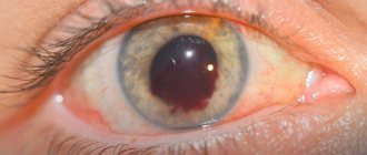
- Hemophthalmos. When hemorrhage occurs in the vitreous area, a small spot of a rich brown hue may appear. It is located behind the lens. In the presence of hemophthalmos, visual acuity may significantly decrease;
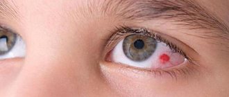
- Subconjunctival hemorrhage. The pathology is accompanied by the appearance of a bright red spot in the area of the eyeball;
- Preretinal hemorrhage, which occurs between the posterior part of the vitreous and the nerve endings located next to it.
Causes and types of retinal hemorrhages
Retinal hemorrhages occur due to the following reasons:
- Mechanical trauma (blunt and penetrating wounds, contusions);
- Closure of the lumen of blood vessels with a thrombus or atherosclerotic plaque;
- Their pathological fragility and high permeability (including against the background of systemic diseases);
- Hypertensive and diabetic crises.
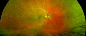
Fig. 1 Different types of retinal hemorrhages
Ophthalmologists distinguish several types of retinal hemorrhages, depending on the location and appearance when examining the fundus:
- Streak-shaped (“tongues of flame”) - the cut extends into a layer rich in nerve fibers - they resemble strokes, and at the macula (the central point of the retina at which rays of light intersect) and the disc they look like radial stripes;
- Round hemorrhage - located, as a rule, in the middle layers of the retina - looks like small circles of bright purple or red color with clear edges;
- Preretinal - located between the posterior hyaloid membrane of the vitreous body and the layer of nerve fibers. Looks like a big puddle. Its size is equal to 4-5 diameters of the optic nerve head. It determines the horizontal level of separation of formed elements and blood plasma;
- Subretinal - localized between the layers of the neuroepithelium and retinal pigment epithelium. It is darker than retinal hemorrhage, without clear contours;
- Choroidal – has a dark red color with a bluish tint;
- Retrochoroidal - arterial bleeding, which is called expulsive hemorrhage.
Diagnostic procedures in the presence of hemorrhage
To establish an accurate diagnosis, the following diagnostic techniques can be used:
- Retinoscopy and ophthalmoscopy, necessary to examine the fundus of the patient;
- Ultrasonography;
- Fluorescein angiography of the retina. During a detailed examination of the relevant vessels, a contrast agent is injected into the patient’s body, which makes it possible to detect the source of bleeding;
- Determination of visual acuity to identify the degree of visual impairment;
- Optical coherence tomography. Using this scanning technique, high-precision images of the retina can be obtained.
Ways to combat retinal hemorrhage
If there is excessive bleeding, surgery (vitrectomy) may be performed. During surgery, the affected areas of the vitreous body and blood clots are removed. In most cases, vision recovery occurs in the first 2-4 weeks after surgery. If there are irreversible changes in the structure of the retina, vision may still remain insufficiently clear.
In case of minor hemorrhage, not caused by chronic diseases, it is recommended to provide rest to the eyes. You can sit for a while with your eyes closed. This will normalize blood circulation.
For hemorrhage in the retina of the eye, medications that have a hemostatic and vascular strengthening effect can be used. Such drugs include “Emoxipin”. This medication is recommended to be used under the supervision of a physician.
In some cases, the use of products containing vitamins K and E is indicated. These products have a beneficial effect on the walls of blood vessels and help improve blood clotting.
If there is retinal hemorrhage, the following medications may also be used:
- Corticosteroids;
- Medicines with anti-inflammatory properties;
- Medicines belonging to the group of diuretics;
- Vitamin and mineral preparations rich in antioxidants.
In some situations, in the presence of retinal hemorrhage, calcium therapy is performed. The procedure promotes blood resorption, helps relieve tissue swelling, and reduces inflammation.
How to treat bleeding in the eye
Subconjunctival hemorrhage in most cases does not require treatment and goes away on its own.
If you experience pain and discomfort, your doctor may prescribe decongestant and anti-inflammatory drops. For concomitant eye infections, antibacterial or antiviral drugs are prescribed. As a rule, subconjunctival hemorrhage resolves within 2 weeks, without complications.
In other cases of hemorrhages in the eye, immediate treatment is required in an eye hospital.
How to get rid of “floaters” before your eyes using folk remedies?
With hemorrhage in the retina of the eye, “floaters” often appear in front of the eyes. They can be combated not only with medications, but also with traditional methods.
If you have complaints about “floaters” in your eyes, your daily menu should include dishes that contain a lot of retinol and ascorbic acid. It is recommended to include hemp oil in the diet: the product is enriched with fatty acids.
To reduce the likelihood of retinal detachment, you can prepare an infusion of mistletoe. The medicinal plant has many beneficial properties. Products containing mistletoe can reduce intraocular pressure. To prepare an infusion of mistletoe, add 5 grams of the plant to 200 ml of boiled water. It is recommended to take 100 ml twice a day.
Berries such as blueberries and blueberries are effective in treating various eye diseases. Products are enriched with antioxidants. Berries help improve vision at night. Blueberries and blueberries are actively used in the complex therapy of myopia, glaucoma, and retinal detachment. The berries are used both fresh and in the form of a medicinal product (capsules). Blueberries help fight eye fatigue. The berries contain antioxidants that help strengthen fiber vessels.
Blackberries contain large amounts of ascorbic acid. It is endowed with a pronounced anti-inflammatory effect. With regular consumption of healthy berries, fluid outflow improves and intraocular pressure decreases.
Hawthorn is also useful for eye diseases. Products made on its basis improve blood circulation.
The process of preparing an elderberry-based infusion is quite simple:
- 20 grams of plant material should be poured with a liter of water;
- The resulting mixture is infused for 40 minutes;
- After the specified time, the product is filtered.
The finished infusion is used to treat the eyes. The procedure should be performed twice a day.
If there is hemorrhage in the retina of the eye, you can also take an infusion made according to the recipe below:
- 5 grams of buckwheat flowers are poured into 0.2 liters of hot water;
- The resulting mixture must be infused for three hours;
- After the specified time, the product is filtered.
It is recommended to consume 50 ml of infusion three times a day. The recommended course duration is 7 days.
Symptoms and signs
A person should be wary if he begins to see double.
Retinal hemorrhage shows clear signs that get worse with each stage. The disease primarily causes vision problems. Visual functions also depend on the localization of the process; macular hemorrhage is considered the most dangerous. First, blurriness and double vision appear, then visual acuity decreases. Symptoms can be rapid or take a long time to develop. During the active phase, the patient exhibits the following symptoms:
- redness of the eyes of various localizations;
- pressing pain in the eyes;
- increased eye pressure;
- the effect of “fly spots” before the eyes;
- change in color perception.
It should be noted that retinal hemorrhages often occur in only one eye. A person feels this by a sharp deterioration in visual acuity. Carrying out ophthalmoscopy allows the ophthalmologist to identify internal symptoms of the pathological process, which can manifest themselves in the form of tortuosity or dilation of veins, vascular microaneurysms.
- Perimetry,
- Ophthalmoscopy,
- Visometry,
- Fluorescein angiography of the retina,
- Computed tomography of the retina,
- Blood pressure measurement;
- ECG.
In addition, the patient is prescribed urine and blood tests for sugar and RW. A consultation with a therapist is mandatory.
The disease is widespread and can be caused either by mechanical trauma to the upper body or as a complication of other diseases. Any manifestation of pathology requires immediate intervention by specialists in order to avoid retinal detachment.
Retinal hemorrhage is bleeding into the retinal tissue resulting from damage to the walls of the eye vessels.
- Causes
- Symptoms
- Treatment
One of the main causes of hemorrhage is trauma to the head or visual organs. Hemorrhage into the organs of vision has three degrees of severity.
Lightweight
With a mild degree of damage, pronounced symptoms do not appear. The structure of the eyeball does not change its shape. Sometimes swelling may occur in the cornea area. At this stage, vision almost does not lose its sharpness.
Average
At the middle stage, certain tissues of the eyeball are affected. Blood in the vessels can spread under the conjunctival layer and cause it to rupture. There is a partial deterioration in vision. Irritation occurs when the light is too bright.
Heavy
Severe damage is characterized by changes in the structure of the eye tissues. In some areas of the retina, tears and detachments may form. There is a small chance of vitreous displacement. The quality of vision is rapidly deteriorating.
Even the most minor injury to the upper body can lead to the development of serious pathologies in the eyeball. In some cases, the obvious symptoms of the disease may not correspond to the real threat. Very often, hemorrhage is accompanied by a concussion.
Also, retinal hemorrhage can be a complication of the following diseases:
- Hypertension. Constantly high blood pressure leads to the fact that the vascular system cannot cope with the large amount of incoming substances. Its walls become thinner, and fluid begins to accumulate under the retina.
- Atherosclerosis. With this disease, damaged vessels change their structure. Changes occurring in the vascular system make it less elastic, which affects its susceptibility to any external influences.
- Diabetes. During this chronic disease, blood sugar increases several times. This leads to the fact that the vessels begin to enlarge, and their walls become more weakened.
- Connective tissue diseases also play a large role in the general condition of the vascular system. With the development of such diseases, the risk of fluid accumulation in the retina, which is passed through the walls of blood vessels, increases.
- Diseases associated with the circulatory system. Due to pathologies that result in changes in the composition of the blood and its coagulability, the functioning of the vascular system is disrupted.
Retinal hemorrhage is quite common and can be a consequence of both mechanical trauma and certain diseases.
In addition, the cause of hemorrhage in the retina of the eye can be diseases of the visual organs. The formation of tumors inside the eyeball can lead to compression of the vascular and venous system, resulting in hemorrhage. In addition, a high degree of myopia also affects the formation of pathology.
Recipe for a folk remedy for subconjunctival bleeding
If the patient has subconjunctival bleeding, you can use the recipe below:
- 30 grams of carefully crushed chicory roots are poured into 1.5 liters of water;
- The product should be brought to a boil over medium heat;
- After this, the drink is infused for 30 minutes.
It is recommended to take 0.1 liter of the product three times a day. A medicinal infusion prepared from chicory roots is used to apply lotions to the affected eye. The average course duration depends on the severity of the pathology.
The importance of preventive measures
To prevent hemorrhage into the tissue, you must follow the recommendations listed below:
- You should visit an ophthalmologist regularly;
- It is necessary to promptly treat the patient’s cardiovascular diseases;
- Patients who have been diagnosed with diabetes need to monitor their blood glucose levels;
- It is recommended to give up bad habits and eliminate junk food from your diet.
In order to reduce the likelihood of hemorrhage in the retinal area, you should adjust your diet. Foods rich in fatty acids have a beneficial effect on the condition of the visual organs. You can compensate for the deficiency of relevant substances using the following products:
- Olive oils;
- Orekhov.
Products containing ginkgo biloba help strengthen the retina and neutralize the activity of free radicals. They improve vision and activate blood circulation in the area of the optic nerve.
It is worth paying special attention to foods rich in vitamin A. Large amounts of this substance are present in palm oil, carrots, and fish oil.
Prevention
Retinal hemorrhage can be prevented. To do this, follow simple preventive recommendations, which include:
- exclusion of exposure to provoking factors;
- weight normalization;
- sufficient physical activity;
- rational nutrition with limitation of fatty, fried foods;
- giving up bad habits, including smoking and systematic drinking of alcohol;
- reducing the load on the organ of vision.
If a person knows that the strength of his vascular walls is reduced, then he needs to avoid sudden movements, turning his head, and bending. At the same time, for preventive purposes, it is recommended to take vitamin C and rutin, which strengthen the walls of blood vessels.
Hemorrhage in the retina can be dangerous for vision, so to prevent vision problems, you should consult a doctor at the first sign of changes.
Author of the article: Igor Mikhailovich Krivoguz, specialist for the website glazalik.ru Share your experience and opinion in the comments.